



Suprafenacine

Based on 1 Customer Validation
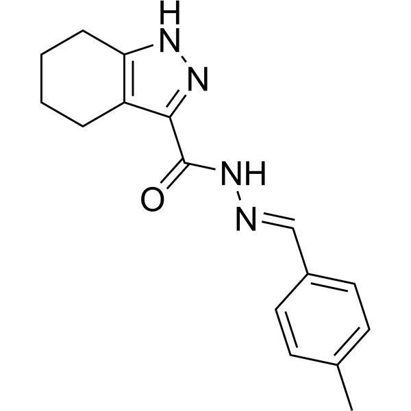
Suprafenacine is a cell permeable, tubulin-destabilizing molecule which bind microtubules at the colchicine-binding site and inhibit polymerization. Suprafenacine can induce G2/M cell cycle arrest and apoptosis, and can be used for cancer research.
For research use only. We do not sell to patients.
- Purity : 98.0%
- CAS No.: 1477482-50-0
- Formula: C16H18N4O
- Molecular Weight:282.34
-
Storage:Powder -20°C, 3 years ; In solvent -80°C, 6 months , -20°C, 1 month

To place orders, for customer services and technical support, please contact: MedChemExpress USA
Tel: 609-228-6898 E-mail: [email protected] [email protected]


-
 Biological Activity
-
Chemical Information
-
Solvent & Solubility
- Protocols
- Purity & Documentation
- References
-
 Help & FAQs


-
HY-135960
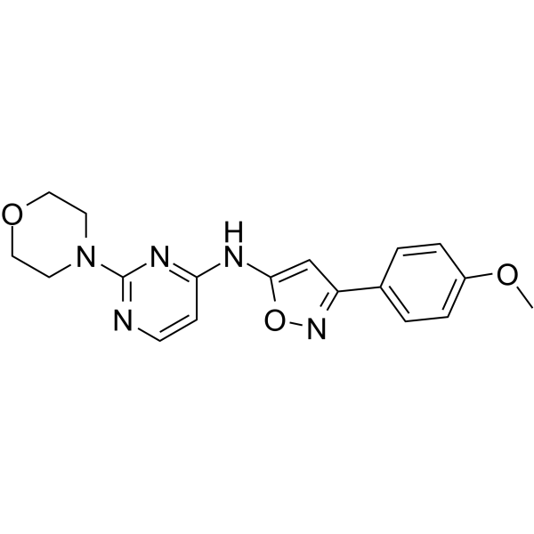
BO-264TACC3 Inhibitor -
HY-120786
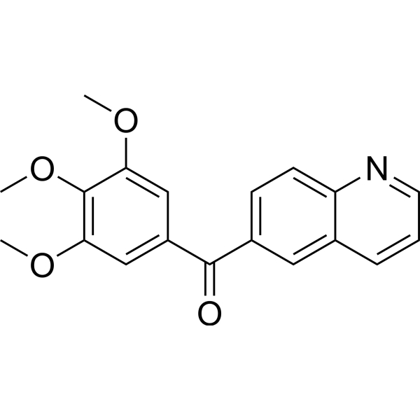
MPT0B014Tubulin Polymerization Inhibitor -
HY-N0282
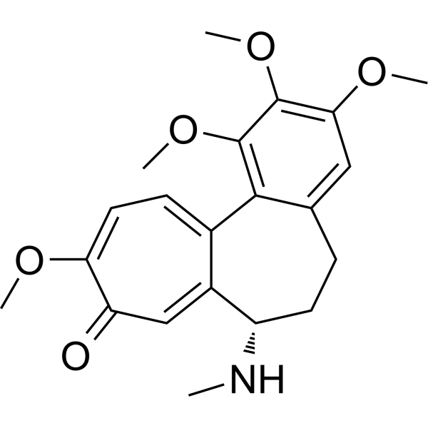
DemecolcineTubulin Inhibitor -

HY-N0282R
Demecolcine (Standard)Analytical Standard -
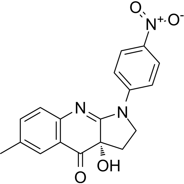
HY-120870
para-NitroblebbistatinMyosin II Inhibitor -
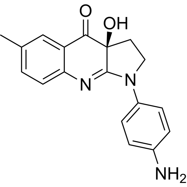
HY-111474
Para-aminoblebbistatinMyosin II Inhibitor -
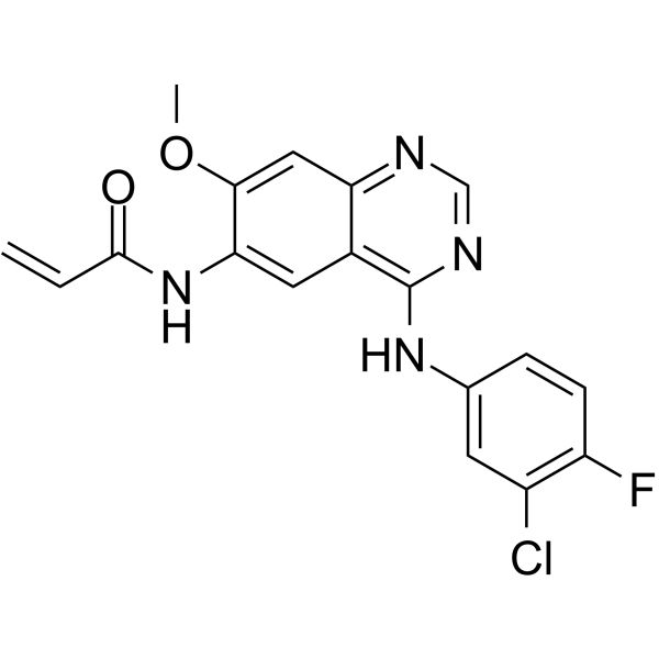
HY-101450
PF-6274484EGFR Inhibitor -
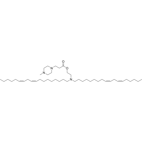
HY-150115
Lipid 10ionisable aminolipid -

HY-133885
S-(-)-7-Desmethyl-8-nitro blebbistatin(-)-Blebbistatin Analog -

HY-137139
10'-DesmethoxystreptonigrinAntibiotic -

HY-133885A
R-(-)-7-Desmethyl-8-nitro blebbistatin -

HY-129452
(S)-3'-Hydroxy blebbistatinMyosin II Inhibitor


Biological Activity
Description
In Vitro
Suprafenacine (10 μM, 24 h) causes a complete collapse of all the cells at the G1 phase and increases the G2/M population of the Hela cells while 7 h of pretreatment induces rapid apoptosis in proliferating Hela cells[1].
MedChemExpress (MCE) has not independently confirmed the accuracy of these methods. They are for reference only.
In Vivo
MedChemExpress (MCE) has not independently confirmed the accuracy of these methods. They are for reference only.


Chemical Information
-
CAS No. 1477482-50-0
-
Appearance Solid
-
Molecular Weight 282.34
-
Formula C16H18N4O
-
Color White to off-white
-
SMILES
O=C(N/N=C/C1=CC=C(C=C1)C)C2=NNC3=C2CCCC3
-
Synonyms
SRF
-
Shipping
Room temperature in continental US; may vary elsewhere.
-
Storage
Powder -20°C 3 years In solvent -80°C 6 months -20°C 1 month


Solvent & Solubility
In Vitro:
DMSO : 10 mg/mL (35.42 mM; Need ultrasonic and warming; Hygroscopic DMSO has a significant impact on the solubility of product, please use newly opened DMSO)
Please refer to the solubility information to select the appropriate solvent. Once prepared, please aliquot and store the solution to prevent product inactivation from repeated freeze-thaw cycles.
Storage method and period of stock solution: -80°C, 6 months; -20°C, 1 month. When stored at -80°C, please use it within 6 months. When stored at -20°C, please use it within 1 month.
Please refer to the solubility information to select the appropriate solvent. Once prepared, please aliquot and store the solution to prevent product inactivation from repeated freeze-thaw cycles.
Storage method and period of stock solution: -80°C, 6 months; -20°C, 1 month. When stored at -80°C, please use it within 6 months. When stored at -20°C, please use it within 1 month.
Concentration (start) × Volume (start) = Concentration (final) × Volume (final)


Purity & Documentation
-
Data Sheet (273 KB)
-
SDS (251 KB)
- English - EN (251 KB)
- Français - FR (251 KB)
- Deutsch - DE (251 KB)
- Norwegian - NO (251 KB)
- Español - ES (251 KB)
- Swedish - SV (251 KB)
- Italian - IT (251 KB)
- Korean - KR (251 KB)
- Portuguese - PT (251 KB)
-
Handling Instructions (2659 KB)
References


Complete Stock Solution Preparation Table
Please refer to the solubility information to select the appropriate solvent. Once prepared, please aliquot and store the solution to prevent product inactivation from repeated freeze-thaw cycles.
Storage method and period of stock solution: -80°C, 6 months; -20°C, 1 month. When stored at -80°C, please use it within 6 months. When stored at -20°C, please use it within 1 month.
| Optional Solvent | Concentration Solvent Mass | 1 mg | 5 mg | 10 mg | 25 mg |
|---|---|---|---|---|---|
| DMSO | 1 mM | 3.5418 mL | 17.7091 mL | 35.4183 mL | 88.5457 mL |
| 5 mM | 0.7084 mL | 3.5418 mL | 7.0837 mL | 17.7091 mL | |
| 10 mM | 0.3542 mL | 1.7709 mL | 3.5418 mL | 8.8546 mL | |
| 15 mM | 0.2361 mL | 1.1806 mL | 2.3612 mL | 5.9030 mL | |
| 20 mM | 0.1771 mL | 0.8855 mL | 1.7709 mL | 4.4273 mL | |
| 25 mM | 0.1417 mL | 0.7084 mL | 1.4167 mL | 3.5418 mL | |
| 30 mM | 0.1181 mL | 0.5903 mL | 1.1806 mL | 2.9515 mL |


Suprafenacine Related Classifications
HY-118748 Related Classifications
 Powered by Bioz