



Calcitroic acid

Based on 1 Customer Validation
Calcitroic acid is a water-soluble terminal vitamin D metabolite and also a ligand for vitamin D receptor (VDR). Calcitroic acid binds to the ligand-binding domain of VDR, forms a retinoic X receptor complex, recruits the coactivator peptide MED1, mediates partial VDR-dependent transcription, inhibits Calcitriol (HY-10002)-induced VDR activation, and reduces the transcription level of CYP24A1 in the presence of 1α,25-dihydroxyvitamin D3. Calcitroic acid exerts selective activating effects on VDR, upregulates the expression of CYP24A1 and CYP3A4, decreases the transcription levels of iNOS and IL-1β, reduces the secretion of nitric oxide and IL-1β, and possesses metabolic stability due to its resistance to phase I oxidation and hepatic glucuronidation. Calcitroic acid can be used in research related to colon cancer, inflammatory bowel disease and rickets.
For research use only. We do not sell to patients.
- Purity : 85.0%
- CAS No.: 71204-89-2
- Formula: C23H34O4
- Molecular Weight:374.51
-
Storage:
-80°C, protect from light, stored under nitrogen


To place orders, for customer services and technical support, please contact: MedChemExpress USA
Tel: 609-228-6898 E-mail: [email protected] [email protected]


-
 Biological Activity
-
Chemical Information
- Protocols
- Purity & Documentation
- References
-
 Help & FAQs


-
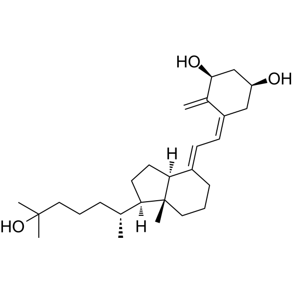
HY-10002A
(1S)-Calcitriol1α,25(OH)2D3 Metabolite -
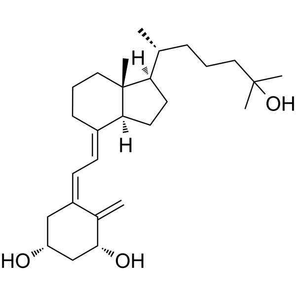
HY-13292
Impurity B of CalcitriolDiastereomer -
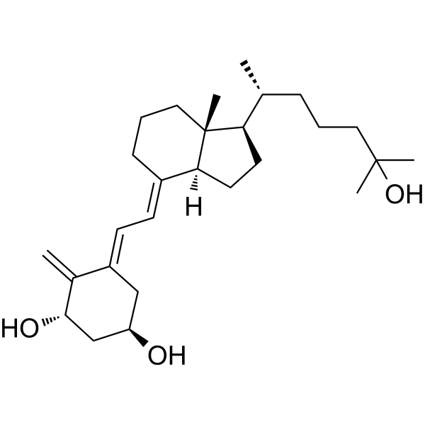
HY-75041
Calcitriol Impurities AImpurity Of Calcitriol -
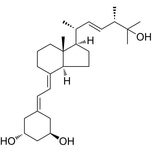
HY-50919
ParicalcitolVitamin D Receptor Agonist -
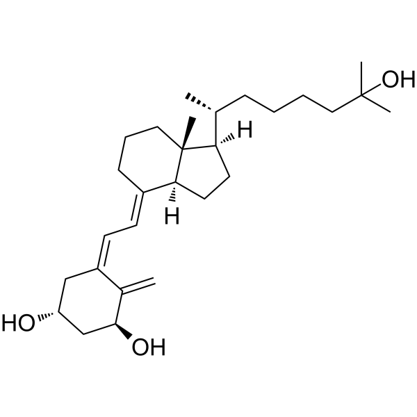
HY-77274
Calcitriol Impurities DVD/VDR Activator -
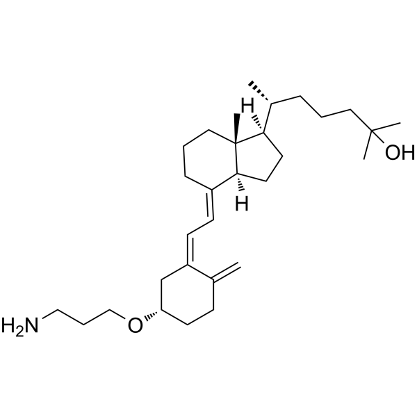
HY-15254
3-O-(2-Aminoethyl)-25-hydroxyvitamin D3Vitamin D3 Derivative -
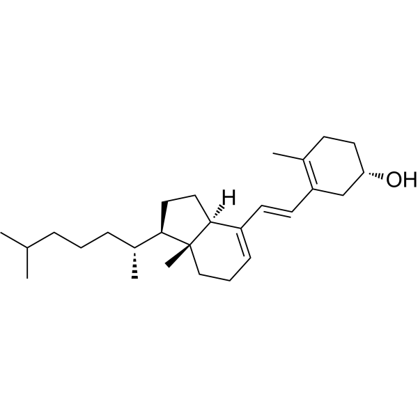
HY-130705A
Tachysterol 3Vitamin D3 Analog -
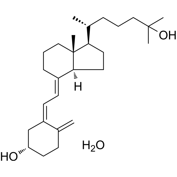
HY-32351A
Calcifediol monohydrateVDR Ligand -
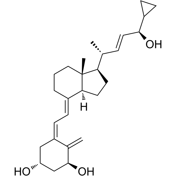
HY-15266
24R-CalcipotriolVitamin D Analog -
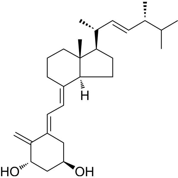
HY-75992
trans-DoxercalciferolVitamin D Receptor Activator -

HY-13292R
Impurity B of Calcitriol (Standard)VD/VDR Activator -
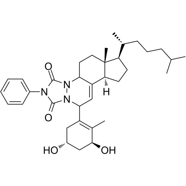
HY-13294
Impurity C of AlfacalcidolAlfacalcidol Impurity


Biological Activity
Description
Cellular Effect
|
Cell Line

|
Type | Value | Description | References |
|---|---|---|---|---|
| HEK-293T | EC50 |
0.87 μM
Compound: Calcitroic Acid
|
Agonist activity at VP16 tagged-VDR-LBD (unknown origin) expressed in HEK293T cells assessed as SRC1 coactivator peptide recruitment after 16 hrs by luciferase reporter gene based two hybrid assay
Agonist activity at VP16 tagged-VDR-LBD (unknown origin) expressed in HEK293T cells assessed as SRC1 coactivator peptide recruitment after 16 hrs by luciferase reporter gene based two hybrid assay
|
[PMID: 26774929] |
| HEK-293T | IC50 |
>100 μM
Compound: Calcitroic Acid
|
Antagonist activity against VP16 tagged-VDR-LBD (unknown origin) expressed in HEK293T cells assessed as inhibition of 1,25-dihydroxyvitamin D3-induced SRC1 coactivator peptide recruitment after 16 hrs by luciferase reporter gene based two hybrid assay
Antagonist activity against VP16 tagged-VDR-LBD (unknown origin) expressed in HEK293T cells assessed as inhibition of 1,25-dihydroxyvitamin D3-induced SRC1 coactivator peptide recruitment after 16 hrs by luciferase reporter gene based two hybrid assay
|
[PMID: 26774929] |
In Vitro
Calcitroic acid (up to 150 μM; 30 min) potently inhibits VDR-LBD interaction with SRC2-3 coactivator peptide in a cell-free system with an IC50 of 2.29 μM, but does not act as an agonist for this interaction[1].
Calcitroic acid (up to 150 μM; 16 h) activates VDR-mediated recruitment of SRC1 in HEK-293T cells with an EC50 of 0.87 μM and 55% efficacy relative to 10 nM 1,25(OH)2D3[1].
Calcitroic acid (up to 150 μM; 18 h) is non-toxic to HEK-293T cells at concentrations up to 150 μM, with an LD50 >100 μM[1].
Calcitroic acid (7.5 μM; 18 h) induces VDR-mediated CYP24A1 gene expression in DU145 prostate cancer cells when administered at 7.5 μM for 18 hours[1].
Calcitroic acid binds to VDR LBD in an agonistic conformation that supports coactivator peptide recruitment, with hydrogen bonding interactions largely matching those of 1,25(OH)2D3 except for the absence of a His333 interaction[2].
Calcitroic acid (18 h) acts as a VDR agonist in HEK293 cells with an EC50 of 2.89 μM, and its apparent antagonistic activity in the presence of 1,25(OH)2D3 is due to cytotoxicity rather than receptor antagonism[2].
Calcitroic acid (1-50 μM; 16 h for LXRα/β, RARα assays) selectively activates the vitamin D receptor and does not interact with 14 other tested nuclear receptors at concentrations up to 50 μM[2].
Calcitroic acid (10 μM; 18 h) upregulates the VDR target gene CYP24A1 in Caco2 cells with similar efficacy as 20 nM 1,25(OH)2D3, confirming its activity as a full VDR agonist[2].
Calcitroic acid (1 h) is stable against phase I oxidative metabolism by human and mouse liver microsomes, confirming it is the true end product of the vitamin D 25-oxidative pathway[3].
Calcitroic acid (10 μM; 2 h) is stable against phase II glucuronidation by human and mouse liver microsomes, suggesting it does not undergo this conjugation pathway in hepatic tissue[3].
Calcitroic acid (1 h) binds to recombinant vitamin D receptor ligand-binding domain with an IC50 of 8.5 μM, competing with the agonist LG190178 (HY-117182)[4].
Calcitroic acid (16 h) acts as a partial agonist/antagonist of vitamin D receptor-mediated transcription in HEK293T cells, with an EC50 of 2.5 μM for agonism and an IC50 of 3.2 μM for antagonism in the presence of calcitriol[4].
Calcitroic acid (6 μM; 24 hours) specifically upregulates CYP3A4 mRNA expression in Caco2 human colon cancer cells[4].
Calcitroic acid (10 μM; 24 hours) upregulates both CYP3A4 and CYP24A1 mRNA expression in HIEC-6 human normal intestinal epithelial cells[4].
Calcitroic acid (20 μM; 0-120 minutes) is stable to phase I metabolism by human liver microsomes over 2 hours and does not inhibit Calcitriol (HY-10002) metabolism[4].
Calcitroic acid (1.40 μM; 96 h) is identified as a major terminal water-soluble metabolite produced from 1α,25-(OH)2D2 metabolism in human HPK1A-ras keratinocyte cells, confirmed via chromatographic comigration and mass spectral matching[6].
Calcitroic acid (5-20 μM; 18 h) reduces nitric oxide production, pro-inflammatory gene transcription, and IL-1β protein secretion in IFNγ/LPS-activated RAW264.7 macrophages, with anti-inflammatory effects similar to 20 nM 1,25(OH)2D3 and no cytotoxicity in this cell line[2].
Calcitroic acid (20 μM; 18 h) reduces nitric oxide production in IFNγ- and LPS-stimulated RAW264.7 murine macrophages, indicating anti-inflammatory activity[4].
MedChemExpress (MCE) has not independently confirmed the accuracy of these methods. They are for reference only. Further protocols information, click here.
-
Cell Line:HEK-293T kidney cells
-
Concentration:Up to 150 μM
-
Incubation Time:18 h
-
Result:Showed no significant cytotoxicity in HEK-293T cells, with an LD50 >100 μM.
-
Cell Line:DU145 prostate cancer cells
-
Concentration:7.5 μM
-
Incubation Time:18 h
-
Result:Induced expression of the VDR target gene CYP24A1 in DU145 cells, with a higher transcriptional activation magnitude than LCA.
-
Cell Line:Caco2
-
Concentration:10 μM
-
Incubation Time:18 h
-
Result:Induced CYP24A1 gene expression to a similar level as 20 nM 1,25(OH)2D3, with fold induction comparable to the positive control and significantly higher than lithocholic acid at 10 μM.
Did not antagonize the induction of CYP24A1 by 1,25(OH)2D3.
-
Cell Line:Caco2 human colon cancer cells
-
Concentration:6 μM
-
Incubation Time:24 h
-
Result:Specifically upregulated CYP3A4 mRNA expression, with no significant effect on CYP2B6 or CYP2C9 mRNA levels.
-
Cell Line:HIEC-6 human normal intestinal epithelial cells
-
Concentration:10 μM
-
Incubation Time:24 h
-
Result:Upregulated both CYP3A4 and CYP24A1 mRNA expression, with a more pronounced effect than 10 μM lithocholic acid.
In Vivo
Calcitroic acid (25-50 ng; s.c.; three times daily; 7 days) exhibits weak antirachitic activity, with a statistically significant epiphyseal plate calcification score of 1.75 at a 50 ng per dose, three-times-daily subcutaneous regimen for 7 days in vitamin D-deficient, rachitic rats[7].
Calcitroic acid (52-2000 ng; i.v.; single dose) induces weak bone calcium mobilization, with significant serum calcium elevation above control values at 6 hours post-dose following single intrajugular doses of 500 ng and 2000 ng, but no effect on intestinal calcium transport in vitamin D-deficient, hypocalcemic rats[7].
MedChemExpress (MCE) has not independently confirmed the accuracy of these methods. They are for reference only.
-
Animal Model:unspecified[4]
-
Dosage:50 ng/animal
-
Administration:s.c.; daily; 7 days
-
Result:Induced calcification of the epiphyseal plate, which was 3 times less potent than the effect of 5 ng/animal calcitriol.
-
Animal Model:Holtzman rats (male, weanling; maintained on low phosphorus vitamin D-deficient diet for 2 weeks to induce rickets)[7]
-
Dosage:25 ng; 50 ng
-
Administration:s.c.; three times daily; 7 days
-
Result:Resulted in a calcification score of 0.64, which was not statistically significant compared to controls.
Resulted in a statistically significant calcification score of 1.75.
-
Animal Model:Holtzman rats (male, weanling; maintained on low calcium vitamin D-deficient diet for 3 weeks to induce hypocalcemia)[7]
-
Dosage:52 ng; 500 ng; 2000 ng
-
Administration:i.v.; single dose
-
Result:Did not significantly elevate serum calcium at 6 or 24 hours post-dose, nor did it increase intestinal calcium transport.
Significantly elevated serum calcium to 4.5 mg/100 mL at 6 hours post-dose (control: 4.3 mg/100 mL).
Significantly elevated serum calcium to 4.4 mg/100 mL at 6 hours post-dose, and to 4.4 mg/100 mL at 4 hours post-dose (control: 4.0 mg/100 mL); no significant increase in intestinal calcium transport was observed.


Chemical Information
-
CAS No. 71204-89-2
-
Appearance Solid
-
Molecular Weight 374.51
-
Formula C23H34O4
-
Color White to light yellow
-
SMILES
C[C@@]12[C@](CC[C@]2([H])[C@H](C)CC(O)=O)([H])/C(CCC1)=C/C=C3C([C@H](C[C@@H](C\3)O)O)=C
-
Structure Classification
-
Initial Source
-
Shipping
Shipping with dry ice.
-
Storage
-80°C, protect from light, stored under nitrogen

Protocols
-
RNA extraction experimental
By lysing cells, releasing RNA, and removing impurities such as proteins and DNA, high-purity RNA products are finally obtained. The commonly used traditional method is the guanidine isothiocyanate/phenol/chloroform method (Trizol), which is suitable for a variety of animal materials including animal tissues, microorganisms, cultured cells, etc., and most plant materials.
-
DSS-Induced Colitis
Dextran sulfate sodium (DSS)-induced colitis is generated by administering DSS in mouse drinking water, producing epithelial injury, barrier disruption, weight loss, diarrhea, fecal blood, colon shortening, histologic mucosal damage, and inflammatory mediator changes; the model is mainly used to study acute or chronic intestinal inflammation resembling selected features of ulcerative colitis. DSS injury is interpreted through clinical and tissue readouts rather than a single molecular endpoint: daily body weight, stool consistency, and bleeding are combined into a disease activity index, while colon length, histology, cytokines, myeloperoxidase activity, intestinal permeability, and tight-junction markers provide complementary measures of inflammation and barrier damage.
-
TNBS-Induced Colitis
TNBS-induced colitis is produced by intrarectal delivery of 2,4,6-trinitrobenzene sulfonic acid in ethanol, where ethanol disrupts the mucosal barrier and TNBS haptenates colonic proteins, generating immune-mediated colonic inflammation with weight loss, diarrhea, ulceration, transmural injury, inflammatory-cell infiltration, and cytokine responses. The model is used as an experimental intestinal inflammation model with Crohn’s disease–like features, especially when Th1-type responses, IL-12–dependent inflammation, chronic relapsing inflammation, or fibrosis-related endpoints are studied.
-
Real Time qPCR (Q-PCR)
Real-time quantitative PCR (qPCR) quantifies an amplifiable nucleic-acid target by monitoring fluorescence during PCR cycling rather than measuring product only after amplification. The increase in fluorescence tracks accumulation of PCR product, and the quantification cycle (Cq; historically also Ct/CP) is related to the initial amount of target: samples containing more starting target generally reach the defined fluorescence threshold in fewer cycles.
-
Pyroptosis Solutions
Pyroptosis is a lytic inflammatory cell-death pathway executed by gasdermin pores, most classically through inflammasome-mediated activation of caspase-1, cleavage of gasdermin D, membrane pore formation, LDH release, and secretion of IL-1β and IL-18. The canonical pathway is commonly modeled by priming cells with an inflammatory signal such as LPS to induce pro-IL-1β and inflammasome components, followed by an activation signal such as ATP or nigericin to activate NLRP3, ASC speck formation, caspase-1 cleavage, GSDMD cleavage, cytokine release, and pyroptotic membrane rupture. The non-canonical pathway is triggered when cytosolic LPS activates mouse caspase-11 or human caspase-4/5, leading to GSDMD cleavage and pyroptosis, and this can secondarily activate NLRP3-dependent IL-1β release. Pyroptosis is linked to inflammatory injury, infection, cancer, liver disease, ocular disease, placental inflammation, and other disease phenotypes, but unresolved questions include which gasdermin fam


Purity & Documentation
-
Data Sheet (310 KB)
-
SDS (251 KB)
- English - EN (251 KB)
- Français - FR (251 KB)
- Deutsch - DE (251 KB)
- Norwegian - NO (251 KB)
- Español - ES (251 KB)
- Swedish - SV (251 KB)
- Italian - IT (251 KB)
- Korean - KR (251 KB)
- Portuguese - PT (251 KB)
-
Handling Instructions (2659 KB)
References
[1]. Teske KA, et al. Synthesis and evaluation of vitamin D receptor-mediated activities of cholesterol and vitamin D metabolites. European journal of medicinal chemistry. 2016 Feb 15;109:238-46. [Content Brief]
[2]. Yu OB, et al. Biological evaluation and synthesis of calcitroic acid. Bioorg Chem. 2021 Nov;116:105310. [Content Brief]
[5]. Yu OB, et al. Synthesis and biological evaluation of calcioic acid. Steroids. 2020 Feb;154:108536. [Content Brief]
[6]. Zimmerman DR, et al. Calcitroic acid is a major catabolic metabolite in the metabolism of 1 alpha-dihydroxyvitamin D(2). Arch Biochem Biophys. 2001 Aug 1;392(1):14-22. [Content Brief]

Calculators
Concentration (start) × Volume (start) = Concentration (final) × Volume (final)


Calcitroic acid Related Classifications
HY-N10508 Related Classifications
 Powered by Bioz