



Precemtabart

Based on 1 Customer Validation
Precemtabart is a humanized IgG1 antibody targeting CEACAM5, with selectivity for CEACAM5-positive cells. Precemtabart binds to CEACAM5 on tumor cells to deliver the topoisomerase 1 inhibitor Exatecan (HY-13631), and induces cell death by inhibiting the function of TOP1. Precemtabart inhibits the growth of CEACAM5-positive cancer cells. Precemtabart causes dose-dependent hematolymphoid and intestinal-related effects in cynomolgus monkeys. Precemtabart can be used in research related to metastatic colorectal cancer, metastatic pancreatic ductal carcinoma, advanced gastric cancer, advanced non-small cell lung cancer, and advanced pancreatic adenocarcinoma.
Nur für Forschungszwecke. Wir verkaufen nicht an Patienten.
- Reinheit : 99.00%
- CAS. Nr.: 2883118-92-9
- Molecular Weight:144.471 kDa
-
Speicherung:
Please store the product under the recommended conditions in the Certificate of Analysis.


To place orders, for customer services and technical support, please contact: MedChemExpress USA
Tel: 609-228-6898 E-mail: [email protected] [email protected]


-
 Biologische Aktivität
-
Chemical Information
- Protokoll
- Reinheit & Dokumentation
- Verweise
-
 Help & FAQs


-

HY-P99054
TusamitamabAnti-CEACAM5 Antibody -

HY-P99843
DatopotamabAnti-TROP2 Antibody -

HY-P99001
Human IgG1 kappa, Isotype ControlIgG1κ Isotype Control -

HY-P99058
ZolbetuximabClaudin-18.2 Inhibitor -
HY-153179
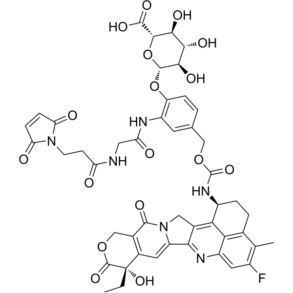
Mal-Gly-PAB-Exatecan-D-glucuronic acidADC Intermediate Compound -

HY-P990764
SigvotatugAnti-ITGB6 Antibody -

HY-P991218
Anti-IL11 Antibody (X203)Anti-IL11 Antibody -

HY-P991488
BI-765423Anti-IL-11 mAb
Alle Topoisomerase Isoform-spezifische Produkte anzeigen
More

Biologische Aktivität
Beschreibung
Isotype
Human IgG1 kappa
Recommend Isotype Controls
Species Reactivity
Human
IC50 & Target
CEACAM5/CEA/CD66e
In Vitro
Precemtabart (Serial dilutions; 144 h) potently inhibits CEACAM5-positive SK-CO-1 (IC50 = 0.09 nM) and MKN-45 (IC50 = 0.63 nM) cell viability, but has minimal activity against CEACAM5-negative MDA-MB-231 cells at concentrations below 10 nM[1].
Precemtabart (1 nM; 6 days) exhibits a potent bystander effect, inducing cell death in CEACAM5-negative MDA-MB-231 cells when co-cultured with CEACAM5-positive SK-CO-1 cells[1].
Precemtabart (Serial dilutions; 144 h) efficacy is unaffected by MDR1 inhibition in LS513 cancer cells[1].
MedChemExpress (MCE) has not independently confirmed the accuracy of these methods. They are for reference only.
-
Cell Line:CEACAM5-positive SK-CO-1, CEACAM5-positive MKN-45, CEACAM5-negative MDA-MB-231
-
Concentration:Serial dilutions
-
Incubation Time:144 h
-
Result:Potently inhibited cell growth in CEACAM5-positive SK-CO-1 and MKN-45 cell lines, with IC50 values of 0.09 nM and 0.63 nM, respectively.
Had minimal effects on the CEACAM5-negative MDA-MB-231 cell line, requiring concentrations greater than 10 nM for any considerable inhibition.
-
Cell Line:LS513
-
Concentration:Serial dilutions
-
Incubation Time:144 h
-
Result:Showed efficacy not affected by MDR1 inhibition in LS513 cell lines.
Clinical Trial
| NCT Number | Sponsor | Condition | Start Date |
Phase

|
|---|---|---|---|---|
| NCT01329991 | Plexxikon| | 2011-05 | PHASE1 |
Gene ID
Accession
Conjugated
Unconjugated
Reconsititution
The product can be reconstituted/diluted with sterile PBS or saline.
Format
-
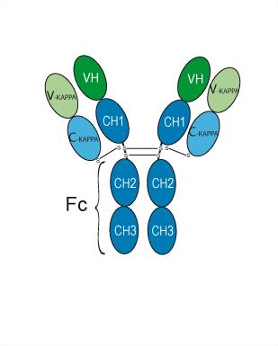
IgG1-kappa
Anwendung
ELISA, FACS, Functional assay
Verified Bioactivity
-
 Loaded Precemtabart on AHC2 biosensor, can bind CEACAM5 Protein, Human (HEK293, His, HY-P70675) with an affinity constant of 3.214E-09 M as determined in BLI assay.
 -
 Flow cytometric analysis of 1X10⁶ MCF-7 cells with Precemtabart (HY-P990940, red). Cells were fixed with 4% paraformaldehyde. Then stained with the primary antibody at 1/200 dilution for an hour at 4℃. Alexa Fluor 488-conjugated AffiniPure Goat Anti-Human IgG H&L (AF488) (HY-P83776) was used as the secondary antibody at 1/1,000 dilution for 30 minutes at 4℃. Human IgG1 kappa (HY-P99001, blue) was used as the isotype control, cells without incubation with primary antibody were used as the unlabeled control (black).


Chemical Information
-
CAS. Nr. 2883118-92-9
-
Appearance Liquid
-
Molecular Weight 144.471 kDa
-
Color Colorless to light yellow
-
SMILES
[Precemtabart]
-
Synonyms
MBE-748; M-9140 Antibody; MBE-2882 Antibody
-
Versand
Shipping with dry ice.
-
Formulation
Please refer to the lot-specific COA for specific buffer information.
-
Speicherung
Please store the product under the recommended conditions in the Certificate of Analysis.


Reinheit & Dokumentation
-
Data Sheet (260 KB)
-
SDS (252 KB)
- English - EN (252 KB)
- Français - FR (252 KB)
- Deutsch - DE (252 KB)
- Norwegian - NO (252 KB)
- Español - ES (252 KB)
- Swedish - SV (252 KB)
- Italian - IT (252 KB)
- Korean - KR (252 KB)
- Portuguese - PT (252 KB)
-
Inhibitory Antibodies User Guide (603 KB)
Verweise

Calculators
Konzentration (Stammlösung) × Volumen (Stammlösung) = Konzentration (Ziellösung) × Volumen (Ziellösung)


Precemtabart Related Classifications
HY-P990940 Related Classifications
 Powered by Bioz