



SZM679

Based on 1 Customer Validation
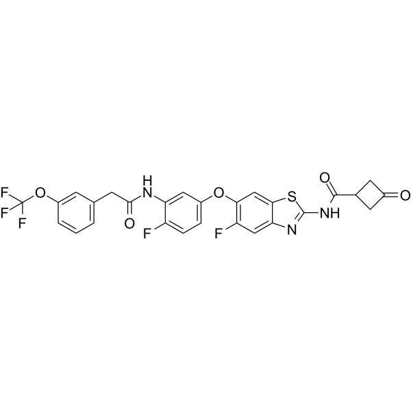
SZM679 is a potent, orally active and selective RIPK1 inhibitor with Kd values of 8.6 nM and >5000 nM for RIPK1 and RIPK3, respectively. SZM679 reverses the tumor necrosis factor-induced systemic inflammatory response. SZM679 decreases the Tau hyperphosphorylation, neuroinflammation, and the RIPK1 phosphorylation level in the hippocampus and cortex. SZM679 can be used in research of Alzheimer's disease (AD).
Nur für Forschungszwecke. Wir verkaufen nicht an Patienten.
- Reinheit : 99.59%
- CAS. Nr.: 3027645-52-6
- Formel: C27H18F5N3O5S
- Molecular Weight:591.51
-
Speicherung:Powder -20°C, 3 years ; In solvent -80°C, 6 months , -20°C, 1 month

To place orders, for customer services and technical support, please contact: MedChemExpress USA
Tel: 609-228-6898 E-mail: [email protected] [email protected]


-
 Biologische Aktivität
-
Chemical Information
-
Lösungsmittel & Löslichkeit
- Protokoll
- Reinheit & Dokumentation
- Verweise
-
 Help & FAQs




Biologische Aktivität
Beschreibung
IC50 & Target
[1]|
RIPK1 8.6 nM (Kd) |
RIPK3 >5000 nM (Kd) |
In Vitro
SZM679 (0-10 μM; 24 h; necrotic L929 and HT-29 cells) has an anti-necrosis activity by Inhibiting the RIPK1 pathway with an EC50 value of 2 nM. SZM679 also protects against TNF-α, cycloheximide, and z-VAD-fmk (TCZ)-induced necroptosis. SZM679 protects against necroptosis induced by TZ in a dose dependent manner[1].
SZM679 (1 μM; 6 h; necrotic HT-29 cells) selectively inhibits the expression of RIPK1 but not RIPK3 or MLKL. SZM679 blocks necrosome formation by inhibiting TSZ-induced phosphorylation of RIPK1[1].
MedChemExpress (MCE) has not independently confirmed the accuracy of these methods. They are for reference only. Further protocols information, click here.
-
Cell Line:Necrotic HT-29 cells
-
Concentration:1 μM
-
Incubation Time:6 hours
-
Result:Inhibited the phosphorylation of RIPK1 at 1 μM, resulting in the inhibition of the downstream phosphorylation of RIPK3 and MLKL.
In Vivo
SZM679 (1 mg/kg; administered intragastrically; once daily for 7 days) improves cognitive function in STZ-induced AD mice[1].
SZM679 (1 mg/kg; administered intragastrically; once daily for 7 days) rescues brain structure damage with no obvious toxicity and decreases AD biomarkers and decreases the expression levels of inflammatory cytokines and inhibits RIPK1 phosphorylation in the brain tissues of AD mice[1].
MedChemExpress (MCE) has not independently confirmed the accuracy of these methods. They are for reference only.
-
Animal Model:Male C57BL/6 J mice with TNF-induced SIRS models[1]
-
Dosage:10, 20, and 40 mg/kg
-
Administration:Intraperitoneal injection
-
Result:Protected mice in a dose-dependent manner from hypothermia and death.
-
Animal Model:Male C57BL/6 J mice with AD models[1]
-
Dosage:1 mg/kg
-
Administration:Administered intragastrically; once daily for 7 days
-
Result:Improved the anxiety, behavior, and exploratory ability of AD mice.
Improved the learning and memory ability of AD mice.
-
Animal Model:Male C57BL/6 J mice with AD models[1]
-
Dosage:1 mg/kg
-
Administration:Administered intragastrically; once daily for 7 days
-
Result:Rescued the damaged hippocampal structure of AD mice and restored the cell number and morphology.
Down-regulated the expression of the inflammatory cytokines, the IL-1β and TNF-α levels.


Chemical Information
-
CAS. Nr. 3027645-52-6
-
Appearance Solid
-
Molecular Weight 591.51
-
Formel C27H18F5N3O5S
-
Color Off-white to light yellow
-
SMILES
O=C(C1CC(C1)=O)NC2=NC3=CC(F)=C(OC4=CC=C(F)C(NC(CC5=CC=CC(OC(F)(F)F)=C5)=O)=C4)C=C3S2
-
Versand
Room temperature in continental US; may vary elsewhere.
-
Speicherung
Powder -20°C 3 years In solvent -80°C 6 months -20°C 1 month


Lösungsmittel & Löslichkeit
In Vitro:
DMSO : 125 mg/mL (211.32 mM; Need ultrasonic; Hygroscopic DMSO has a significant impact on the solubility of product, please use newly opened DMSO)
Please refer to the solubility information to select the appropriate solvent. Once prepared, please aliquot and store the solution to prevent product inactivation from repeated freeze-thaw cycles.
Storage method and period of stock solution: -80°C, 6 months; -20°C, 1 month. When stored at -80°C, please use it within 6 months. When stored at -20°C, please use it within 1 month.
Please refer to the solubility information to select the appropriate solvent. Once prepared, please aliquot and store the solution to prevent product inactivation from repeated freeze-thaw cycles.
Storage method and period of stock solution: -80°C, 6 months; -20°C, 1 month. When stored at -80°C, please use it within 6 months. When stored at -20°C, please use it within 1 month.
Konzentration (Stammlösung) × Volumen (Stammlösung) = Konzentration (Ziellösung) × Volumen (Ziellösung)

Protokoll
-
Kinase activity and phosphorylation assays
Kinase activity assays measure the ability of kinases to transfer phosphate groups from ATP to specific substrates, while phosphorylation assays detect the presence and levels of phosphorylated proteins. Common methods include radiolabeled ATP incorporation (e. g. ,), ADP release detection via bioluminescence (e. g. ,[3]), enzyme-linked immunosorbent assays (ELISA) for phospho-specific epitopes (e. g. ,[6]), and microtiter-based formats for high-throughput screening (e. g. ,[8]). The ADP-Glo assay quantifies kinase activity by measuring ADP produced during phosphorylation using a luciferase-based system. Radiometric assays involve autoradiography or scintillation counting after incorporation of 32P-labeled ATP into substrate proteins. ELISA-based approaches rely on phospho-specific antibodies to detect activated kinases in cell lysates or purified samples.
-
Western Blot
Western blotting (WB) is a commonly used experimental method in molecular biology, biochemistry, and immunogenetics for identifying and quantifying target proteins. It combines gel electrophoresis with immunoassay, enabling researchers to analyze protein expression, post-translational modifications, and molecular weight.
-
Research Protocol for Inflammation-related Diseases
The NLRP3 inflammasome is a cytosolic innate immune signaling platform that integrates priming signals and danger-signal activation to promote caspase-1 activation, maturation of IL-1β and IL-18, and gasdermin D-mediated pyroptotic cell death. The core experimental logic is to determine whether inflammatory disease phenotypes are driven by increased NLRP3 expression, ASC-containing inflammasome assembly, caspase-1 cleavage, GSDMD cleavage, and extracellular release of IL-1β/IL-18 rather than by nonspecific cell injury alone. The pathway is strongly linked to inflammation-related disease phenotypes because monosodium urate crystals activate NALP3/NLRP3 inflammasome signaling in gout-like crystal inflammation, cholesterol crystals activate NLRP3 inflammasomes in atherogenesis models, and DSS-induced intestinal inflammation has been reported to involve NLRP3 inflammasome activity. However, experimental colitis studies also show context-dependent protective effects of NLRP3 inflammasome co
-
Protocol for Kinase activity and phosphorylation assays
Kinase activity assays measure transfer of phosphate from ATP to a protein or peptide substrate, generating phosphorylated substrate, ADP, or incorporated radiolabeled phosphate as the readout; phosphorylation assays measure site-specific phosphorylation in cells or tissues as a proxy for kinase-pathway activation, inhibition, or substrate regulation. Phosphorylation can be detected by phospho-specific Western blot, immunoprecipitation kinase assay, phospho-immunofluorescence, phospho-flow cytometry, luminescent ADP detection, radiolabeled ATP incorporation, or reporter-based pathway assays, and these readouts can be applied to cancer cells, primary neurons, mouse tumors, organoids, inflammatory macrophages, ferroptosis studies, and mitophagy studies when the kinase target is biologically relevant.
-
Research Protocol for Neurological Diseases
PINK1/Parkin-mediated mitophagy pathway is a mitochondrial quality-control signaling axis in which mitochondrial depolarization stabilizes PINK1 on damaged mitochondria, activates Parkin recruitment and E3 ubiquitin ligase activity, promotes ubiquitination of outer mitochondrial membrane proteins, recruits selective autophagy adaptors, and drives lysosomal degradation of damaged mitochondria. In neurological disease research, this pathway is experimentally important because neurons, especially dopaminergic neurons, are highly dependent on mitochondrial integrity, and defective mitochondrial turnover can lead to mitochondrial dysfunction, oxidative stress, impaired neuronal survival, α-synuclein accumulation, and neuroinflammatory damage-associated signals. The genetic disease link is strongest in Parkinson’s disease because mutations in PRKN/parkin cause autosomal recessive juvenile parkinsonism, mutations in PINK1 cause hereditary early-onset Parkinson’s disease, and Drosophila studie
-
Alzheimer’s Disease Modeling
Alzheimer’s Disease (AD) is a neurodegenerative disorder characterized by a progressive decline in cognitive functions and loss of specific types of neurons and synapses. Alzheimer's symptoms can be simulated in mice by injecting drugs (such as Aβ) or genetically modified.


Reinheit & Dokumentation
-
Data Sheet (274 KB)
-
SDS (251 KB)
- English - EN (251 KB)
- Français - FR (251 KB)
- Deutsch - DE (251 KB)
- Norwegian - NO (251 KB)
- Español - ES (251 KB)
- Swedish - SV (251 KB)
- Italian - IT (251 KB)
- Korean - KR (251 KB)
- Portuguese - PT (251 KB)
-
Handling Instructions (2659 KB)
Verweise


Complete Stock Solution Preparation Table
Please refer to the solubility information to select the appropriate solvent. Once prepared, please aliquot and store the solution to prevent product inactivation from repeated freeze-thaw cycles.
Storage method and period of stock solution: -80°C, 6 months; -20°C, 1 month. When stored at -80°C, please use it within 6 months. When stored at -20°C, please use it within 1 month.
| Optional Solvent | Concentration Solvent Mass | 1 mg | 5 mg | 10 mg | 25 mg |
|---|---|---|---|---|---|
| DMSO | 1 mM | 1.6906 mL | 8.4529 mL | 16.9059 mL | 42.2647 mL |
| 5 mM | 0.3381 mL | 1.6906 mL | 3.3812 mL | 8.4529 mL | |
| 10 mM | 0.1691 mL | 0.8453 mL | 1.6906 mL | 4.2265 mL | |
| 15 mM | 0.1127 mL | 0.5635 mL | 1.1271 mL | 2.8176 mL | |
| 20 mM | 0.0845 mL | 0.4226 mL | 0.8453 mL | 2.1132 mL | |
| 25 mM | 0.0676 mL | 0.3381 mL | 0.6762 mL | 1.6906 mL | |
| 30 mM | 0.0564 mL | 0.2818 mL | 0.5635 mL | 1.4088 mL | |
| 40 mM | 0.0423 mL | 0.2113 mL | 0.4226 mL | 1.0566 mL | |
| 50 mM | 0.0338 mL | 0.1691 mL | 0.3381 mL | 0.8453 mL | |
| 60 mM | 0.0282 mL | 0.1409 mL | 0.2818 mL | 0.7044 mL | |
| 80 mM | 0.0211 mL | 0.1057 mL | 0.2113 mL | 0.5283 mL | |
| 100 mM | 0.0169 mL | 0.0845 mL | 0.1691 mL | 0.4226 mL |


SZM679 Related Classifications
HY-151873 Related Classifications
 Powered by Bioz