



Agatolimod
Based on 2 publication(s) in Google Scholar
Agatolimod ((ODN 2006; PF-3512676; CpG 7909)) is a TLR9 agonist and immunomodulator with an EC50 of 180 nM against human TLR9. Agatolimod activates and upregulates the expression of both TLR9 and TLR6, and mediates downstream signaling pathways via IRAK4, IRF5, IRF7. Agatolimod induces Th1-type innate and adaptive immune responses, activates various immune cells and promotes antigen presentation, regulates antibody levels and immune cell infiltration, upregulates the secretion of multiple cytokines, induces apoptosis and cell cycle arrest, enhances cytotoxicity, and clears intracellular Salmonella. Agatolimod is applicable to research on COVID-19, breast cancer, lung adenocarcinoma, HPV-related tumors, melanoma, and salmonellosis.
For research use only. We do not sell to patients.
- Purity : 98.84%
- CAS No.: 525625-52-9
- Molecular Weight:7699.00
-
Storage:
-20°C, sealed storage, away from moisture
* In solvent : -80°C, 6 months; -20°C, 1 month (sealed storage, away from moisture)


To place orders, for customer services and technical support, please contact: MedChemExpress USA
Tel: 609-228-6898 E-mail: [email protected] [email protected]


-
 Biological Activity
-
Chemical Information
-
Solvent & Solubility
- Protocols
- Purity & Documentation
- References
-
 Help & FAQs


-
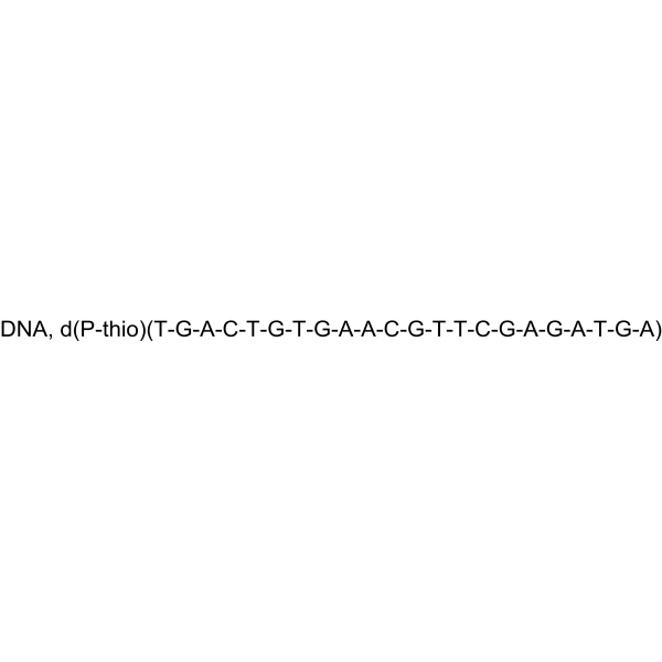
HY-150724
ODN 1018TLR9 Agonist -
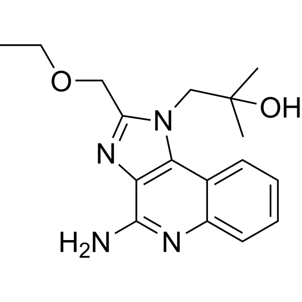
HY-13740
ResiquimodTLR7/8 Agonist -

HY-150741
ODN 2216TLR9 Ligand -

HY-146245
ODN 1826TLR9 Agonist -
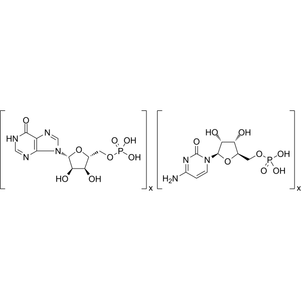
HY-107202
Polyinosinic-polycytidylic acidTLR-3 Agonist -

HY-10219
RapamycinmTOR Inhibitor -
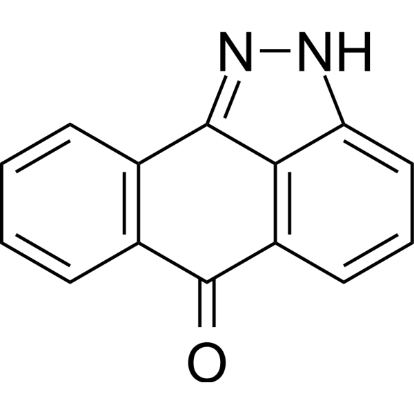
HY-12041
SP600125JNK Inhibitor -
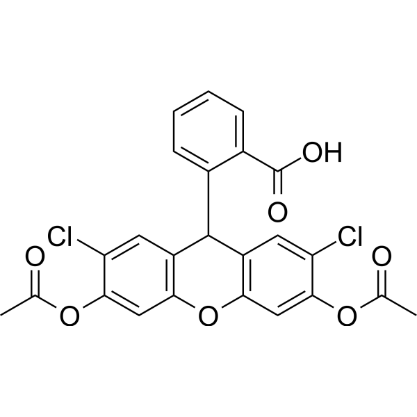
HY-D0940
H2DCFDAROS-Sensitive Probe -
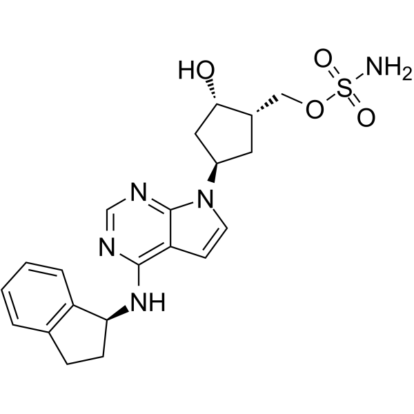
HY-70062
PevonedistatNAE Inhibitor -

HY-13259
MG-132Proteasome Inhibitor -
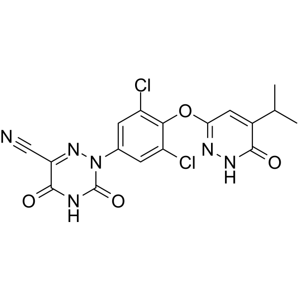
HY-12216
ResmetiromTHR-β Agonist -
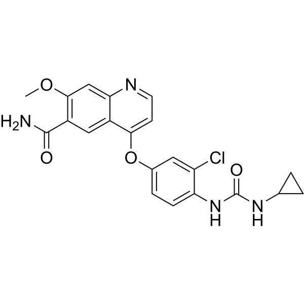
HY-10981
LenvatinibVEGFR/FGFR Inhibitor
Publications Citing Use of MedChemExpress (MCE) Agatolimod
More

Biological Activity
Description
IC50 & Target
|
TLR9 |
iNOS |
In Vitro
Agatolimod (10 μM) enhances expression of immunostimulatory cytokine IL-12p70 and chemoattractant cytokines in THP-1 cells, supporting its adjuvant activity[2].
Agatolimod (10 μg/mL; 14 days) increases the radiosensitivity of R-A549 cells, as evidenced by a reduced D0 value of 1.33 Gy in clonogenic assays when combined with radiation[3].
Agatolimod stimulates TLR9 signaling in HEK-Blue-hTLR9 cells with an EC50 of 180 nM[4].
Agatolimod (1-6 μM; 24 h) increases K55 cell surface marker expression in HD11 chicken macrophage cells in a dose-dependent manner (1-3 μM) but does not affect MHC class I or II expression[5].
MedChemExpress (MCE) has not independently confirmed the accuracy of these methods. They are for reference only. Further protocols information, click here.
-
Cell Line:AU565, THP-1, activated CD8+ T cells (from healthy HLA-A2+ human donors)
-
Concentration:10 μM (THP-1 pre-treatment)
-
Incubation Time:16 h (THP-1 pre-treatment); 5 d (CD8+ T cell stimulation); 48 h (cytotoxicity assay)
-
Result:Enhanced MamA-specific tetramer staining of CD8+ T cells to 11.1%. Induced 69 ± 10% specific lysis of AU565 cells at an effector-to-target ratio of 40:1.
Observed no significant cytotoxicity on MCF7 cells.
-
Cell Line:radioresistant human lung adenocarcinoma R-A549 cells
-
Concentration:10 μg/mL (for combined treatment with radiation)
-
Incubation Time:12-72 hours
-
Result:Lowered the proliferation rate of R-A549 cells; extended the doubling time to 39.0 hours.
Increased the proportion of R-A549 cells in the G2/M phase to 18.64 ± 0.98% at 24 hours and 28.58 ± 1.34% at 48 hours.
In Vivo
Agatolimod (5 nmol; s.c.; days 7 and 14 after tumor inoculation) enhances the anti-tumor effect of the TRP2 peptide vaccine against TRP2-expressing B16F10 melanoma in mice, though less potently than S-540956[4].
MedChemExpress (MCE) has not independently confirmed the accuracy of these methods. They are for reference only.
-
Animal Model:C57BL/6J mice bearing Melanoma (female, 6-8 weeks old)[4]
-
Dosage:5 nmol
-
Administration:s.c.; days 7 and 14 after tumor inoculation
-
Result:Showed lower adjuvanticity than S-540956 for enhancing the anti-tumor effect of the TRP2 peptide vaccine; Did not exhibit anti-tumor effect when used alone.
-
Animal Model:C57BL/6J mice bearing Cervical cancer (female, 6-8 weeks old)[4]
-
Dosage:5 nmol
-
Administration:i.m.; days 9 and 16 after tumor inoculation
-
Result:Showed lower adjuvanticity than S-540956 for enhancing the anti-tumor effect of the HPV-E7 peptide vaccine.


Chemical Information
-
CAS No. 525625-52-9
-
Appearance Solid
-
Molecular Weight 7699.00
-
Color White to off-white
-
SMILES
[Agatolimod]
-
Synonyms
ODN 2006; PF-3512676; CpG 7909; ODN 7909
-
Sequence
DNA, d(P-thio)(T-C-G-T-C-G-T-T-T-T-G-T-C-G-T-T-T-T-G-T-C-G-T-T)
-
Shipping
Room temperature in continental US; may vary elsewhere.
-
Storage
-20°C, sealed storage, away from moisture
* In solvent : -80°C, 6 months; -20°C, 1 month (sealed storage, away from moisture)


Publications (2)
-
Journal Impact Factor
-
Most Recent
-
-
J Colloid Interface Sci
Hafnium hydride Nanosheets with reactive oxygen and nitrogen species and cell-free DNA-dual scavenging capabilities for inflammatory bowel disease treatment. [Abstract]2025 Jun 17;699(Pt 2):138222. PMID: 40544617


Solvent & Solubility
In Vitro:
H2O : ≥ 125 mg/mL (16.24 mM)
* "≥" means soluble, but saturation unknown.
Please refer to the solubility information to select the appropriate solvent. Once prepared, please aliquot and store the solution to prevent product inactivation from repeated freeze-thaw cycles.
Storage method and period of stock solution: -80°C, 6 months; -20°C, 1 month (sealed storage, away from moisture). When stored at -80°C, please use it within 6 months. When stored at -20°C, please use it within 1 month.
* Note: If you choose water as the stock solution, please dilute it to the working solution, then filter and sterilize it with a 0.22 μm filter before use.
Please refer to the solubility information to select the appropriate solvent. Once prepared, please aliquot and store the solution to prevent product inactivation from repeated freeze-thaw cycles.
Storage method and period of stock solution: -80°C, 6 months; -20°C, 1 month (sealed storage, away from moisture). When stored at -80°C, please use it within 6 months. When stored at -20°C, please use it within 1 month.
* Note: If you choose water as the stock solution, please dilute it to the working solution, then filter and sterilize it with a 0.22 μm filter before use.
Concentration (start) × Volume (start) = Concentration (final) × Volume (final)

Protocols
-
RNA extraction experimental
By lysing cells, releasing RNA, and removing impurities such as proteins and DNA, high-purity RNA products are finally obtained. The commonly used traditional method is the guanidine isothiocyanate/phenol/chloroform method (Trizol), which is suitable for a variety of animal materials including animal tissues, microorganisms, cultured cells, etc., and most plant materials.
-
Multiplex immunofluorescence IHC
Multiplex immunofluorescence IHC detects multiple protein biomarkers in one tissue section by sequential antibody staining, HRP-mediated tyramide fluorophore deposition, heat-mediated antibody stripping, nuclear counterstaining, multispectral imaging, spectral unmixing, and digital cell phenotyping; TSA deposits fluorophore near the antigen so the fluorescence signal remains after primary and secondary antibodies are removed, enabling repeated staining cycles, including with antibodies from the same host species. Classic FFPE tumor immune-profiling applications use panels such as CD3, CD8, CD68/CD163, FOXP3, PD-1, PD-L1, pancytokeratin, Ki67, and DAPI to identify tumor cells, immune-cell subsets, checkpoint-marker expression, co-expression phenotypes, cell density, and spatial relationships in the tumor microenvironment.
-
Mammalian live/dead viability and cytotoxicity staining
Live/dead viability and cytotoxicity staining assays are based on the simultaneous detection of intracellular esterase activity in metabolically active (viable) cells and membrane integrity loss in non-viable cells. In commonly used dual-staining approaches, membrane-permeant fluorogenic substrates are converted by intracellular esterases into fluorescent products in live cells, while impermeant DNA-binding dyes selectively enter cells with compromised plasma membranes and label nucleic acids in dead or dying cells, enabling discrimination between viable and non-viable populations by fluorescence microscopy or flow cytometry.
-
Naïve CD4+ T-cell subset differentiation/polarization
Naïve CD4+ T-cell subset differentiation/polarization is an in vitro assay in which purified naïve CD4+ T cells are activated through TCR and CD28 costimulation and cultured with defined cytokines and neutralizing antibodies to generate Th0, Th1, Th2, Th17, or induced Treg-like populations. Differentiation is detected by subset-associated cytokines and transcription factors: IFN-γ/T-bet for Th1, IL-4/GATA3 for Th2, IL-17A/RORγt for Th17, and Foxp3 for induced Treg cells. The assay readout is usually generated by intracellular cytokine staining after restimulation, transcription-factor staining by flow cytometry, ELISA of secreted cytokines, or gene-expression analysis. The result reflects cytokine-directed lineage commitment or polarization rather than antigen-specific immune protection by itself.
-
Annexin V plus membrane-impermeant dye apoptosis staining
Annexin V-based apoptosis assays rely on the detection of phosphatidylserine (PS) externalization from the inner leaflet of the plasma membrane to the outer leaflet, an early biochemical hallmark of apoptosis. Fluorescently labeled Annexin V binds PS in a calcium-dependent manner, enabling identification of early apoptotic cells by flow cytometry or fluorescence microscopy. When combined with a membrane-impermeant DNA-binding dye (e. g. , propidium iodide), this approach allows discrimination between viable (Annexin V−/dye−), early apoptotic (Annexin V+/dye−), and late apoptotic or necrotic (Annexin V+/dye+) cell populations by assessing membrane integrity and PS exposure.
-
BrdU Incorporation Assay
Bromodeoxyuridine (BrdU) incorporation assay is based on the principle that BrdU, a thymidine analog, is incorporated into newly synthesized DNA during the S phase of the cell cycle, thereby serving as a marker of DNA replication and cellular proliferation. Incorporated BrdU can be detected using anti-BrdU antibodies following DNA denaturation, enabling visualization or quantification of proliferating cells through immunochemical detection methods such as immunofluorescence or immunohistochemistry.
-
Multiplex immunohistochemistry
Multiplex immunohistochemistry (mIHC), also known as tyramide dignal amplification (TSA), is an enzymatic detection method that uses horseradish peroxidase (HRP) to perform high-density in-situ labeling of target proteins or nucleic acids.
-
Protocol for Cell Cycle
Cell-cycle analysis by flow cytometry measures DNA content in single cells to estimate the fraction of cells in G0/G1, S, and G2/M phases. Propidium iodide intercalates into DNA, and after RNA removal with RNase, fluorescence intensity reflects cellular DNA content: 2N cells are assigned to G0/G1, cells between 2N and 4N to S phase, and 4N cells to G2/M. DNA-content analysis alone cannot reliably separate G0 from G1 or G2 from M. Ki-67 can distinguish quiescent G0 cells from cycling cells, EdU or BrdU incorporation marks active DNA synthesis in S phase, and phospho-histone H3 staining identifies mitotic cells within the 4N population.
-
Breast Cancer Modeling
Breast cancer is a heterogeneous cancer, and it has been distinguished into four subtypes: luminal A, luminal B, HER2-positive and basal-like. Molecular mutations, epigenetic alterations, hormone exposure and immune microenvironment are related to the progression of breast cancer.
-
Research Protocol for Cancer Immunology
Cancer immunology studies how the immune system recognizes, suppresses, edits, or fails to eliminate malignant cells through tumor antigen release, antigen presentation, T-cell priming, immune trafficking, tumor-cell killing, and feedback inhibition in the tumor microenvironment. The cancer-immunity cycle links tumor antigenicity, dendritic-cell priming, CD8+ T-cell infiltration, cytotoxic function, and immune-checkpoint regulation to tumor rejection or immune escape. Immune-checkpoint pathways such as PD-1/PD-L1 and CTLA-4 suppress antitumor T-cell activity and can be therapeutically blocked, but many tumors remain resistant because of poor antigen presentation, weak T-cell infiltration, suppressive myeloid cells, regulatory T cells, and tumor-intrinsic immune-exclusion programs. Unresolved questions include which immune-cell states predict response, how tumor-intrinsic pathways exclude immune cells, how myeloid suppression limits checkpoint blockade, and which combination strategies
-
Cell Viability Determination by MTT Colorimetric Assay
The following protocol uses the MTT colorimetric assay as a classic literature-established method for assessing cell viability/metabolic activity in cultured mammalian cells. MTT[3-(4,5-dimethylthiazol-2-yl)-2,5-diphenyltetrazolium bromide] is reduced by metabolically active cells to a colored formazan product; the amount of formazan is quantified spectrophotometrically and provides an indirect measure of metabolically active viable cells. Importantly, MTT reduction reflects cellular oxidoreductase/metabolic activity rather than an absolute direct count of living cells, so changes in cellular metabolism can alter the signal independently of cell number.
-
Apoptosis
Apoptosis, also called programmed cell death, is generally characterized by distinct morphological characteristics.
-
TUNEL staining for apoptotic DNA fragmentation
TUNEL staining detects DNA strand breaks by using terminal deoxynucleotidyl transferase to add labeled nucleotides to exposed 3′-OH DNA termini, generating either microscopic staining in fixed cells or tissue sections, or fluorescence/cytometric signal in cell suspensions. TUNEL positivity reflects DNA fragmentation but should not be interpreted alone as definitive apoptosis, because TUNEL can also label necrotic, autolytic, mechanically damaged, or DNA-repair-associated DNA breaks.
-
Flow cytometric DNA-content cell-cycle staining
Flow cytometric DNA-content cell-cycle staining measures the fluorescence intensity of DNA-bound fluorochromes in single cells or nuclei to estimate DNA content distributions, allowing assignment of populations to G0/G1, S, and G2/M phases by DNA histogram deconvolution. Propidium iodide (PI) intercalates into DNA, and PI fluorescence is proportional to cellular DNA content when staining is performed under conditions that make DNA accessible and minimize non-DNA signal. Cells with G2/M DNA content are expected to show approximately twice the fluorescence intensity of G0/G1 cells, while S-phase cells occupy intermediate fluorescence values. PI-based DNA-content analysis can also detect cells with fractional DNA content, often reported as sub-G1, when DNA fragmentation and extraction during staining reduce retained DNA signal in apoptotic cells. DAPI is an alternative DNA fluorochrome for univariate DNA-content analysis, while bivariate approaches combining DNA content with proliferation
-
Cell Cytotoxicity Assay
Cytotoxicity assays are usually based on the assessment of cell membrane damage, which can also be indirectly detected by measuring cell viability. Detection methods include MTT assay, CKK-8 assay, LDH assay and ATP assay, etc.
-
Apoptosis Solutions
Apoptosis is a regulated, generally non-lytic cell-death pathway that removes unwanted, damaged, infected, or abnormal cells through coordinated morphological changes, caspase activation, DNA fragmentation, and membrane remodeling. The intrinsic apoptosis pathway is controlled mainly by mitochondrial outer membrane permeabilization, BCL-2 family proteins, cytochrome c release, apoptosome formation, caspase-9 activation, and downstream executioner caspase-3/7 activation. The extrinsic apoptosis pathway is initiated by death receptors such as Fas, TNFR, and TRAIL receptors, which recruit adaptor proteins and activate caspase-8 before engaging executioner caspases or mitochondrial amplification through BID cleavage. Apoptosis is linked to many phenotypes, including cancer cell killing, tissue homeostasis, immune regulation, neurodegeneration, infection response, and treatment-induced cytotoxicity; unresolved questions include how apoptosis interacts with necroptosis, pyroptosis, ferroptos


Purity & Documentation
-
Data Sheet (280 KB)
-
SDS (252 KB)
- English - EN (252 KB)
- Français - FR (252 KB)
- Deutsch - DE (252 KB)
- Norwegian - NO (252 KB)
- Español - ES (252 KB)
- Swedish - SV (252 KB)
- Italian - IT (252 KB)
- Korean - KR (252 KB)
- Portuguese - PT (252 KB)
-
Handling Instructions (2242 KB)
References
[1]. Sonoyama T, et al. Results from a preclinical study in rodents and a Phase 1/2, randomized, double-blind, placebo-controlled, parallel-group study of COVID-19 vaccine S-268019-a in Japanese adults. Vaccine. 2023;41(11):1834-1847. [Content Brief]
[2]. Babaer D, et al. Oligodeoxynucleotides ODN 2006 and M362 Exert Potent Adjuvant Effect through TLR-9/-6 Synergy to Exaggerate Mammaglobin-A Peptide Specific Cytotoxic CD8+T Lymphocyte Responses against Breast Cancer Cells. Cancers (Basel). 2019;11(5):672. Published 2019 May 14. [Content Brief]
[3]. Yan L, et al. CpG-ODN 7909 increases radiation sensitivity of radiation-resistant human lung adenocarcinoma cell line by overexpression of Toll-like receptor 9. Cancer Biother Radiopharm. 2013;28(7):559-564. [Content Brief]
[4]. Nakagawa T, et al. S-540956, a CpG Oligonucleotide Annealed to a Complementary Strand With an Amphiphilic Chain Unit, Acts as a Potent Cancer Vaccine Adjuvant by Targeting Draining Lymph Nodes. Front Immunol. 2021;12:803090. Published 2021 Dec 23. [Content Brief]
[5]. Xie H, et al. CpG-induced immunomodulation and intracellular bacterial killing in a chicken macrophage cell line. Dev Comp Immunol. 2003;27(9):823-834. [Content Brief]


Complete Stock Solution Preparation Table
Please refer to the solubility information to select the appropriate solvent. Once prepared, please aliquot and store the solution to prevent product inactivation from repeated freeze-thaw cycles.
Storage method and period of stock solution: -80°C, 6 months; -20°C, 1 month (sealed storage, away from moisture). When stored at -80°C, please use it within 6 months. When stored at -20°C, please use it within 1 month.
| Optional Solvent | Concentration Solvent Mass | 1 mg | 5 mg | 10 mg | 25 mg |
|---|---|---|---|---|---|
| H2O | 1 mM | 0.1299 mL | 0.6494 mL | 1.2989 mL | 3.2472 mL |
| 5 mM | 0.0260 mL | 0.1299 mL | 0.2598 mL | 0.6494 mL | |
| 10 mM | 0.0130 mL | 0.0649 mL | 0.1299 mL | 0.3247 mL | |
| 15 mM | 0.0087 mL | 0.0433 mL | 0.0866 mL | 0.2165 mL |
* Note: If you choose water as the stock solution, please dilute it to the working solution, then filter and sterilize it with a 0.22 μm filter before use.


Agatolimod Related Classifications
HY-146244 Related Classifications
 Powered by Bioz
Keywords
- Agatolimod
- 525625-52-9
- ODN 2006
- PF-3512676
- CpG 7909
- ODN 7909
- ODN2006
- ODN-2006
- PF3512676
- PF 3512676
- PF-3512676
- CpG7909
- CpG 7909
- CpG-7909
- ODN7909
- ODN 7909
- ODN-7909
- Toll-like Receptor (TLR)
- Bacterial
- NO Synthase
- Apoptosis
- TLR9
- TC-1 tumors
- Salmonella enteritidis
- IRAK4
- COVID-19
- TLR6
- lung adenocarcinoma
- IRF5
- breast cancer
- IRF7
- Inhibitor
- inhibitor
- inhibit