



Clofibric acid-d4
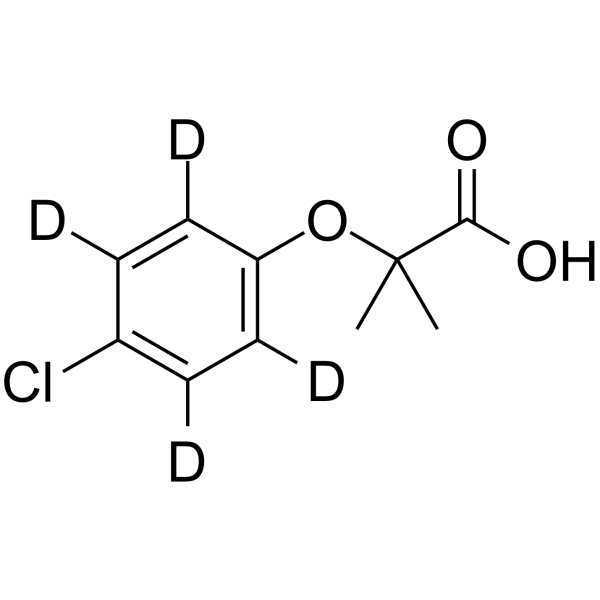
Clofibric acid-d4 is the deuterium labeled Clofibric acid (HY-B1415). Clofibric acid also is an herbicideClofibric acid (Chlorofibrinic acid) is an orally active PPARα agonist. Clofibric acid inhibits the fimbriation of Escherichia coli. Clofibric acid increases SOD activity. Clofibric acid lowers blood lipids and prevents experimental pyelonephritis. Clofibric acid has anticancer activity against ovarian cancer. Clofibric acid is also a herbicide. Clofibric acid is used in ovarian cancer, liver cancer, obesity, and urinary tract infection research.
For research use only. We do not sell to patients.
- CAS No.: 1184991-14-7
- Formula: C10H7D4ClO3
- Molecular Weight:218.67
-
Storage:Powder -20°C, 3 years , 4°C, 2 years ; In solvent -80°C, 6 months , -20°C, 1 month

To place orders, for customer services and technical support, please contact: MedChemExpress USA
Tel: 609-228-6898 E-mail: [email protected] [email protected]


-
 Biological Activity
-
Chemical Information
- Protocols
- Purity & Documentation
- References
-
 Help & FAQs


-
HY-101559
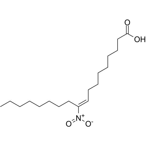
10-Nitrooleic acidNitro Fatty Acid -
HY-121457
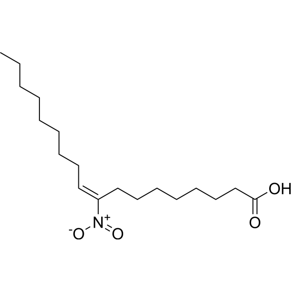
9-NitrooleateNitrated Fatty Acid -
HY-17004S1
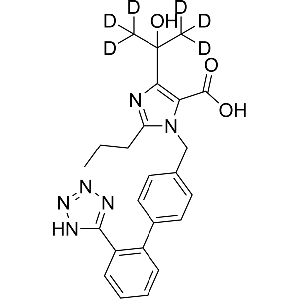
Olmesartan-d6Stable Isotope -
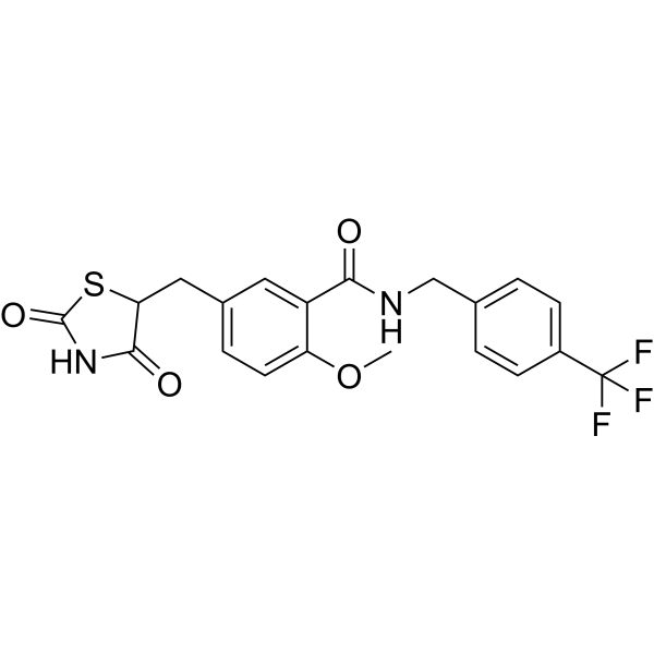
HY-119248
KRP-297PPARα/γ agonist -

HY-113162A
Bovinic acid sodiumFatty Acid -
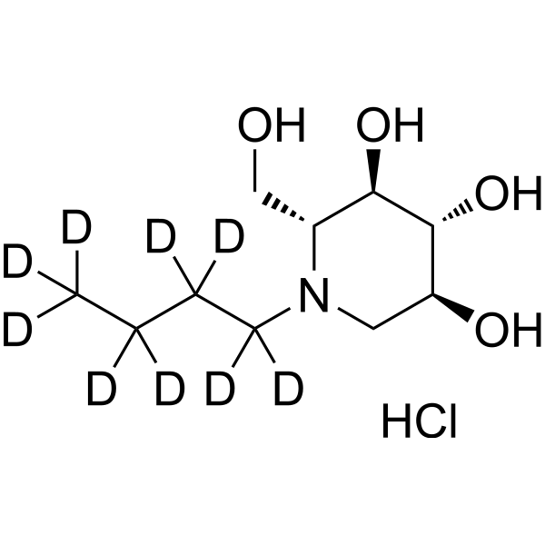
HY-110363
Miglustat-d9 hydrochlorideStable Isotope -
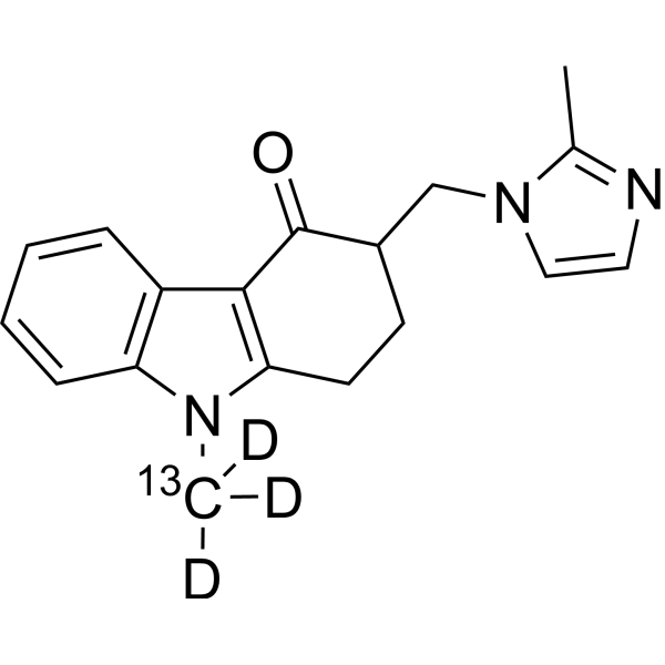
HY-B0002BS2
Ondansetron-13C,d3Stable Isotope -
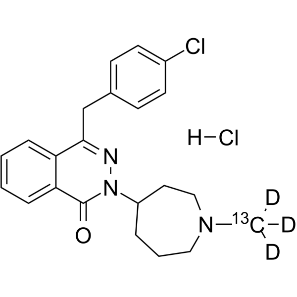
HY-B0462S
Azelastine-13C,d3 hydrochlorideStable Isotope


Biological Activity
Description
In Vitro
Stable heavy isotopes of hydrogen, carbon, and other elements have been incorporated into drug molecules, largely as tracers for quantitation during the drug development process. Deuteration has gained attention because of its potential to affect the pharmacokinetic and metabolic profiles of drugs[1].
MedChemExpress (MCE) has not independently confirmed the accuracy of these methods. They are for reference only. Further protocols information, click here.
Application
1. This compound can be used as a tracer
2. This compound can be used as an internal standard for quantitative analysis by NMR, GC-MS, or LC-MS.


Chemical Information
-
CAS No. 1184991-14-7
-
Unlabeled CAS 882-09-7
-
Appearance Solid
-
Molecular Weight 218.67
-
Formula C10H7D4ClO3
-
Color White to off-white
-
SMILES
OC(C(C)(C)OC1=C([2H])C([2H])=C(C([2H])=C1[2H])Cl)=O
-
Synonyms
Chlorofibrinic acid-d4
-
Shipping
Room temperature in continental US; may vary elsewhere.
-
Storage
Powder -20°C 3 years 4°C 2 years In solvent -80°C 6 months -20°C 1 month

Protocols
-
Research Protocol for Infectious Diseases
Infectious-disease experiments test how pathogens interact with host barriers, innate immune receptors, inflammatory signaling, pathogen replication, and tissue injury; pattern-recognition receptors such as TLRs, RIG-I-like receptors, NOD-like receptors, and inflammasomes detect microbial molecules and activate NF-κB, interferon, and cytokine responses. The central hypothesis is that infection severity reflects the balance between pathogen burden and host response: protective inflammation restricts pathogen growth, whereas excessive or mislocalized inflammation contributes to tissue damage and disease phenotype. Unresolved questions include which host pathways are protective versus pathogenic, why some infection models fail to translate to human disease, and which combined readouts best predict clinically relevant infection outcomes.
-
Liver Cancer Modeling
Liver cancer can be classified into primary liver cancer and secondary liver cancer. Secondary liver cancer is the metastatic liver cancer. Primary liver cancer includes hepatocellular carcinoma (HCC), intrahepatic cholangiocarcinoma (ICC) and fibrolamellar HCC, of which HCC is the most common form, accounting for approximately 90% of primary liver cancers[1]. HCC mouse models include chemical agent-induced models, transplanted tumor models, and genetic engineered models.
-
Research Protocol for Metabolic Diseases
AMP-activated protein kinase, AMPK, is a conserved cellular energy sensor that responds to reduced cellular energy status and coordinates metabolism by increasing ATP-generating catabolic pathways while suppressing ATP-consuming anabolic processes. In metabolic disease research, the AMPK pathway is experimentally relevant because it regulates hepatic lipid synthesis, fatty acid oxidation, glucose production, skeletal-muscle glucose disposal, mTORC1-linked biosynthesis, autophagy, mitochondrial homeostasis, and whole-body energy balance. The central pathway logic is that energy stress, metformin, exercise-like stimulation, or direct AMPK activators increase AMPKα Thr172 phosphorylation and downstream substrate phosphorylation, including ACC and RAPTOR. Phosphorylation of ACC suppresses lipogenesis and supports fatty acid oxidation, whereas phosphorylation of RAPTOR suppresses mTORC1 signaling and links cellular energy status to growth and protein synthesis control. The pathway is linked


Purity & Documentation
-
Data Sheet (285 KB)
-
SDS (393 KB)
- English - EN (393 KB)
- Français - FR (393 KB)
- Deutsch - DE (393 KB)
- Norwegian - NO (393 KB)
- Español - ES (393 KB)
- Swedish - SV (393 KB)
- Italian - IT (393 KB)
- Korean - KR (393 KB)
- Portuguese - PT (393 KB)
-
Handling Instructions (2659 KB)
References
[1]. Forman BM, et, al. Hypolipidemic drugs, polyunsaturated fatty acids, and eicosanoids are ligands for peroxisome proliferator-activated receptors alpha and delta. Proc Natl Acad Sci U S A. 1997 Apr 29;94(9):4312-7. [Content Brief]
[2]. Salgado R, et, al. Biodegradation of clofibric acid and identification of its metabolites. J Hazard Mater. 2012 Nov 30;241-242:182-9. [Content Brief]
[3]. Kawashima Y, et, al. Increased activity of stearoyl-CoA desaturation in liver from rat fed clofibric acid. Biochim Biophys Acta. 1982 Dec 13;713(3):622-8. [Content Brief]
[4]. Yokoyama Y, et al. Clofibric acid, a peroxisome proliferator-activated receptor alpha ligand, inhibits growth of human ovarian cancer. Mol Cancer Ther. 2007 Apr;6(4):1379-86. [Content Brief]
[5]. Bécuwe P, et al. Effects of the peroxisome proliferator clofibric acid on superoxide dismutase expression in the human HepG2 hepatoma cell line. Biochem Pharmacol. 1999 Sep 15;58(6):1025-33. [Content Brief]
[7]. Cleary MP, et al. Effect of long-term clofibric acid treatment on serum and tissue lipid and cholesterol levels in obese Zucker rats. Atherosclerosis. 1987 Jul;66(1-2):107-12. [Content Brief]
[8]. Balagué CE, et al. Clofibric and ethacrynic acids prevent experimental pyelonephritis by Escherichia coli in mice. FEMS Immunol Med Microbiol. 2004 Nov 1;42(3):313-9. [Content Brief]
[9]. Yamakawa Y, et al. A single pretreatment with clofibric acid attenuates carbon tetrachloride-induced necrosis, but not steatosis, in rat liver. Food Chem Toxicol. 2020 Nov;145:111591. [Content Brief]

Calculators
Concentration (start) × Volume (start) = Concentration (final) × Volume (final)


Clofibric acid-d4 Related Classifications
HY-B1415S Related Classifications
 Powered by Bioz