



Cyclic GMP
Based on 1 publication(s) in Google Scholar
Cyclic GMP (cGMP), an important second messenger, is a major intracellular mediator of extracellular signals such as nitric oxide (NO) and natriuretic peptides (NPs). Effects of Cyclic GMP occur through three main groups of cellular targets: cGMP-dependent protein kinases (PKGs), cGMP-gated cation channels, and PDEs. Cyclic GMP can inhibit both platelet adhesion and aggregation. cGAMP (Cyclic-GMP-AMP) (HY-12512), a conjugate of Cyclic GMP and AMP, can induce IRF3 phosphorylation and nuclear translocation, enhancing antiviral immune responses.
For research use only. We do not sell to patients.
- Purity : 99.77%
- CAS No.: 7665-99-8
- Formula: C10H12N5O7P
- Molecular Weight:345.21
-
Storage:Powder -20°C, 3 years ; In solvent -80°C, 6 months , -20°C, 1 month


To place orders, for customer services and technical support, please contact: MedChemExpress USA
Tel: 609-228-6898 E-mail: [email protected] [email protected]


-
 Biological Activity
-
Chemical Information
-
Solvent & Solubility
- Protocols
- Purity & Documentation
- References
-
 Help & FAQs


-
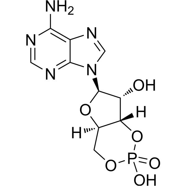
HY-B1511
Cyclic AMPSecond Messenger -

HY-113469R
Cyclic GMP (Standard)Standard of Cyclic GMP -
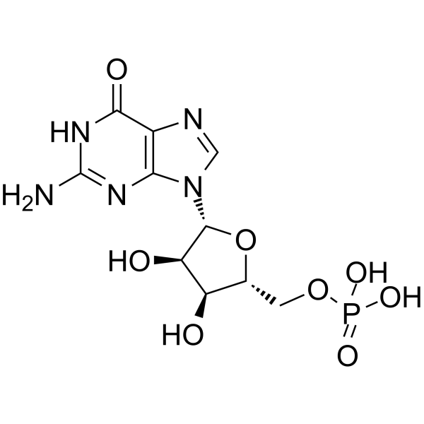
HY-N5134
5'-Guanylic acidNucleotide -
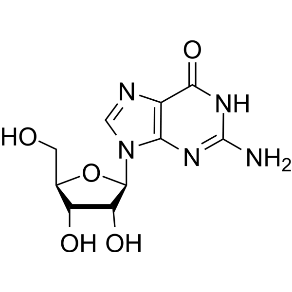
HY-N0097
GuanosineHSV Inhibitor -
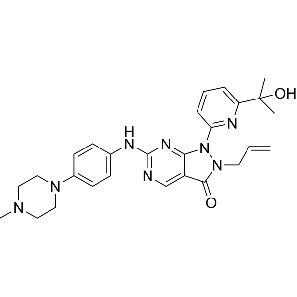
HY-10993
AdavosertibWee1 Inhibitor -

HY-A0181R
Adenosine monophosphate (Standard)Adenosine A1 Receptor Agonist -

HY-B1511R
Cyclic AMP (Standard)Second Messenger -

HY-B2176R
ATP (Standard)Reference Standard -
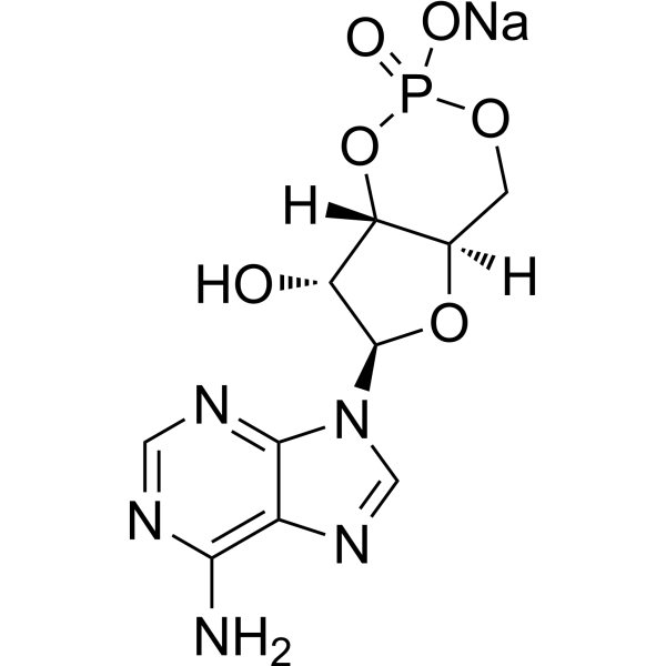
HY-B1511A
Cyclic AMP sodiumSecond Messenger -
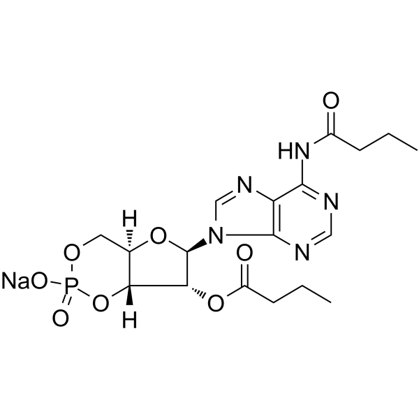
HY-B0764
Bucladesine sodiumcAMP Analog -
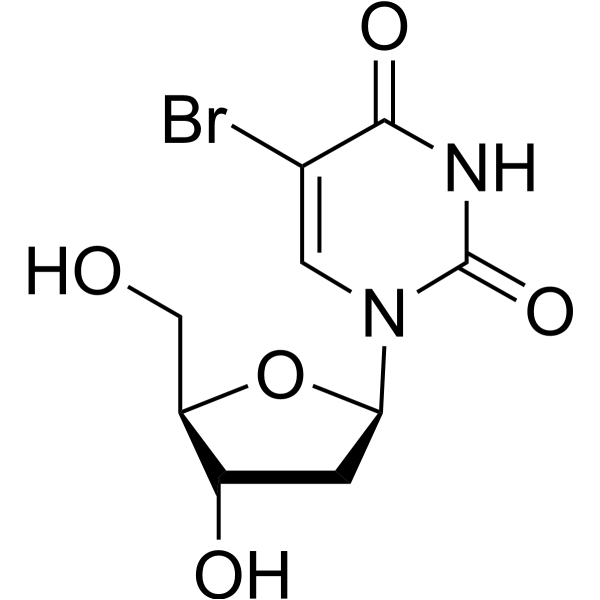
HY-15910
5-BrdUThymidine Analog -
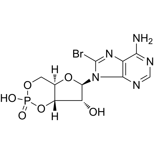
HY-12306A
8-Bromo-cAMPPKA Activator
Publications Citing Use of MedChemExpress (MCE) Cyclic GMP
MoreAll Endogenous Metabolite Isoforms
More

Biological Activity
Description
IC50 & Target
|
Human Endogenous Metabolite |
In Vitro
ACh can stimulate the accumulation of cyclic GMP (cGMP), thereby activating cGMP-mediated protein kinases. Increased cGMP causes a decrease in Ca ion influx, which shortens the action potential and inhibits ICa. Cyclic GMP (1 μM; 3-5 min) can activate cGMP-stimulated phosphodiesterases when acting synergistically with Isoprenaline (HY-B0468), reversing cAMP-mediated enhancement or inhibiting cAMP-induced inward calcium current (ICa)[3].
MedChemExpress (MCE) has not independently confirmed the accuracy of these methods. They are for reference only. Further protocols information, click here.


Chemical Information
-
CAS No. 7665-99-8
-
Appearance Solid
-
Molecular Weight 345.21
-
Formula C10H12N5O7P
-
Color White to off-white
-
SMILES
O[C@@H]([C@H](N1C=NC2=C1N=C(N)NC2=O)O[C@@H]3CO4)[C@@H]3OP4(O)=O
-
Structure Classification
-
Initial Source
-
Shipping
Room temperature in continental US; may vary elsewhere.
-
Storage
Powder -20°C 3 years In solvent -80°C 6 months -20°C 1 month


Publications (1)
-
Journal Impact Factor
-
Most Recent
-
Acta Neuropathol Commun
Hyperoside mitigates photoreceptor degeneration in part by targeting cGAS and suppressing DNA-induced microglial activation. [Abstract]2024 May 16;12(1):76. PMID: 38755736


Solvent & Solubility
In Vitro:
H2O : 5 mg/mL (14.48 mM; ultrasonic and warming and heat to 60°C)
Please refer to the solubility information to select the appropriate solvent. Once prepared, please aliquot and store the solution to prevent product inactivation from repeated freeze-thaw cycles.
Storage method and period of stock solution: -80°C, 6 months; -20°C, 1 month. When stored at -80°C, please use it within 6 months. When stored at -20°C, please use it within 1 month.
* Note: If you choose water as the stock solution, please dilute it to the working solution, then filter and sterilize it with a 0.22 μm filter before use.
Please refer to the solubility information to select the appropriate solvent. Once prepared, please aliquot and store the solution to prevent product inactivation from repeated freeze-thaw cycles.
Storage method and period of stock solution: -80°C, 6 months; -20°C, 1 month. When stored at -80°C, please use it within 6 months. When stored at -20°C, please use it within 1 month.
* Note: If you choose water as the stock solution, please dilute it to the working solution, then filter and sterilize it with a 0.22 μm filter before use.
Concentration (start) × Volume (start) = Concentration (final) × Volume (final)

Protocols
-
Kinase activity and phosphorylation assays
Kinase activity assays measure the ability of kinases to transfer phosphate groups from ATP to specific substrates, while phosphorylation assays detect the presence and levels of phosphorylated proteins. Common methods include radiolabeled ATP incorporation (e. g. ,), ADP release detection via bioluminescence (e. g. ,[3]), enzyme-linked immunosorbent assays (ELISA) for phospho-specific epitopes (e. g. ,[6]), and microtiter-based formats for high-throughput screening (e. g. ,[8]). The ADP-Glo assay quantifies kinase activity by measuring ADP produced during phosphorylation using a luciferase-based system. Radiometric assays involve autoradiography or scintillation counting after incorporation of 32P-labeled ATP into substrate proteins. ELISA-based approaches rely on phospho-specific antibodies to detect activated kinases in cell lysates or purified samples.
-
Western Blot
Western blotting (WB) is a commonly used experimental method in molecular biology, biochemistry, and immunogenetics for identifying and quantifying target proteins. It combines gel electrophoresis with immunoassay, enabling researchers to analyze protein expression, post-translational modifications, and molecular weight.
-
Cytoplasmic-Nuclear Fractionated Protein Extraction
Cytoplasmic-nuclear fractionated protein extraction separates soluble cytoplasmic proteins from nuclear-enriched proteins by mild plasma-membrane permeabilization, differential centrifugation, washing of nuclei, and extraction of nuclear proteins for downstream immunoblotting or related molecular analysis. The readout is the relative abundance of a protein in cytoplasmic and nuclear fractions, commonly assessed by western blotting together with compartment markers such as tubulin or pyruvate kinase for cytoplasm and lamin, nucleoporin, hnRNP, H2AX, or Lamin B for nuclear fractions.
-
Research Protocol for Infectious Diseases
Infectious-disease experiments test how pathogens interact with host barriers, innate immune receptors, inflammatory signaling, pathogen replication, and tissue injury; pattern-recognition receptors such as TLRs, RIG-I-like receptors, NOD-like receptors, and inflammasomes detect microbial molecules and activate NF-κB, interferon, and cytokine responses. The central hypothesis is that infection severity reflects the balance between pathogen burden and host response: protective inflammation restricts pathogen growth, whereas excessive or mislocalized inflammation contributes to tissue damage and disease phenotype. Unresolved questions include which host pathways are protective versus pathogenic, why some infection models fail to translate to human disease, and which combined readouts best predict clinically relevant infection outcomes.
-
Protocol for Kinase activity and phosphorylation assays
Kinase activity assays measure transfer of phosphate from ATP to a protein or peptide substrate, generating phosphorylated substrate, ADP, or incorporated radiolabeled phosphate as the readout; phosphorylation assays measure site-specific phosphorylation in cells or tissues as a proxy for kinase-pathway activation, inhibition, or substrate regulation. Phosphorylation can be detected by phospho-specific Western blot, immunoprecipitation kinase assay, phospho-immunofluorescence, phospho-flow cytometry, luminescent ADP detection, radiolabeled ATP incorporation, or reporter-based pathway assays, and these readouts can be applied to cancer cells, primary neurons, mouse tumors, organoids, inflammatory macrophages, ferroptosis studies, and mitophagy studies when the kinase target is biologically relevant.


Purity & Documentation
-
Data Sheet (276 KB)
-
SDS (393 KB)
- English - EN (393 KB)
- Français - FR (393 KB)
- Deutsch - DE (393 KB)
- Norwegian - NO (393 KB)
- Español - ES (393 KB)
- Swedish - SV (393 KB)
- Italian - IT (393 KB)
- Korean - KR (393 KB)
- Portuguese - PT (393 KB)
-
Handling Instructions (2659 KB)
References
[1]. Francis SH, et al. cGMP-dependent protein kinases and cGMP phosphodiesterases in nitric oxide and cGMP action. Pharmacol Rev. 2010 Sep;62(3):525-63. [Content Brief]
[2]. Friebe A, et al. cGMP: a unique 2nd messenger molecule - recent developments in cGMP research and development. Naunyn Schmiedebergs Arch Pharmacol. 2020 Feb;393(2):287-302. [Content Brief]
[3]. Hartzell HC, et al. Opposite effects of cyclic GMP and cyclic AMP on Ca2+ current in single heart cells. Nature. 1986 Sep 18-24;323(6085):273-5. [Content Brief]
[4]. Chintala MS, et al. Cyclic GMP but not cyclic AMP prevents renal platelet accumulation after ischemia-reperfusion in anesthetized rats. J Pharmacol Exp Ther. 1994 Dec;271(3):1203-8. [Content Brief]


Complete Stock Solution Preparation Table
Please refer to the solubility information to select the appropriate solvent. Once prepared, please aliquot and store the solution to prevent product inactivation from repeated freeze-thaw cycles.
Storage method and period of stock solution: -80°C, 6 months; -20°C, 1 month. When stored at -80°C, please use it within 6 months. When stored at -20°C, please use it within 1 month.
| Optional Solvent | Concentration Solvent Mass | 1 mg | 5 mg | 10 mg | 25 mg |
|---|---|---|---|---|---|
| H2O | 1 mM | 2.8968 mL | 14.4839 mL | 28.9679 mL | 72.4197 mL |
| 5 mM | 0.5794 mL | 2.8968 mL | 5.7936 mL | 14.4839 mL | |
| 10 mM | 0.2897 mL | 1.4484 mL | 2.8968 mL | 7.2420 mL |
* Note: If you choose water as the stock solution, please dilute it to the working solution, then filter and sterilize it with a 0.22 μm filter before use.


Cyclic GMP Related Classifications
HY-113469 Related Classifications
 Powered by Bioz