



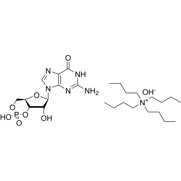
Cyclic GMP TBAOH
Based on 2 publication(s) in Google Scholar
Cyclic GMP (cGMP) TBAOH, an important second messenger, is a major intracellular mediator of extracellular signals such as nitric oxide (NO) and natriuretic peptides (NPs). Effects of Cyclic GMP TBAOH occur through three main groups of cellular targets: cGMP-dependent protein kinases (PKGs), cGMP-gated cation channels, and PDEs. Cyclic GMP can inhibit both platelet adhesion and aggregation. cGAMP (Cyclic-GMP-AMP) (HY-12512), a conjugate of Cyclic GMP and AMP, can induce IRF3 phosphorylation and nuclear translocation, enhancing antiviral immune responses.
For research use only. We do not sell to patients.
- Formula: C26H49N6O8P
- Molecular Weight:604.68
-
Storage:
-20°C, stored under nitrogen, away from moisture
* In solvent : -80°C, 6 months; -20°C, 1 month (stored under nitrogen, away from moisture)

To place orders, for customer services and technical support, please contact: MedChemExpress USA
Tel: 609-228-6898 E-mail: [email protected] [email protected]


-
 Biological Activity
-
Chemical Information
-
Solvent & Solubility
- Protocols
- Purity & Documentation
- References
-
 Help & FAQs


Publications Citing Use of MedChemExpress (MCE) Cyclic GMP TBAOH
MoreAll Endogenous Metabolite Isoforms
More

Biological Activity
Description
In Vitro
ACh can stimulate the accumulation of cyclic GMP (cGMP) TBAOH, thereby activating Cyclic GMP TBAOH-mediated protein kinases. Increased cGMP causes a decrease in Ca ion influx, which shortens the action potential and inhibits ICa. Cyclic GMP TBAOH (1 μM; 3-5 min) can activate Cyclic GMP TBAOH-stimulated phosphodiesterases when acting synergistically with Isoprenaline (HY-B0468), reversing cAMP-mediated enhancement or inhibiting cAMP-induced inward calcium current (ICa)[3].
MedChemExpress (MCE) has not independently confirmed the accuracy of these methods. They are for reference only.


Chemical Information
-
Appearance Solid
-
Molecular Weight 604.68
-
Formula C26H49N6O8P
-
Color White to off-white
-
SMILES
CCCC[N+](CCCC)(CCCC)CCCC.O[C@@H]([C@H](N1C=NC2=C1N=C(N)NC2=O)O[C@@H]3CO4)[C@@H]3OP4(O)=O.[OH-]
-
Shipping
Room temperature in continental US; may vary elsewhere.
-
Storage
-20°C, stored under nitrogen, away from moisture
* In solvent : -80°C, 6 months; -20°C, 1 month (stored under nitrogen, away from moisture)


Publications (2)
-
Journal Impact Factor
-
Most Recent
-
-
Acta Neuropathol Commun
Hyperoside mitigates photoreceptor degeneration in part by targeting cGAS and suppressing DNA-induced microglial activation. [Abstract]2024 May 16;12(1):76. PMID: 38755736


Solvent & Solubility
In Vitro:
DMSO : 100 mg/mL (165.38 mM; Need ultrasonic; Hygroscopic DMSO has a significant impact on the solubility of product, please use newly opened DMSO)
Please refer to the solubility information to select the appropriate solvent. Once prepared, please aliquot and store the solution to prevent product inactivation from repeated freeze-thaw cycles.
Storage method and period of stock solution: -80°C, 6 months; -20°C, 1 month (stored under nitrogen, away from moisture). When stored at -80°C, please use it within 6 months. When stored at -20°C, please use it within 1 month.
Please refer to the solubility information to select the appropriate solvent. Once prepared, please aliquot and store the solution to prevent product inactivation from repeated freeze-thaw cycles.
Storage method and period of stock solution: -80°C, 6 months; -20°C, 1 month (stored under nitrogen, away from moisture). When stored at -80°C, please use it within 6 months. When stored at -20°C, please use it within 1 month.
Concentration (start) × Volume (start) = Concentration (final) × Volume (final)


Purity & Documentation
-
Data Sheet (272 KB)
-
SDS (252 KB)
- English - EN (252 KB)
- Français - FR (252 KB)
- Deutsch - DE (252 KB)
- Norwegian - NO (252 KB)
- Español - ES (252 KB)
- Swedish - SV (252 KB)
- Italian - IT (252 KB)
- Korean - KR (252 KB)
- Portuguese - PT (252 KB)
-
Handling Instructions (2659 KB)
References
[1]. Francis SH, et al. cGMP-dependent protein kinases and cGMP phosphodiesterases in nitric oxide and cGMP action. Pharmacol Rev. 2010 Sep;62(3):525-63. [Content Brief]
[2]. Friebe A, et al. cGMP: a unique 2nd messenger molecule - recent developments in cGMP research and development. Naunyn Schmiedebergs Arch Pharmacol. 2020 Feb;393(2):287-302. [Content Brief]
[3]. Hartzell HC, et al. Opposite effects of cyclic GMP and cyclic AMP on Ca2+ current in single heart cells. Nature. 1986 Sep 18-24;323(6085):273-5. [Content Brief]
[4]. Chintala MS, et al. Cyclic GMP but not cyclic AMP prevents renal platelet accumulation after ischemia-reperfusion in anesthetized rats. J Pharmacol Exp Ther. 1994 Dec;271(3):1203-8. [Content Brief]


Complete Stock Solution Preparation Table
Please refer to the solubility information to select the appropriate solvent. Once prepared, please aliquot and store the solution to prevent product inactivation from repeated freeze-thaw cycles.
Storage method and period of stock solution: -80°C, 6 months; -20°C, 1 month (stored under nitrogen, away from moisture). When stored at -80°C, please use it within 6 months. When stored at -20°C, please use it within 1 month.
| Optional Solvent | Concentration Solvent Mass | 1 mg | 5 mg | 10 mg | 25 mg |
|---|---|---|---|---|---|
| DMSO | 1 mM | 1.6538 mL | 8.2688 mL | 16.5377 mL | 41.3442 mL |
| 5 mM | 0.3308 mL | 1.6538 mL | 3.3075 mL | 8.2688 mL | |
| 10 mM | 0.1654 mL | 0.8269 mL | 1.6538 mL | 4.1344 mL | |
| 15 mM | 0.1103 mL | 0.5513 mL | 1.1025 mL | 2.7563 mL | |
| 20 mM | 0.0827 mL | 0.4134 mL | 0.8269 mL | 2.0672 mL | |
| 25 mM | 0.0662 mL | 0.3308 mL | 0.6615 mL | 1.6538 mL | |
| 30 mM | 0.0551 mL | 0.2756 mL | 0.5513 mL | 1.3781 mL | |
| 40 mM | 0.0413 mL | 0.2067 mL | 0.4134 mL | 1.0336 mL | |
| 50 mM | 0.0331 mL | 0.1654 mL | 0.3308 mL | 0.8269 mL | |
| 60 mM | 0.0276 mL | 0.1378 mL | 0.2756 mL | 0.6891 mL | |
| 80 mM | 0.0207 mL | 0.1034 mL | 0.2067 mL | 0.5168 mL | |
| 100 mM | 0.0165 mL | 0.0827 mL | 0.1654 mL | 0.4134 mL |


Cyclic GMP TBAOH Related Classifications
HY-113469B Related Classifications
 Powered by Bioz