



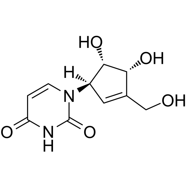
Cyclopentenyl uracil

Based on 1 Customer Validation
Cyclopentenyl uracil is a non-cytotoxic Uridine-Cytidine Kinase inhibitor and an antagonist of EIDD-1931 (NHC) (HY-125033) bioactivation. Cyclopentenyl uracil blocks NHC phosphorylation by inhibiting UCK1/UCK2, reduces intracellular NHC-TP accumulation, and depletes UTP/CTP pools, thereby antagonizing the antiviral activity of NHC and inhibiting dengue virus replication. Cyclopentenyl uracil is used in research on SARS-CoV-2 infection, dengue virus infection, leukemia, and solid tumors.
For research use only. We do not sell to patients.
- Purity : 99.84%
- CAS No.: 90597-20-9
- Formula: C10H12N2O5
- Molecular Weight:240.22
-
Storage:Powder -20°C, 3 years ; In solvent -80°C, 6 months , -20°C, 1 month

To place orders, for customer services and technical support, please contact: MedChemExpress USA
Tel: 609-228-6898 E-mail: [email protected] [email protected]


-
 Biological Activity
-
Chemical Information
-
Solvent & Solubility
- Protocols
- Purity & Documentation
- References
-
 Help & FAQs




Biological Activity
Description
IC50 & Target
[2]|
UCK1 |
UCK2 |
Cellular Effect
|
Cell Line

|
Type | Value | Description | References |
|---|---|---|---|---|
| CCRF-CEM | IC50 |
> 100 μM
Compound: 52
|
In vitro cytotoxicity in CEM cells.
In vitro cytotoxicity in CEM cells.
|
[PMID: 11689085] |
| Vero | IC50 |
> 100 μM
Compound: 52
|
In vitro cytotoxicity in vero cells.
In vitro cytotoxicity in vero cells.
|
[PMID: 11689085] |
In Vitro
Cyclopentenyl uracil (96 h) effectively antagonizes the cytotoxicity induced by Molnupiravir (HY-135853)/EIDD-1931 (NHC) (HY-125033) in HL-60 and THP-1 cell lines, indicating that UCKs are the primary kinases regulating the intracellular response to Molnupiravir/NHC[1].
Cyclopentenyl uracil (CPEU) (10-50 µM) inhibits the phosphorylation of Zebularine (HY-13420) in T24 cells, indicating that uridine-cytidine kinase catalyzes the initial phosphorylation step[5].
Cyclopentenyl uracil (CPE-U) (300 μM; 48 h) shows no cytotoxicity against L1210 cells and effectively blocks the Uridine (HY-B1449) salvage pathway, restoring the growth inhibitory effect of PALA in the presence of plasma concentrations of uridine[6].
Cyclopentenyl uracil (25 μM; 24 h) restores the normal growth rate of MOLT-4 lymphoblasts exposed to 200 nM CPEC[7].
Cyclopentenyl uracil (4-24 h), when added within 4 hr after 200 nM CPEC, allows normal proliferation of MOLT-4 lymphoblasts, but loses its detoxifying activity when added later[7].
Cyclopentenyl uracil (CPU) (5 mM; 24 h) acts as a substrate for human CMPK1, leading to the formation of CPU-DP[4].
Cyclopentenyl uracil (500 µM; 48-72 h) in combination with GSK983 (HY-119098) significantly reduces DENV-2 infection in A549 cells in the presence of physiological uridine concentrations[4].
Cyclopentenyl uracil (125-1000 µM; 48 h) in combination with GSK983 selectively and dose-dependently inhibits DENV-2 replication in A549 cells[4].
Cyclopentenyl uracil (125-500 µM; 48 h) in combination with GSK983 enhances the antiviral activity of the RdRp inhibitor R1479 (HY-10444) in A549 cells[4].
Cyclopentenyl uracil (250 µM; 48 h) in combination with GSK983 significantly enhances the antiviral activity of R1479 in the DENV-2 replicon assay in A549 cells, reducing its EC50 from approximately 90 µM to approximately 19 µM[4].
Cyclopentenyl uracil selectively inhibits UCK, reducing uridine salvage synthesis and, to a lesser extent, cytidine salvage synthesis in various cancers[8].
MedChemExpress (MCE) has not independently confirmed the accuracy of these methods. They are for reference only. Further protocols information, click here.
-
Cell Line:HL-60 and THP-1
-
Concentration:100 µM
-
Incubation Time:96 h
-
Result:Protected both HL-60 and THP-1 cells from molnupiravir-induced cytotoxicity.
-
Cell Line:A549
-
Concentration:500 µM
-
Incubation Time:48, 72 h
-
Result:In the presence of 20 µM exogenous uridine, combination of GSK983 with 500 µM CPU caused significant reductions in viral infection.
Appeared to have a workable index at this concentration and time point.
-
Cell Line:A549
-
Concentration:250 µM, 1 mM
-
Incubation Time:48 h
-
Result:Combining 0.2 µM GSK983 and 250 µM CPU inhibited ~50% of virus replication.
At a CPU dose of 1 mM, virus replication was suppressed almost completely, with much less effect on A549 cell growth.
-
Cell Line:L1210
-
Concentration:300 μM (alone); 300 μM (combination with 1 mM PALA and 0-20 μM uridine)
-
Incubation Time:48 h
-
Result:Increased cell number by 1.4 × 10⁵ cells/well in the absence of added uridine.
Increased cell number by 0.02 × 10⁵ cells/well in combination with 1 mM PALA and 5 μM uridine.
Increased cell number by 0.07 × 10⁵ cells/well in combination with 1 mM PALA and 20 μM uridine.
-
Cell Line:MOLT-4 human T-lymphoblasts
-
Concentration:25 μM
-
Incubation Time:24 hr
-
Result:Maintained logarithmic growth of MOLT-4 lymphoblasts with a doubling-time of 32 hr.\nRestored normal growth rate in cells exposed to 200 nM CPEC.
In Vivo
Cyclopentenyl uracil (1 g/kg; i.p.; every 8 h; 5 days) is a potent inhibitor of the uridine salvage pathway in intact mice, but frequent dosing (every 8 h) is required to maintain > 64-79% sustained inhibition, and it is nontoxic to mice during the 5-day treatment period[6].
Cyclopentenyl uracil (1 g/kg; i.p.; every 8 h; 5 days) alone is non-toxic and does not affect body weight gain, but its combination with Polyalanine peptide (PALA) (HY-P10546) (200 mg/kg, once daily for 5 days) is lethal in mice, indicating that circulating uridine can alter the toxicity of drugs acting on the de novo pyrimidine synthesis pathway[6].
MedChemExpress (MCE) has not independently confirmed the accuracy of these methods. They are for reference only.
-
Animal Model:BALB/c × DBA/2 (CDF1) (male, bearing P388 ascites)[6]
-
Dosage:1 g/kg
-
Administration:i.p.; twice at 12-hr intervals
-
Result:Inhibited uridine salvage by 77% in ascites, 74% in kidney, 64% in liver, 79% in spleen, and 79% in intestine.
-
Animal Model:BALB/c × DBA/2 (CDF1) (male)[6]
-
Dosage:1 g/kg
-
Administration:i.p.; every 8 hr; 5 days (sustained inhibition); i.p.; single injection (time course)
-
Result:Inhibited uridine salvage by > 64-79% with multiple doses of 1 g/kg given every 8 hr.
Showed no signs of toxicity and continued weight gain over 14 days.
-
Animal Model:BALB/c × DBA/2 (CDF1) (male)[6]
-
Dosage:1 g/kg
-
Administration:i.p.; every 8 hr; 5 days
-
Result:Showed no decrease in weight gain compared with control animals over 14 days.
Resulted in death of all mice during the 5-day period when combined with PALA (200 mg/kg daily for 5 days).


Chemical Information
-
CAS No. 90597-20-9
-
Appearance Solid
-
Molecular Weight 240.22
-
Formula C10H12N2O5
-
Color White to off-white
-
SMILES
O[C@H]1[C@@](N2C(NC(C=C2)=O)=O)([H])C=C([C@H]1O)CO
-
Shipping
Room temperature in continental US; may vary elsewhere.
-
Storage
Powder -20°C 3 years In solvent -80°C 6 months -20°C 1 month


Solvent & Solubility
In Vitro:
DMSO : 100 mg/mL (416.29 mM; Need ultrasonic; Hygroscopic DMSO has a significant impact on the solubility of product, please use newly opened DMSO)
Please refer to the solubility information to select the appropriate solvent. Once prepared, please aliquot and store the solution to prevent product inactivation from repeated freeze-thaw cycles.
Storage method and period of stock solution: -80°C, 6 months; -20°C, 1 month. When stored at -80°C, please use it within 6 months. When stored at -20°C, please use it within 1 month.
Please refer to the solubility information to select the appropriate solvent. Once prepared, please aliquot and store the solution to prevent product inactivation from repeated freeze-thaw cycles.
Storage method and period of stock solution: -80°C, 6 months; -20°C, 1 month. When stored at -80°C, please use it within 6 months. When stored at -20°C, please use it within 1 month.
Concentration (start) × Volume (start) = Concentration (final) × Volume (final)

Protocols
-
Kinase activity and phosphorylation assays
Kinase activity assays measure the ability of kinases to transfer phosphate groups from ATP to specific substrates, while phosphorylation assays detect the presence and levels of phosphorylated proteins. Common methods include radiolabeled ATP incorporation (e. g. ,), ADP release detection via bioluminescence (e. g. ,[3]), enzyme-linked immunosorbent assays (ELISA) for phospho-specific epitopes (e. g. ,[6]), and microtiter-based formats for high-throughput screening (e. g. ,[8]). The ADP-Glo assay quantifies kinase activity by measuring ADP produced during phosphorylation using a luciferase-based system. Radiometric assays involve autoradiography or scintillation counting after incorporation of 32P-labeled ATP into substrate proteins. ELISA-based approaches rely on phospho-specific antibodies to detect activated kinases in cell lysates or purified samples.
-
Western Blot
Western blotting (WB) is a commonly used experimental method in molecular biology, biochemistry, and immunogenetics for identifying and quantifying target proteins. It combines gel electrophoresis with immunoassay, enabling researchers to analyze protein expression, post-translational modifications, and molecular weight.
-
Research Protocol for Infectious Diseases
Infectious-disease experiments test how pathogens interact with host barriers, innate immune receptors, inflammatory signaling, pathogen replication, and tissue injury; pattern-recognition receptors such as TLRs, RIG-I-like receptors, NOD-like receptors, and inflammasomes detect microbial molecules and activate NF-κB, interferon, and cytokine responses. The central hypothesis is that infection severity reflects the balance between pathogen burden and host response: protective inflammation restricts pathogen growth, whereas excessive or mislocalized inflammation contributes to tissue damage and disease phenotype. Unresolved questions include which host pathways are protective versus pathogenic, why some infection models fail to translate to human disease, and which combined readouts best predict clinically relevant infection outcomes.
-
Protocol for Kinase activity and phosphorylation assays
Kinase activity assays measure transfer of phosphate from ATP to a protein or peptide substrate, generating phosphorylated substrate, ADP, or incorporated radiolabeled phosphate as the readout; phosphorylation assays measure site-specific phosphorylation in cells or tissues as a proxy for kinase-pathway activation, inhibition, or substrate regulation. Phosphorylation can be detected by phospho-specific Western blot, immunoprecipitation kinase assay, phospho-immunofluorescence, phospho-flow cytometry, luminescent ADP detection, radiolabeled ATP incorporation, or reporter-based pathway assays, and these readouts can be applied to cancer cells, primary neurons, mouse tumors, organoids, inflammatory macrophages, ferroptosis studies, and mitophagy studies when the kinase target is biologically relevant.
-
Cell Viability Determination by MTT Colorimetric Assay
The following protocol uses the MTT colorimetric assay as a classic literature-established method for assessing cell viability/metabolic activity in cultured mammalian cells. MTT[3-(4,5-dimethylthiazol-2-yl)-2,5-diphenyltetrazolium bromide] is reduced by metabolically active cells to a colored formazan product; the amount of formazan is quantified spectrophotometrically and provides an indirect measure of metabolically active viable cells. Importantly, MTT reduction reflects cellular oxidoreductase/metabolic activity rather than an absolute direct count of living cells, so changes in cellular metabolism can alter the signal independently of cell number.


Purity & Documentation
-
Data Sheet (297 KB)
-
SDS (252 KB)
- English - EN (252 KB)
- Français - FR (252 KB)
- Deutsch - DE (252 KB)
- Norwegian - NO (252 KB)
- Español - ES (252 KB)
- Swedish - SV (252 KB)
- Italian - IT (252 KB)
- Korean - KR (252 KB)
- Portuguese - PT (252 KB)
-
Handling Instructions (2659 KB)
References
[1]. Shu H, et al. Uridine cytidine kinases govern molnupiravir bioactivation and anti-SARS-CoV-2 activity. PLoS pathogens. 2026 May;22(5):e1014225. [Content Brief]
[2]. Shu H, et al. Uridine cytidine kinases dictate the therapeutic response of molnupiravir via its bioactivation. bioRxiv. 2025 May 14:2025-05. [Content Brief]
[3]. Politi PM, et al. Phase I clinical trial of continuous infusion cyclopentenyl cytosine. Cancer chemotherapy and pharmacology. 1995;36(6):513-23. [Content Brief]
[4]. Liu Q, et al. Enhancing the Antiviral Efficacy of RNA-Dependent RNA Polymerase Inhibition by Combination with Modulators of Pyrimidine Metabolism. Cell chemical biology. 2020 Jun 18;27(6):668-677.e9. [Content Brief]
[5]. Marquez VE, et al. Zebularine: a unique molecule for an epigenetically based strategy in cancer chemotherapy. Annals of the New York Academy of Sciences. 2005 Nov;1058(1):246-54. [Content Brief]
[6]. Cysyk RL, et al. Cyclopentenyl uracil: an effective inhibitor of uridine salvage in vivo. Biochemical pharmacology. 1995 Jan 18;49(2):203-7. [Content Brief]
[7]. Ford H, et al. Reversal by cytidine of cyclopentenyl cytosine-induced toxicity in mice without compromise of antitumor activity. Biochemical pharmacology. 1995 Jan 18;49(2):173-80. [Content Brief]


Complete Stock Solution Preparation Table
Please refer to the solubility information to select the appropriate solvent. Once prepared, please aliquot and store the solution to prevent product inactivation from repeated freeze-thaw cycles.
Storage method and period of stock solution: -80°C, 6 months; -20°C, 1 month. When stored at -80°C, please use it within 6 months. When stored at -20°C, please use it within 1 month.
| Optional Solvent | Concentration Solvent Mass | 1 mg | 5 mg | 10 mg | 25 mg |
|---|---|---|---|---|---|
| DMSO | 1 mM | 4.1629 mL | 20.8143 mL | 41.6285 mL | 104.0713 mL |
| 5 mM | 0.8326 mL | 4.1629 mL | 8.3257 mL | 20.8143 mL | |
| 10 mM | 0.4163 mL | 2.0814 mL | 4.1629 mL | 10.4071 mL | |
| 15 mM | 0.2775 mL | 1.3876 mL | 2.7752 mL | 6.9381 mL | |
| 20 mM | 0.2081 mL | 1.0407 mL | 2.0814 mL | 5.2036 mL | |
| 25 mM | 0.1665 mL | 0.8326 mL | 1.6651 mL | 4.1629 mL | |
| 30 mM | 0.1388 mL | 0.6938 mL | 1.3876 mL | 3.4690 mL | |
| 40 mM | 0.1041 mL | 0.5204 mL | 1.0407 mL | 2.6018 mL | |
| 50 mM | 0.0833 mL | 0.4163 mL | 0.8326 mL | 2.0814 mL | |
| 60 mM | 0.0694 mL | 0.3469 mL | 0.6938 mL | 1.7345 mL | |
| 80 mM | 0.0520 mL | 0.2602 mL | 0.5204 mL | 1.3009 mL | |
| 100 mM | 0.0416 mL | 0.2081 mL | 0.4163 mL | 1.0407 mL |


Cyclopentenyl uracil Related Classifications
HY-145668 Related Classifications
 Powered by Bioz