



Cylindrospermopsin

Based on 1 Customer Validation
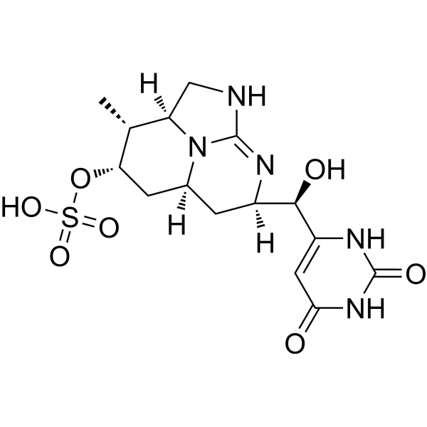
Cylindrospermopsin, a cyanotoxin, is a polycyclic uracil derivative containing guanidine and sulfate groups, which can inhibit protein synthesis and covalently modify DNA or RNA. Cylindrospermopsin induces hepatocellular hypertrophy, renal cellular hypertrophy, intracellular reactive oxygen species (ROS), DNA strand breaks, mitochondrial hyperpolarisation, ultrastructural damage, and altered gene expression in liver, kidney, and intestinal cells. Cylindrospermopsin can be used in research including hepatocellular carcinoma and water quality testing.
For research use only. We do not sell to patients.
- Purity : 98.76%
- CAS No.: 143545-90-8
- Formula: C15H21N5O7S
- Molecular Weight:415.42
-
Storage:Powder -20°C, 3 years ; In solvent -80°C, 6 months , -20°C, 1 month

To place orders, for customer services and technical support, please contact: MedChemExpress USA
Tel: 609-228-6898 E-mail: [email protected] [email protected]


-
 Biological Activity
-
Chemical Information
- Protocols
- Purity & Documentation
- References
-
 Help & FAQs


-
HY-P2040
NodularinHepatotoxin -
HY-W017227
Alloxan hydrateDiabetogenic Agent/Proteasome Inhibitor -
HY-B0526S
Flumequine-13C3Stable Isotope -
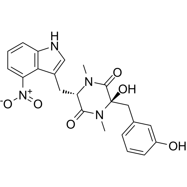
HY-124212
Thaxtomin APhytotoxin -
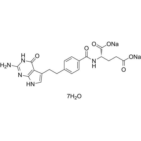
HY-10820B
Pemetrexed disodium heptahydrateAutophagy Inducer -
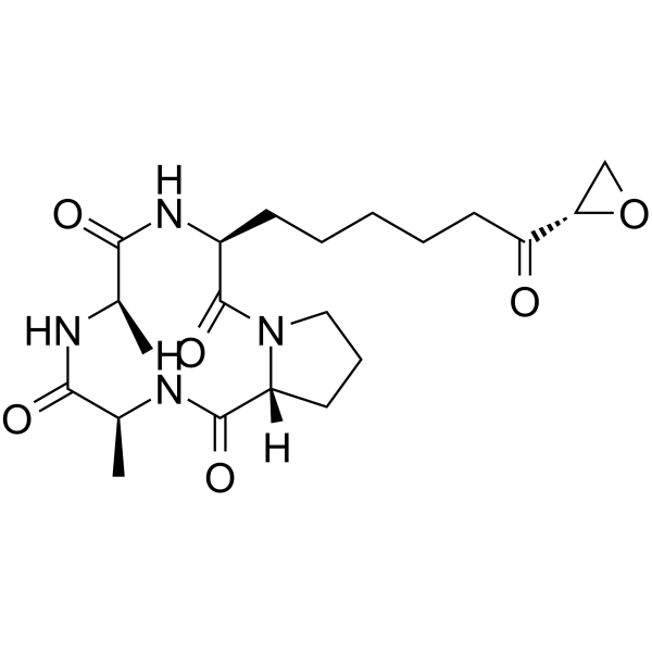
HY-126856
HC-ToxinHDAC Inhibitor -
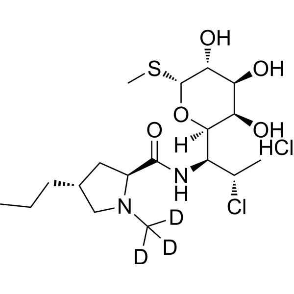
HY-B1455S
Clindamycin-d3 hydrochlorideStable Isotope -

HY-171001
ZM522CD73 Inhibitor -
HY-117313
Fumitremorgin BAntifungal Agent -

HY-125778
Cochlioquinone B -
HY-N10258
(+)-SorokinianinPhytotoxin -

HY-N11952
DihydropyrenophorinMacrolide
All DNA/RNA Synthesis Isoforms
More

Biological Activity
Description
In Vitro
Cylindrospermopsin induces micronucleus formation and whole chromosome loss in transformed human lymphoblastoid cells in vitro, indicating mutagenicity[2].
Cylindrospermopsin (0.3-40 μg/mL; 24-48 h) induces concentration- and time-dependent cytotoxicity in Caco-2 cells, with MTS reduction being the most sensitive endpoint (EC50 of 2.5 μg/mL at 24 h and 0.6 μg/mL at 48 h)[3].
Cylindrospermopsin (0.625-2.5 μg/mL; 24 h) modulates oxidative stress markers in Caco-2 cells, increasing ROS at 1.25 μg/mL and GCS activity and GSH content at 2.5 μg/mL after 24 h exposure[3].
Cylindrospermopsin (0.625-2.5 μg/mL; 24-48 h) induces ultrastructural morphological alterations in Caco-2 cells at both 0.625 μg/mL and 2.5 μg/mL after 24 h and 48 h exposure, including nucleolar segregation, mitochondrial damage, and lipid degeneration[3].
Cylindrospermopsin (0.05-0.5 μg/mL; up to 5 h) induces dose- and time-dependent intracellular ROS formation in HepG2 cells, with a ~5-fold increase at 0.5 μg/mL after 5 h of exposure[4].
Cylindrospermopsin (0.125-0.5 μg/mL; 4, 12, 24 h) induces non-oxidative DNA strand breaks in HepG2 cells at 0.25 and 0.5 μg/mL after 12 and 24 h of exposure, with no oxidative DNA damage detected[4].
Cylindrospermopsin (0.125-0.5 μg/mL; 12, 24 h) does not induce apoptosis or increase cell death in HepG2 cells at concentrations up to 0.5 μg/mL after 12 or 24 h of exposure[4].
Cylindrospermopsin (0.25-0.5 μg/mL; 12, 24 h) induces mitochondrial membrane hyperpolarization in HepG2 cells at 0.25 and 0.5 μg/mL after 12 h, and at 0.5 μg/mL after 24 h of exposure[4].
MedChemExpress (MCE) has not independently confirmed the accuracy of these methods. They are for reference only. Further protocols information, click here.
-
Cell Line:human colon carcinoma Caco-2 cells
-
Concentration:0.3, 0.6, 1.2, 2.5, 5, 10, 20, 40 μg/mL
-
Incubation Time:24 h; 48 h
-
Result:Reduced total protein content by up to 50% at 40 μg/mL (24 h) and above 2.5 μg/mL (48 h). Decreased neutral red uptake by up to 45% at highest concentrations (24 h) and significantly at 5 μg/mL (48 h). Showed progressive concentration-dependent decreases in MTS reduction; reduced viability by about 90% at 40 μg/mL. Reported EC50 values for MTS reduction of 2.5 μg/mL (24 h) and 0.6 μg/mL (48 h).
-
Cell Line:HepG2
-
Concentration:0.125-0.5 μg/mL
-
Incubation Time:12, 24 h
-
Result:Observed no significant differences in total cell death (Annexin V or PI positive cells) or the number of early/late apoptotic cells.
In Vivo
Cylindrospermopsin (500-1500 mg/kg; p.o.; 1-3 doses separated by 2 weeks) shows potential tumour initiation activity in mice, with a relative risk of 6.2 for neoplastic processes[2].
MedChemExpress (MCE) has not independently confirmed the accuracy of these methods. They are for reference only.
-
Animal Model:CD-1 (Swiss-Webster) (male and female, 30 days old at dosing start)[1]
-
Dosage:75, 150, 300 μg/kg/d
-
Administration:p.o.; daily; 90 days
-
Result:Increased absolute and relative liver weights and kidney weights at all doses in males and at 150, 300 μg/kg/d in females; increased absolute and relative testes weights at 300 μg/kg/d in males. Elevated male ALT activity by 64% at 300 μg/kg/d; decreased male BUN at all doses; decreased male cholesterol at 150, 300 μg/kg/d; decreased male triglycerides at 300 μg/kg/d; elevated female ALP activity by 38% at 300 μg/kg/d. Decreased male hematocrit, hemoglobin, and erythrocyte count at 300 μg/kg/d; increased male leukocyte count at 300 μg/kg/d; increased male lymphocyte count at 75, 300 μg/kg/d; increased male monocyte count at 75, 300 μg/kg/d; increased female monocyte count at 300 μg/kg/d. Induced significant liver lesions (hepatic cord distortion, hepatocyte hypertrophy, cell death, inflammation, pigment accumulation) in all treated groups; induced significant kidney lesions (tubule dilation, basophilia, intraluminal protein, epithelial alteration, nuclear crowding, outer medulla thinning) in all male doses and 150, 300 μg/kg/d in females.
-
Animal Model:Swiss Albino (male, 20-30 g)[2]
-
Dosage:1500 mg/kg; 500 mg/kg
-
Administration:p.o.; 1 or 2 doses (1500 mg/kg) separated by 2 weeks; 3 doses (500 mg/kg) separated by 2 weeks
-
Result:Detected neoplastic processes in 5 of 53 treated mice, including 2 hepatocellular dysplastic foci, 1 fibroblastic osteosarcoma, 1 hepatocellular carcinoma, and 1 follicular centre cell lymphoma; calculated a relative risk of 6.2 compared to saline-treated controls.


Chemical Information
-
CAS No. 143545-90-8
-
Appearance Solid
-
Molecular Weight 415.42
-
Formula C15H21N5O7S
-
Color White to light yellow
-
SMILES
O[C@H]([C@]1([H])N=C2N([C@@](CN2)([H])[C@@H](C)[C@@H](OS(=O)(O)=O)C3)[C@@]3([H])C1)C(NC4=O)=CC(N4)=O
-
Initial Source
Cylindrospermopsis raciborskii
-
Shipping
Room temperature in continental US; may vary elsewhere.
-
Storage
Powder -20°C 3 years In solvent -80°C 6 months -20°C 1 month

Protocols
-
RT-PCR
Reverse transcription technology uses RNA as a template to synthesize DNA. RT-PCR is simple, specific and sensitive, and can be used to detect gene expression levels and expression differences in cells; detect RNA virus content; clone cDNA sequences of specific genes.
-
RNA extraction experimental
By lysing cells, releasing RNA, and removing impurities such as proteins and DNA, high-purity RNA products are finally obtained. The commonly used traditional method is the guanidine isothiocyanate/phenol/chloroform method (Trizol), which is suitable for a variety of animal materials including animal tissues, microorganisms, cultured cells, etc., and most plant materials.
-
ROS/oxidative-stress fluorescent staining
ROS/oxidative-stress fluorescent staining uses cell-permeant fluorogenic probes that become fluorescent after oxidation inside cells or tissues; commonly used examples include DCFH-DA/DCFDA for broad cellular oxidant detection, DHE for superoxide-related signal detection, MitoSOX for mitochondrial superoxide-related signal detection, and CellROX probes for oxidative-stress-associated fluorescence readouts. The assay detects probe oxidation rather than a single ROS species unless the probe and analysis method have been chemically validated for that species. DCFH-DA enters cells, is deacetylated by intracellular esterases to DCFH, and produces fluorescent DCF after oxidation, so the readout is used as an operational measure of total cellular oxidative stress rather than a species-specific ROS measurement. DHE and MitoSOX can report superoxide-related oxidation, but red fluorescence alone can include non-specific ethidium-like oxidation products; HPLC or optimized spectral approaches are
-
Liver Cancer Modeling
Liver cancer can be classified into primary liver cancer and secondary liver cancer. Secondary liver cancer is the metastatic liver cancer. Primary liver cancer includes hepatocellular carcinoma (HCC), intrahepatic cholangiocarcinoma (ICC) and fibrolamellar HCC, of which HCC is the most common form, accounting for approximately 90% of primary liver cancers[1]. HCC mouse models include chemical agent-induced models, transplanted tumor models, and genetic engineered models.
-
Real Time qPCR (Q-PCR)
Real-time quantitative PCR (qPCR) quantifies an amplifiable nucleic-acid target by monitoring fluorescence during PCR cycling rather than measuring product only after amplification. The increase in fluorescence tracks accumulation of PCR product, and the quantification cycle (Cq; historically also Ct/CP) is related to the initial amount of target: samples containing more starting target generally reach the defined fluorescence threshold in fewer cycles.


Purity & Documentation
-
Data Sheet (282 KB)
-
SDS (644 KB)
- English - EN (644 KB)
- Français - FR (644 KB)
- Deutsch - DE (644 KB)
- Norwegian - NO (644 KB)
- Español - ES (644 KB)
- Swedish - SV (644 KB)
- Italian - IT (644 KB)
- Korean - KR (644 KB)
- Portuguese - PT (644 KB)
-
Handling Instructions (2659 KB)
References
[1]. Chernoff N, et al. Cylindrospermopsin toxicity in mice following a 90-d oral exposure. J Toxicol Environ Health A. 2018;81(13):549-566. [Content Brief]
[2]. Falconer IR, et al. Preliminary evidence for in vivo tumour initiation by oral administration of extracts of the blue-green alga cylindrospermopsis raciborskii containing the toxin cylindrospermopsin. Environ Toxicol. 2001;16(2):192-195. [Content Brief]
[3]. Gutiérrez-Praena D, et al. Biochemical and pathological toxic effects induced by the cyanotoxin Cylindrospermopsin on the human cell line Caco-2. Water Res. 2012;46(5):1566-1575. [Content Brief]
[4]. Štraser A, et al. The influence of cylindrospermopsin on oxidative DNA damage and apoptosis induction in HepG2 cells. Chemosphere. 2013;92(1):24-30. [Content Brief]

Calculators
Concentration (start) × Volume (start) = Concentration (final) × Volume (final)


Cylindrospermopsin Related Classifications
HY-121360 Related Classifications
 Powered by Bioz