



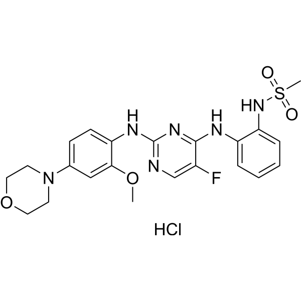
CZC-25146 hydrochloride
CZC-25146 hydrochloride is a potent LRRK2 inhibitor with IC50 values of 4.76 nM and 6.87 nM for wild-type LRRK2 and G2019S LRRK2, respectively. CZC-25146 hydrochloride inhibits PLK4, GAK, TNK1, CAMKK2 and PIP4K2C as well. CZC-25146 hydrochloride prevents mutant LRRK2-induced injury of neurons in vitro. CZC-25146 hydrochloride exhibits relatively favorable pharmacokinetic properties in mice. CZC-25146 hydrochloride can increase normal α-1-antitrypsin (AAT) secretion and reduce inflammatory cytokines. CZC-25146 hydrochloride can be used to research Parkinson's disease and liver diseases.
For research use only. We do not sell to patients.
- CAS No.: 1330003-04-7
- Formula: C22H26ClFN6O4S
- Molecular Weight:525.00
-
Storage:
Please store the product under the recommended conditions in the Certificate of Analysis.

To place orders, for customer services and technical support, please contact: MedChemExpress USA
Tel: 609-228-6898 E-mail: [email protected] [email protected]


-
 Biological Activity
-
Chemical Information
- Protocols
- Purity & Documentation
- References
-
 Help & FAQs


-
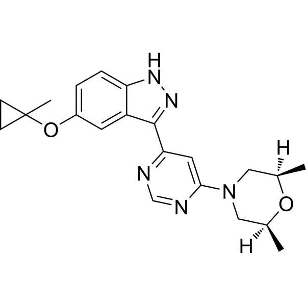
HY-100411
MLi-2LRRK2 Inhibitor -
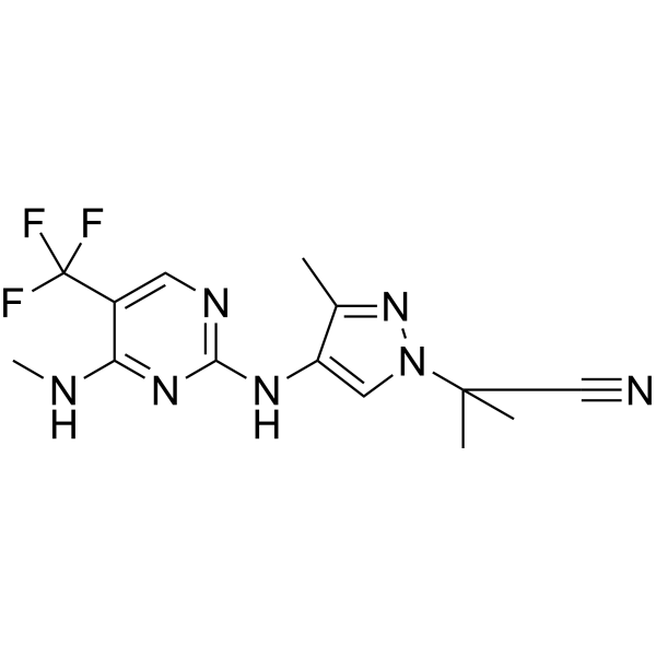
HY-15796
GNE0877LRRK2 Inhibitor -

HY-12477
PF-06447475LRRK2 Inhibitor -
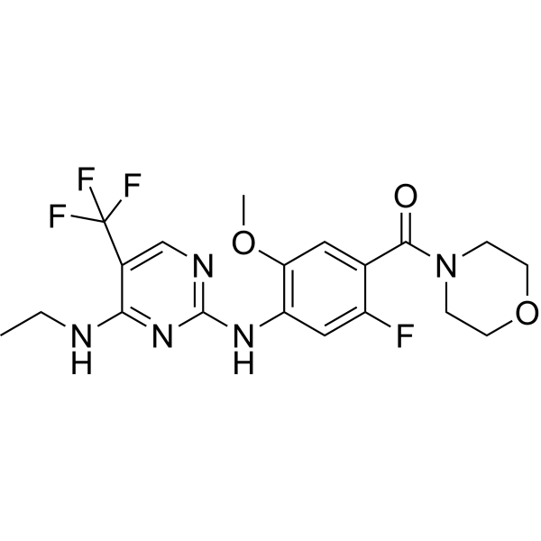
HY-18163
GNE-7915LRRK2 Inhibitor -
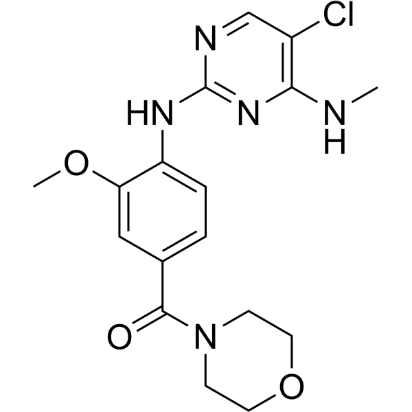
HY-13488
HG-10-102-01LRRK2 Inhibitor -
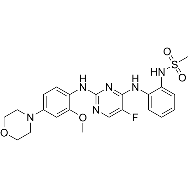
HY-15800A
CZC-25146LRRK2 Inhibitor -
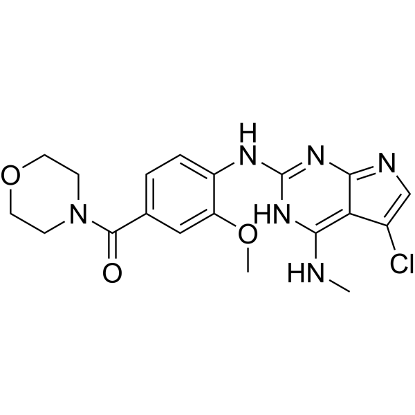
HY-16936
JH-II-127LRRK2 Inhibitor -
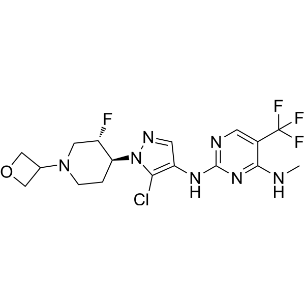
HY-12282
GNE-9605LRRK2 Inhibitor -
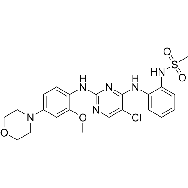
HY-B0792
CZC-54252LRRK2 Inhibitor -
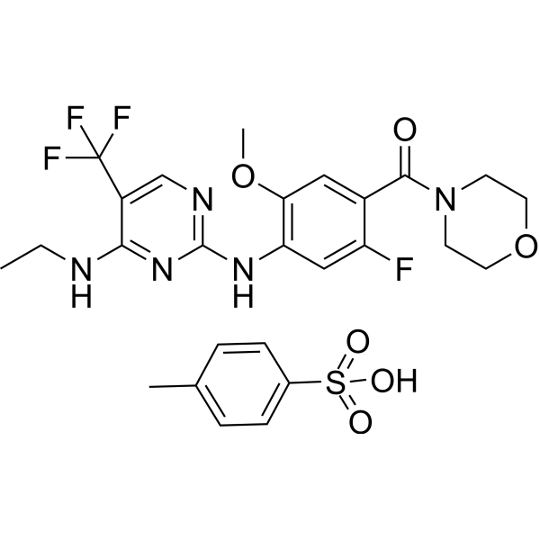
HY-18163A
GNE-7915 tosylateLRRK2 Inhibitor -
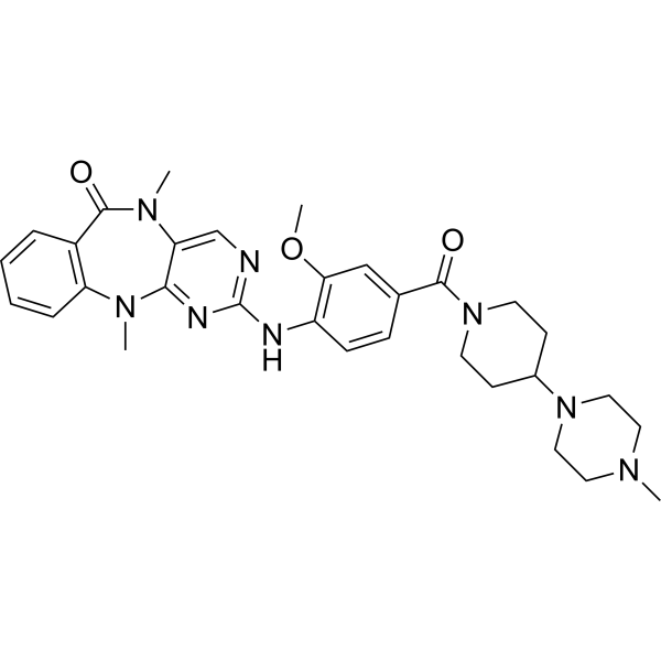
HY-10875
LRRK2-IN-1LRRK2 Inhibitor -
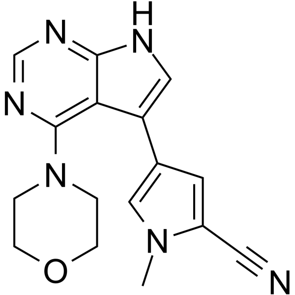
HY-120085
PFE-360LRRK2 Inhibitor


Biological Activity
Description
IC50 & Target
IC50: 4.76 nM (wild-type LRRK2), 6.87 nM (G2019S LRRK2)[1]
In Vitro
CZC-25146 (0.01-5 μM; 7 days) does not cause cytotoxicity in human cortical neurons, nor blocking neuronal development[1].
CZC-25146 (0.01-5 μM; 2 days) potently attenuates G2019S LRRK2-mediated toxicity in primary rodent neurons in a concentration-dependent manner with an EC50 of ~100 nM[1].
CZC-25146 (0.06-1000 nM) rescues LRRK2 G2019S-induced neurite defects in primary human neurons in a dose-dependent manner[1].
CZC-25146 (14.3 and 28.6 μM; 48 h) markedly reduces The mutant AAT encoded by the Z allele (ATZ) polymer load and restores AAT secretion in iPSC-Hepatocyte, without compromising cell viability[3].
MedChemExpress (MCE) has not independently confirmed the accuracy of these methods. They are for reference only. Further protocols information, click here.
-
Cell Line:Human cortical neurons
-
Concentration:0.01, 0.1, 1 and 5 μM
-
Incubation Time:7 days
-
Result:Did not cause cytotoxicity in human cortical neurons at concentrations below 5 μM over a seven-day treatment in culture, nor did it block neuronal development.
In Vivo
CZC-25146 (250 mg/kg; p.o.; 14 days) reduces the ATZ polymer levels in over expressing human polymeric ATZ mice[3].
MedChemExpress (MCE) has not independently confirmed the accuracy of these methods. They are for reference only.
-
Animal Model:Male CD-1 mice[1]
-
Dosage:1 mg/kg for i.v.; 5 mg/kg for p.o.
-
Administration:i.v. and p.o.; single dosage
-
Result:Pharmacokinetic Parameters of CZC-25146 in male CD-1 mice[1].
i.v. (1 mg/kg) p.o. (5 mg/kg) CL (L/h/kg) 2.3 Vss (L/kg) 5.4 t1/2 (h) 1.6 1 tmax (h) 0 0.25 Cmax (ng/mL) 154 1357 AUClast (ng/mL·h) 419 2878 AUCinf (ng/mL·h) 434 2894 F (%) 133
-
Animal Model:Genetically modified male mice (6 weeks; over expressing human polymeric ATZ)[3]
-
Dosage:250 mg/kg
-
Administration:p.o.; 14 days
-
Result:Dramatically and reproducibly reduced the ATZ polymer levels with an overall reduction from 60% in the control group to 37%


Chemical Information
-
CAS No. 1330003-04-7
-
Molecular Weight 525.00
-
Formula C22H26ClFN6O4S
-
SMILES
CS(=O)(NC1=CC=CC=C1NC2=NC(NC3=CC=C(N4CCOCC4)C=C3OC)=NC=C2F)=O.Cl
-
Shipping
Room temperature in continental US; may vary elsewhere.
-
Storage
Please store the product under the recommended conditions in the Certificate of Analysis.

Protocols
-
Cell Cytotoxicity Assay
Cytotoxicity assays are usually based on the assessment of cell membrane damage, which can also be indirectly detected by measuring cell viability. Detection methods include MTT assay, CKK-8 assay, LDH assay and ATP assay, etc.
-
LPS-Induced Endotoxemia/Systemic Inflammation
Lipopolysaccharide (LPS)-induced endotoxemia is a widely used in vivo model of acute systemic inflammation in which LPS, a Gram-negative bacterial endotoxin, activates innate immune signaling primarily through TLR4, leading to rapid and transient induction of pro-inflammatory cytokines such as TNF-α, IL-6, and IL-1β in circulation and tissues. This cytokine surge is commonly used as a measurable readout of systemic inflammatory activation and immune dysregulation, and is typically assessed within hours after intraperitoneal LPS administration in mouse models of endotoxemia. The model captures key features of systemic inflammatory response syndrome, including cytokine release, immune cell activation, and downstream tissue responses, and has been used to evaluate anti-inflammatory interventions such as cytokine modulation, lipid mediators, and immune cell-targeting therapies.
-
Human pluripotent stem cell midbrain dopaminergic neuron differentiation
Human pluripotent stem cells are directed toward midbrain dopaminergic neurons by first inducing a neural floor-plate-like progenitor state, then patterning cells with ventralizing SHH signaling and midbrain/WNT-FGF cues, and finally maturing progenitors into neurons expressing dopaminergic markers such as TH, NURR1/NR4A2, PITX3, DAT/SLC6A3, VMAT2/SLC18A2, GIRK2/KCNJ6, FOXA2, LMX1A, and EN1. The main readouts are loss of pluripotency, acquisition of FOXA2+/LMX1A+ midbrain floor-plate progenitors, emergence of βIII-tubulin+/MAP2+ neurons, and production of TH+ dopaminergic neurons with molecular, dopamine-release, and electrophysiological features of midbrain dopaminergic identity.


Purity & Documentation
References
[1]. Ramsden N, et al. Chemoproteomics-based design of potent LRRK2-selective lead compounds that attenuate Parkinson's disease-related toxicity in human neurons. ACS Chem Biol. 2011 Oct 21;6(10):1021-8. [Content Brief]
[2]. Atashrazm F, et al. LRRK2 inhibitors and their potential in the treatment of Parkinson's disease: current perspectives. Clin Pharmacol. 2016 Oct 20;8:177-189. [Content Brief]

Calculators
Concentration (start) × Volume (start) = Concentration (final) × Volume (final)


CZC-25146 hydrochloride Related Classifications
HY-15800 Related Classifications
 Powered by Bioz