



DC41SMe
DC41SMe, a DC1 derivative, shows cytotoxicity in Ramos, Namalwa, and HL60/s cells with IC50s ranging from 18-25 pM. DC1, a simplified analogue of CC-1065, is an antibody conjugate of cytotoxic DNA alkylators for the targeted treatment of cancer.
For research use only. We do not sell to patients.
- CAS No.: 1354787-71-5
- Formula: C38H36ClN5O4S2
- Molecular Weight:726.31
-
Storage:
Please store the product under the recommended conditions in the Certificate of Analysis.

To place orders, for customer services and technical support, please contact: MedChemExpress USA
Tel: 609-228-6898 E-mail: [email protected] [email protected]


-
 Biological Activity
-
Chemical Information
- Protocols
- Purity & Documentation
- References
-
 Help & FAQs


-
HY-14573
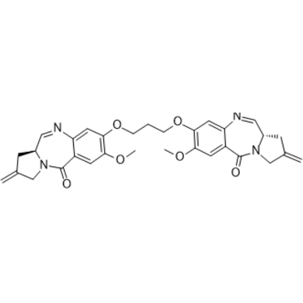
SJG-136DNA Alkylator/Crosslinker -
HY-16503
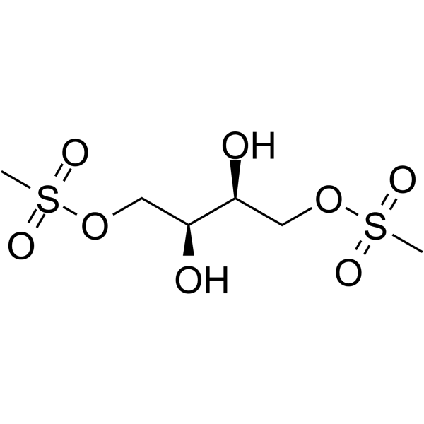
TreosulfanAlkylating Cytotoxic Agent -
HY-17576
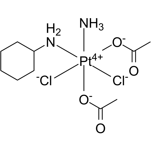
SatraplatinDNA Alkylator -
HY-13585
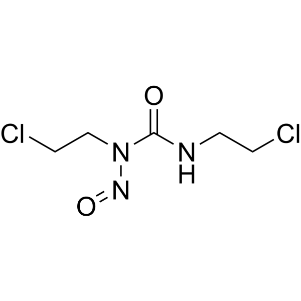
CarmustineDNA/RNA Alkylator -
HY-112898
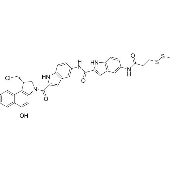
DC1SMeADC Cytotoxin -
HY-112899
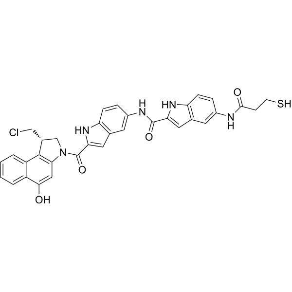
DC1ADC Cytotoxin -
HY-112901
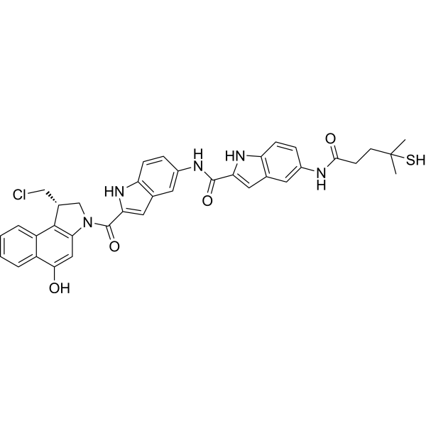
DC41DC1 Derivative -
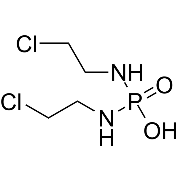
HY-14798
Palifosfamide -
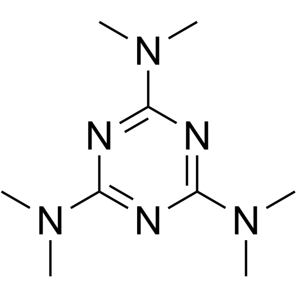
HY-B0181
AltretamineDNA Alkylator/Crosslinker -
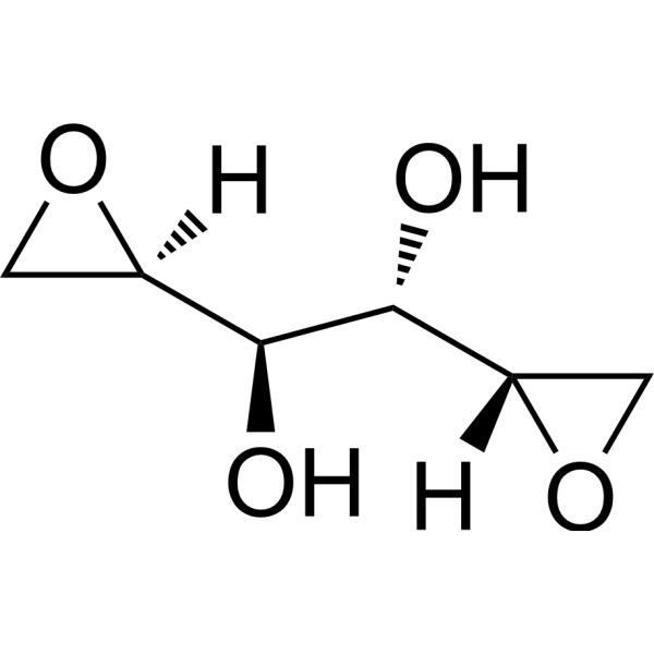
HY-16513
VAL-083Alkylating Agent -
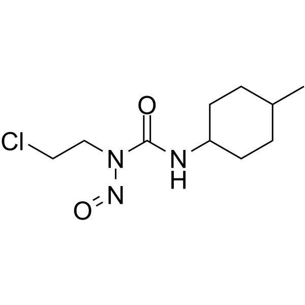
HY-13747
SemustineAntitumor Alkylating Agent -
HY-13733
Procarbazine HydrochlorideAlkylating Agent


Biological Activity
Description
IC50 & Target
|
Duocarmycins |
Cellular Effect
|
Cell Line

|
Type | Value | Description | References |
|---|---|---|---|---|
| COLO 205 | IC50 |
220 pM
Compound: 26, DC41SMe
|
Cytotoxicity against human COLO205 cells assessed as cell survival after 72 hrs by clonogenic assay
Cytotoxicity against human COLO205 cells assessed as cell survival after 72 hrs by clonogenic assay
|
[PMID: 22148292] |
| HL-60 | IC50 |
25 pM
Compound: 26, DC41SMe
|
Cytotoxicity against human HL60 cells assessed as cell survival after 72 hrs by clonogenic assay
Cytotoxicity against human HL60 cells assessed as cell survival after 72 hrs by clonogenic assay
|
[PMID: 22148292] |
| NAMALVA | IC50 |
21 pM
Compound: 26, DC41SMe
|
Cytotoxicity against human NAMALWA cells assessed as cell survival after 72 hrs by clonogenic assay
Cytotoxicity against human NAMALWA cells assessed as cell survival after 72 hrs by clonogenic assay
|
[PMID: 22148292] |
| Ramos | IC50 |
18 pM
Compound: 26, DC41SMe
|
Cytotoxicity against human Ramos cells assessed as cell survival after 72 hrs by clonogenic assay
Cytotoxicity against human Ramos cells assessed as cell survival after 72 hrs by clonogenic assay
|
[PMID: 22148292] |


Chemical Information
-
CAS No. 1354787-71-5
-
Molecular Weight 726.31
-
Formula C38H36ClN5O4S2
-
SMILES
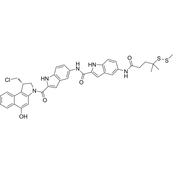
O=C(C(N1)=CC2=C1C=CC(NC(CCC(SSC)(C)C)=O)=C2)NC3=CC4=C(NC(C(N5C[C@@H](CCl)C6=C5C=C(O)C7=CC=CC=C67)=O)=C4)C=C3
-
Shipping
Room temperature in continental US; may vary elsewhere.
-
Storage
Please store the product under the recommended conditions in the Certificate of Analysis.


Purity & Documentation
References
[1]. Zhao RY, et al. Synthesis and biological evaluation of antibody conjugates of phosphate prodrugs of cytotoxic DNA alkylators for the targeted treatment of cancer. J Med Chem. 2012 Jan 26;55(2):766-82. [Content Brief]
[2]. Kovtun YV, et al. Antibody-drug conjugates designed to eradicate tumors with homogeneous and heterogeneous expression of the target antigen. Cancer Res. 2006 Mar 15;66(6):3214-21. [Content Brief]

Calculators
Concentration (start) × Volume (start) = Concentration (final) × Volume (final)


DC41SMe Related Classifications
HY-112900 Related Classifications
 Powered by Bioz