



PP13
PP13 is an antimicrobial peptide, and is active against Gram-negative and Gram-positive bacteria E.coli (MIC: 16.7 uM), B. subtilis (MIC: 13.3 uM), S. aureus (MIC: 23.3 uM), S. lutea (MIC: 8.0 uM), and B. pumilu (MIC: 9.0 uM).
Nur für Forschungszwecke. Wir verkaufen nicht an Patienten.
- CAS. Nr.: 1099486-04-0
- Formel: C121H197N39O29
- Molecular Weight:2662.10
-
Speicherung:
Please store the product under the recommended conditions in the Certificate of Analysis.

To place orders, for customer services and technical support, please contact: MedChemExpress USA
Tel: 609-228-6898 E-mail: [email protected] [email protected]


-
 Biologische Aktivität
-
Chemical Information
- Protokoll
- Reinheit & Dokumentation
- Verweise
-
 Help & FAQs




Biologische Aktivität
Beschreibung


Chemical Information
-
CAS. Nr. 1099486-04-0
-
Molecular Weight 2662.10
-
Formel C121H197N39O29
-
Sequence
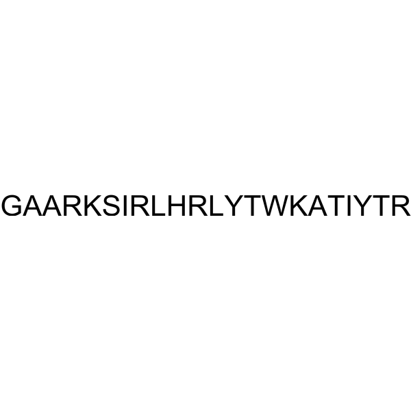
Gly-Ala-Ala-Arg-Lys-Ser-Ile-Arg-Leu-His-Arg-Leu-Tyr-Thr-Trp-Lys-Ala-Thr-Ile-Tyr-Thr-Arg
-
Sequence Shortening
GAARKSIRLHRLYTWKATIYTR
-
Versand
Room temperature in continental US; may vary elsewhere.
-
Speicherung
Please store the product under the recommended conditions in the Certificate of Analysis.


Reinheit & Dokumentation
Verweise

Calculators
Konzentration (Stammlösung) × Volumen (Stammlösung) = Konzentration (Ziellösung) × Volumen (Ziellösung)


PP13 Related Classifications
HY-P5560 Related Classifications
 Powered by Bioz