



VLX600
Based on 5 publication(s) in Google Scholar
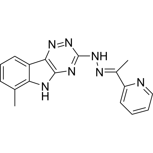
VLX600 is an iron-chelating inhibitor of oxidative phosphorylation (OXPHOS). VLX600 causes mitochondrial dysfunction and induces a strong shift to glycolysis. VLX600 displays selective cytotoxic activity against malignant cell and induces autophagy. Anticancer activity.
For research use only. We do not sell to patients.
- Purity : 99.80%
- CAS No.: 327031-55-0
- Formula: C17H15N7
- Molecular Weight:317.35
-
Storage:Powder -20°C, 3 years , 4°C, 2 years
* The compound is unstable in solutions, freshly prepared is recommended.

To place orders, for customer services and technical support, please contact: MedChemExpress USA
Tel: 609-228-6898 E-mail: [email protected] [email protected]


-
 Biological Activity
-
Chemical Information
-
Solvent & Solubility
- Protocols
- Purity & Documentation
- References
-
 Help & FAQs


-
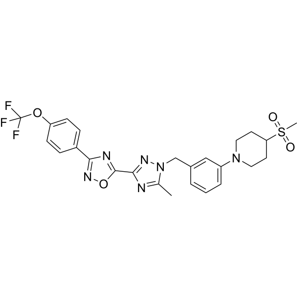
HY-112037
IACS-010759OXPHOS Inhibitor -
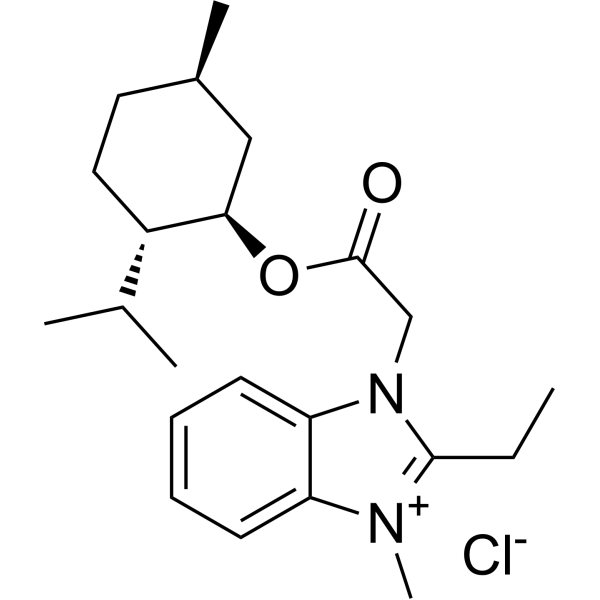
HY-111651
GboxinOXPHOS Inhibitor -

HY-15763
ErastinFerroptosis Inducer -

HY-10219
RapamycinmTOR Inhibitor -
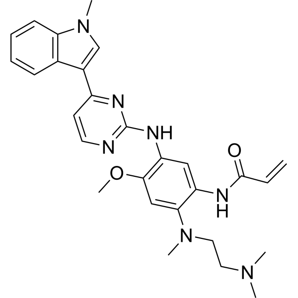
HY-15772
OsimertinibMutant-Selective EGFR Inhibitor -
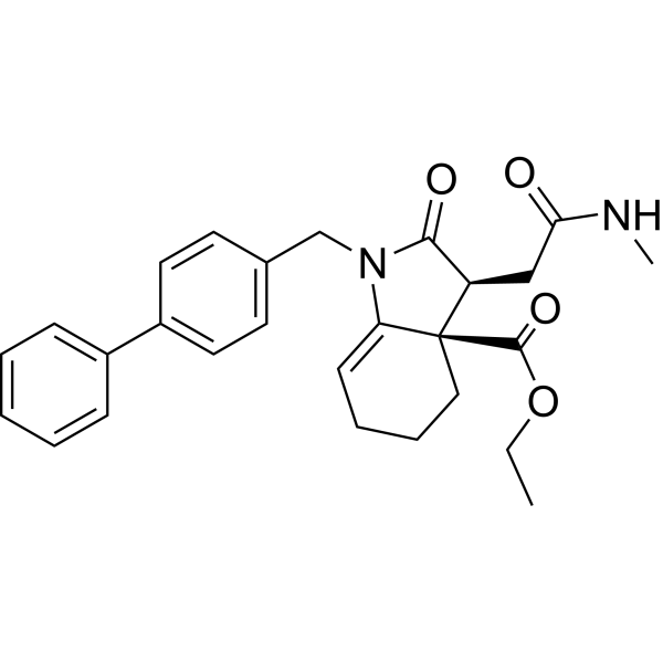
HY-100004
Fumarate hydratase-IN-1Fumarate Hydratase Inhibitor -

HY-B0215
AcetylcysteineMucolytic Agent -

HY-Y0320
Dimethyl sulfoxide, meets analytical specification of Ch.P.Aprotic Solvent -
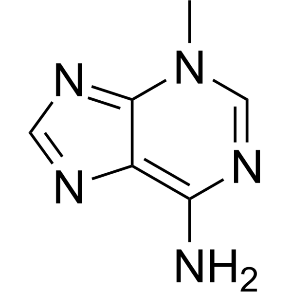
HY-19312
3-MethyladenineAutophagy/PI3K Inhibitor -
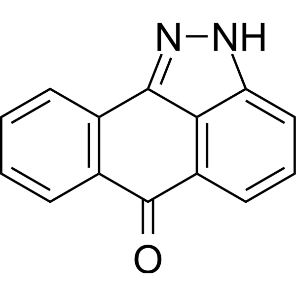
HY-12041
SP600125JNK Inhibitor -

HY-100579
Ferrostatin-1Ferroptosis Inhibitor -
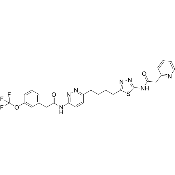
HY-12248
TelaglenastatGLS1 Inhibitor
Publications Citing Use of MedChemExpress (MCE) VLX600
More

Biological Activity
Description
Cellular Effect
|
Cell Line

|
Type | Value | Description | References |
|---|---|---|---|---|
| Caco-2 | CC50 |
0.06 μM
Compound: VLX600
|
Toxicity against Caco-2 cells determined at 48 hours by intracellular ATP concentration using the CellTiter-Glo Luminescent Cell Viability Assay
Toxicity against Caco-2 cells determined at 48 hours by intracellular ATP concentration using the CellTiter-Glo Luminescent Cell Viability Assay
|
10.21203/rs.3.rs-23951/v1 |
| Caco-2 | IC50 |
0.24 μM
Compound: VLX600
|
Determination of IC50 values for inhibition of SARS-CoV-2 induced cytotoxicity of Caco-2 cells after 48 hours by high content imaging
Determination of IC50 values for inhibition of SARS-CoV-2 induced cytotoxicity of Caco-2 cells after 48 hours by high content imaging
|
10.21203/rs.3.rs-23951/v1 |
In Vitro
VLX600 (6 μM; 72 hours) induces an autophagic response[2].
VLX600 is cytotoxic to HCT116 spheroids. VLX600 induces a HIF-1α-dependent glycolytic response. VLX600 inhibits oxygen consumption in HCT116 cells. VLX600 inhibits phosphorylation of the mTOR downstream effectors 4EBP1 and p70-S6K by an HIF-1α-independent mechanism. VLX600 preferentially leads to decreased ATP levels in cancer but not normal cells[2].
MedChemExpress (MCE) has not independently confirmed the accuracy of these methods. They are for reference only.
-
Cell Line:HCT116, HT29, SW620, HT8, DLD and RKO cells
-
Concentration:0.1, 1, 10, 100μM
-
Incubation Time:72 hours
-
Result:Inhibited the proliferation of these cells.
-
Cell Line:HCT116 cells
-
Concentration:6 μM
-
Incubation Time:72 hours
-
Result:LC3-II was induced.
In Vivo
MedChemExpress (MCE) has not independently confirmed the accuracy of these methods. They are for reference only.
-
Animal Model:NMRI nu/nu mice (HCT116 and HT29 colon cancer xenografts)[2]
-
Dosage:16 mg/kg
-
Administration:Intravenously; every third day for 16 days
-
Result:Anti-tumor activity was observed in both HCT116 and HT29 colon cancer xenografts.
- Kinase activity and phosphorylation assays
- Western Blot
- Cell Cytotoxicity Assay
- Mitochondrial membrane-potential and mitochondrial mass staining
- Autophagy
- Protocol for Kinase activity and phosphorylation assays
- Lysosome and acidic-vesicle live-cell staining
- Macroautophagy Solutions
- Cell Viability Determination by MTT Colorimetric Assay
Clinical Trial
| NCT Number | Sponsor | Condition | Start Date |
Phase
|
|---|---|---|---|---|
| NCT01329991 | Plexxikon| | 2011-05 | PHASE1 |


Chemical Information
-
CAS No. 327031-55-0
-
Appearance Solid
-
Molecular Weight 317.35
-
Formula C17H15N7
-
Color Light yellow to yellow
-
SMILES
CC1=C(NC2=C3N=NC(N/N=C(C4=CC=CC=N4)\C)=N2)C3=CC=C1
-
Shipping
Room temperature in continental US; may vary elsewhere.
-
Storage
Powder -20°C 3 years 4°C 2 years * The compound is unstable in solutions, freshly prepared is recommended.


Publications (5)
-
Journal Impact Factor
-
Most Recent
-
-
Adv Sci (Weinh)
Metabolic Imbalance Triggers Adaptive Remodeling to Accelerate Diploidization in Murine Haploid Embryonic Stem Cells. [Abstract]2026 Apr 22:e22570. PMID: 42018132 -
Adv Sci (Weinh)
Single-Mitochondrion ATP Profiling Directs Discovery of Targetable OXPHOS Dependency in Cancers. [Abstract]2026 Apr;13(21):e13341. PMID: 41684301 -
Microbiol Spectr
VLX600, an anticancer iron chelator, exerts antimicrobial effects on Mycobacterium abscessus infections. [Abstract]2025 Aug 5;13(8):e0071925. PMID: 40539807 -
Clin Exp Pharmacol Physiol
Aurora A-Mediated Exosomal Secretion of miR-644a Promotes T-Lymphocyte Apoptosis Through the NOXA/p4E-BP1/MCL-1 Pathway. [Abstract]2026 Jun;53(6):e70132. PMID: 42203488


Solvent & Solubility
In Vitro:
DMSO : 25 mg/mL (78.78 mM; Need ultrasonic; Hygroscopic DMSO has a significant impact on the solubility of product, please use newly opened DMSO)
Please refer to the solubility information to select the appropriate solvent. The compound is unstable in solutions, freshly prepared is recommended.
Please refer to the solubility information to select the appropriate solvent. The compound is unstable in solutions, freshly prepared is recommended.
Concentration (start) × Volume (start) = Concentration (final) × Volume (final)
In Vivo:
Select the appropriate dissolution method based on your experimental animal and administration route.
- For the following dissolution methods, please ensure to first prepare a clear stock solution using an In Vitro approach and then sequentially add co-solvents:
- To ensure reliable experimental results, the clarified stock solution can be appropriately stored based on storage conditions. As for the working solution for In Vivo experiments, it is recommended to prepare freshly and use it on the same day.
- The percentages shown for the solvents indicate their volumetric ratio in the final prepared solution. If precipitation or phase separation occurs during preparation, heat and/or sonication can be used to aid dissolution.
In Vivo Dissolution Calculator
Please enter the basic information of animal experiments:
-
-
-
-
Recommended: Prepare an additional quantity of animals to account for potential losses during experiments.
Please enter your animal formula composition:
-
%DMSO +
Recommended: Keep the proportion of DMSO in working solution below 2% if your animal is weak.
-
%+
-
+%Tween-80 + +
-
%Saline +
The co-solvents required include: DMSO, . All of co-solvents are available by MedChemExpress (MCE). , Tween 80. All of co-solvents are available by MedChemExpress (MCE).
Working solution concentration: 0.22 mg/mL
Method for preparing stock solution: mg drug dissolved in μL DMSO. Stock solution concentration: mg/mL. * The compound is unstable in solutions, freshly prepared is recommended.
1. Take μL DMSO stock solution;
2. Add μL .
μL , mix evenly;
3. Then add μL Tween 80, mix evenly;
4. Then add μL
Please ensure that the stock solution in the first step is dissolved to a clear state, and add co-solvents in sequence. You can use ultrasonic heating (ultrasonic cleaner, recommended frequency 20-40 kHz), vortexing, etc. to assist dissolution.


Purity & Documentation
-
Data Sheet (277 KB)
-
SDS (393 KB)
- English - EN (393 KB)
- Français - FR (393 KB)
- Deutsch - DE (393 KB)
- Norwegian - NO (393 KB)
- Español - ES (393 KB)
- Swedish - SV (393 KB)
- Italian - IT (393 KB)
- Korean - KR (393 KB)
- Portuguese - PT (393 KB)
-
Handling Instructions (2659 KB)
References
[1]. Karlsson H, et al. A novel tumor spheroid model identifies selective enhancement of radiation by an inhibitor of oxidative phosphorylation. Oncotarget. 2019 Sep 3;10(51):5372-5382. [Content Brief]
[2]. Zhang X, et al. Induction of mitochondrial dysfunction as a strategy for targeting tumour cells in metabolically compromised microenvironments. Nat Commun. 2014;5:3295. [Content Brief]


Complete Stock Solution Preparation Table
Please refer to the solubility information to select the appropriate solvent. The compound is unstable in solutions, freshly prepared is recommended.
| Optional Solvent | Concentration Solvent Mass | 1 mg | 5 mg | 10 mg | 25 mg |
|---|---|---|---|---|---|
| DMSO | 1 mM | 3.1511 mL | 15.7555 mL | 31.5109 mL | 78.7774 mL |
| 5 mM | 0.6302 mL | 3.1511 mL | 6.3022 mL | 15.7555 mL | |
| 10 mM | 0.3151 mL | 1.5755 mL | 3.1511 mL | 7.8777 mL | |
| 15 mM | 0.2101 mL | 1.0504 mL | 2.1007 mL | 5.2518 mL | |
| 20 mM | 0.1576 mL | 0.7878 mL | 1.5755 mL | 3.9389 mL | |
| 25 mM | 0.1260 mL | 0.6302 mL | 1.2604 mL | 3.1511 mL | |
| 30 mM | 0.1050 mL | 0.5252 mL | 1.0504 mL | 2.6259 mL | |
| 40 mM | 0.0788 mL | 0.3939 mL | 0.7878 mL | 1.9694 mL | |
| 50 mM | 0.0630 mL | 0.3151 mL | 0.6302 mL | 1.5755 mL | |
| 60 mM | 0.0525 mL | 0.2626 mL | 0.5252 mL | 1.3130 mL |


VLX600 Related Classifications
HY-12406 Related Classifications
 Powered by Bioz