



Di-4-ANEPPDHQ
Based on 1 publication(s) in Google Scholar
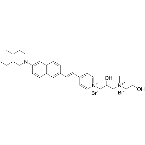
Di-4-ANEPPDHQ is a polarity-sensitive membrane probe with a doubly positive charge that can be used to visualize membrane microdomains in living Arabidopsis cells. Di-4-ANEPPDHQ produces a fluorescence spectrum shift by changing the polarity of the lipid environment after embedding into the cell membrane, thereby quantifying the degree of lipid accumulation. The excitation wavelength of Di-4-ANEPPDHQ is 488 nm, and the emission wavelengths are 560 nm and 650 nm in the liquid ordered and disordered phases, respectively. Di-4-ANEPPDHQ is often used for fluorescence imaging studies of membrane microstructure, lipid order, and membrane tension response in living cells[1][2][3].
For research use only. We do not sell to patients.
- Purity : 98.68%
- CAS No.: 797785-10-5
- Formula: C32H47Br2N3O2
- Molecular Weight:665.54
-
Storage:
4°C, sealed storage, away from moisture and light
* In solvent : -80°C, 6 months; -20°C, 1 month (sealed storage, away from moisture and light)

To place orders, for customer services and technical support, please contact: MedChemExpress USA
Tel: 609-228-6898 E-mail: [email protected] [email protected]


-
 Biological Activity
-
Chemical Information
-
Solvent & Solubility
- Protocols
- Purity & Documentation
- References
-
 Help & FAQs



Publications Citing Use of MedChemExpress (MCE) Di-4-ANEPPDHQ
More

Biological Activity
Description
In Vitro
Guide (The following is our recommended protocol. This protocol is only a guide and should be modified according to your specific needs).
I. Arabidopsis thaliana live cell staining[1]
Steps:
1. Sample preparation: 3-day-old Arabidopsis seedlings were placed in 1/2 MS medium, and root hairs and epidermal cells were taken for staining.
2. Staining operation: Add 5 μM Di-4-ANEPPDHQ (dissolved in 1/2 MS medium), incubate on ice for 5 minutes, and wash with cold 1/2 MS medium for 1 minute.
3. Plasmolysis verification: Treat with 650 mM Sorbitol (HY-B0400) for 5 minutes to confirm that the dye labels the cell membrane rather than the cell wall.
4. Fluorescence detection: using a confocal microscope (Leica SP5), 488 nm laser excitation, dual-channel acquisition: green channel 500-580 nm, red channel 620-750 nm.
5. GP value calculation: using ImageJ macro program to process images.
II. Detection of A549 cell membrane tension response[2]
Steps:
1. Cell culture: A549 cells were cultured to confluence or non-confluence, digested with trypsin (HY-B2118) and plated on glass-bottom culture dishes.
2. Osmotic pressure treatment:
Hyperosmotic pressure: Add medium containing 100 mM sucrose and incubate for 10 minutes;
Hypoosmotic pressure: Mild hypotonic pressure (25% 1 mM CaCl2+75% medium) or severe hypotonic pressure (50% 1 mM CaCl2 + 50% medium) treatment for 10 minutes.
3. Staining operation: Add 1-5 μM Di-4-ANEPPDHQ and incubate at room temperature for 1-30 minutes (short-term labeling of cell membranes, long-term visualization of endosomes).
4. Fluorescence detection: Excitation wavelength 488 nm, emission channels 500-580 nm and 620-750 nm, use Zeiss 780 microscope to collect spectral images.
5. Data analysis: Analyze lipid accumulation by changes in GP value. The decrease in GP value after hypotonic treatment indicates that lipids are loosely arranged.
III. Liposomes and giant plasma membrane vesicles (GPMVs)[3]
Steps:
1. Liposome preparation:
POPC and cholesterol are dissolved in chloroform in proportion, evaporated into film under nitrogen flow, and vacuum dried overnight;
HEPES buffer (10 mM HEPES (HY-D0857), 150 mM NaCl, pH 7.0) is added for hydration for 30 minutes, vortexed and extruded through a 100 nm polycarbonate membrane, with a lipid concentration of 0.5 mM.
2. Dye incorporation: Di-4-ANEPPDHQ (aqueous solution) was added to the hydrated liposomes at a molar ratio of 1:100, with a final concentration of about 10 μM.
3. GPMVs preparation: RBL-2H3 cells were treated with 25 mM paraformaldehyde (HY-Y0333) + 10 mM DTT (HY-15917) at 37°C for 1 hour, collected and stained with 250 nM Di-4-ANEPPDHQ for 30 minutes.
4. Spectral detection:
Steady-state spectroscopy: Fluorolog-3 spectrometer, 488 nm excitation, emission range 500-700 nm;
Time-resolved spectroscopy (TRES): 470 nm pulsed laser excitation, detection of 540-700 nm emission, analysis of fluorescence decay kinetics.
MedChemExpress (MCE) has not independently confirmed the accuracy of these methods. They are for reference only. Further protocols information, click here.
Emission (Em)
635
Excitation (Ex)
465


Chemical Information
-
CAS No. 797785-10-5
-
Appearance Solid
-
Molecular Weight 665.54
-
Formula C32H47Br2N3O2
-
Color Brown to reddish brown
-
SMILES
CCCCN(C1=CC(C=C2)=C(C=C1)C=C2/C=C/C(C=C3)=CC=[N+]3CC(O)C[N+](C)(C)CCO)CCCC.[Br-].[Br-]
-
Shipping
Room temperature in continental US; may vary elsewhere.
-
Storage
4°C, sealed storage, away from moisture and light
* In solvent : -80°C, 6 months; -20°C, 1 month (sealed storage, away from moisture and light)


Publications (1)
-
Journal Impact Factor
-
Most Recent
-
Food Funct
Phytosterol-mediated disturbance of the lipid packing order in the plasma membrane regulates inflammatory response. [Abstract]2025 Jul 28;16(15):6150-6159. PMID: 40654019


Solvent & Solubility
In Vitro:
DMSO : 50 mg/mL (75.13 mM; Need ultrasonic; Hygroscopic DMSO has a significant impact on the solubility of product, please use newly opened DMSO)
Please refer to the solubility information to select the appropriate solvent. Once prepared, please aliquot and store the solution to prevent product inactivation from repeated freeze-thaw cycles.
Storage method and period of stock solution: -80°C, 6 months; -20°C, 1 month (sealed storage, away from moisture and light). When stored at -80°C, please use it within 6 months. When stored at -20°C, please use it within 1 month.
Please refer to the solubility information to select the appropriate solvent. Once prepared, please aliquot and store the solution to prevent product inactivation from repeated freeze-thaw cycles.
Storage method and period of stock solution: -80°C, 6 months; -20°C, 1 month (sealed storage, away from moisture and light). When stored at -80°C, please use it within 6 months. When stored at -20°C, please use it within 1 month.
Concentration (start) × Volume (start) = Concentration (final) × Volume (final)

Protocols
-
3T3-L1 preadipocyte-to-adipocyte differentiation
3T3-L1 preadipocytes are induced to differentiate after growth arrest using adipogenic media containing insulin, dexamethasone, and IBMX; differentiation is assessed by lipid-droplet accumulation, triglyceride increase, Oil Red O staining, and adipocyte-marker induction such as PPARγ and C/EBPα.
-
Bioluminescent/Fluorescent Imaging Xenograft
Bioluminescent and fluorescent imaging xenograft models use tumor cells engineered to express optical reporters so tumor engraftment, growth, dissemination, and treatment response can be monitored longitudinally in living animals and validated ex vivo. Bioluminescence imaging usually measures luciferase activity after substrate administration and is commonly used as a surrogate for viable reporter-expressing tumor burden, while fluorescence imaging measures reporter or probe emission and can support tumor localization, ex vivo confirmation, or complementary multimodal analysis.
-
Lipid Droplets: Oil Red O/Sudan Dye Lipid Staining
Lipid droplets are intracellular organelles with a neutral-lipid core that stores triacylglycerols and sterol esters, and Oil Red O or Sudan dyes detect these hydrophobic lipid deposits by partitioning into retained lipids in fresh or frozen specimens. Oil Red O stains neutral triglycerides and lipids in frozen tissue sections or air-dried cytologic preparations, while Sudan Black B has also been used as a histochemical fat stain for lipid-rich tissue structures.


Purity & Documentation
-
Data Sheet (275 KB)
-
SDS (252 KB)
- English - EN (252 KB)
- Français - FR (252 KB)
- Deutsch - DE (252 KB)
- Norwegian - NO (252 KB)
- Español - ES (252 KB)
- Swedish - SV (252 KB)
- Italian - IT (252 KB)
- Korean - KR (252 KB)
- Portuguese - PT (252 KB)
-
Handling Instructions (2659 KB)
References
[1]. Xiaoyu Zhao, et al. Di-4-ANEPPDHQ, a fluorescent probe for the visualisation of membrane microdomains in living Arabidopsis thaliana cells. Plant Physiol Biochem. 2015 Feb;87:53-60. [Content Brief]
[3]. Amaro M, et al. Laurdan and Di-4-ANEPPDHQ probe different properties of the membrane. J Phys D Appl Phys. 2017 Apr 5;50(13):134004. [Content Brief]


Complete Stock Solution Preparation Table
Please refer to the solubility information to select the appropriate solvent. Once prepared, please aliquot and store the solution to prevent product inactivation from repeated freeze-thaw cycles.
Storage method and period of stock solution: -80°C, 6 months; -20°C, 1 month (sealed storage, away from moisture and light). When stored at -80°C, please use it within 6 months. When stored at -20°C, please use it within 1 month.
| Optional Solvent | Concentration Solvent Mass | 1 mg | 5 mg | 10 mg | 25 mg |
|---|---|---|---|---|---|
| DMSO | 1 mM | 1.5025 mL | 7.5127 mL | 15.0254 mL | 37.5635 mL |
| 5 mM | 0.3005 mL | 1.5025 mL | 3.0051 mL | 7.5127 mL | |
| 10 mM | 0.1503 mL | 0.7513 mL | 1.5025 mL | 3.7563 mL | |
| 15 mM | 0.1002 mL | 0.5008 mL | 1.0017 mL | 2.5042 mL | |
| 20 mM | 0.0751 mL | 0.3756 mL | 0.7513 mL | 1.8782 mL | |
| 25 mM | 0.0601 mL | 0.3005 mL | 0.6010 mL | 1.5025 mL | |
| 30 mM | 0.0501 mL | 0.2504 mL | 0.5008 mL | 1.2521 mL | |
| 40 mM | 0.0376 mL | 0.1878 mL | 0.3756 mL | 0.9391 mL | |
| 50 mM | 0.0301 mL | 0.1503 mL | 0.3005 mL | 0.7513 mL | |
| 60 mM | 0.0250 mL | 0.1252 mL | 0.2504 mL | 0.6261 mL |


Di-4-ANEPPDHQ Related Classifications
HY-D1428 Related Classifications
 Powered by Bioz