



Diallyl disulfide
Based on 1 publication(s) in Google Scholar
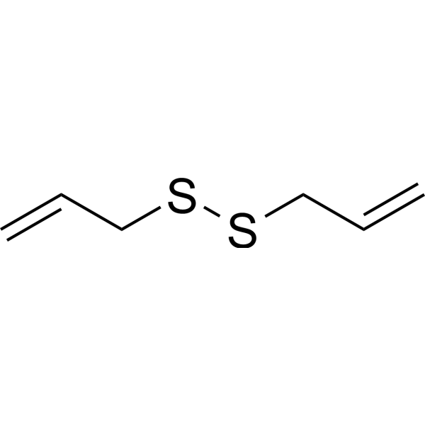
Diallyl disulfide is an orally active decomposition product of allicin and has a pungent odor. Diallyl disulfide can act as a hydrogen sulfide donor. Diallyl disulfide has inhibitory activity against squalene monooxygenase with an IC50 of 400 μM. Diallyl disulfide can alleviate the cytotoxicity and apoptosis of intestinal and liver cells induced by Ethyl Carbamate (HY-B1207). Diallyl disulfide can prevent emphysema induced by cigarette smoke. Diallyl disulfide also has anti-colon cancer and anti-inflammatory neurogenic activities, and can reverse the depressive-like behavior of mice.
For research use only. We do not sell to patients.
- Purity : 99.8%
- CAS No.: 2179-57-9
- Formula: C6H10S2
- Molecular Weight:146.27
-
Storage:
4°C, stored under nitrogen
* In solvent : -80°C, 6 months; -20°C, 1 month (stored under nitrogen)

To place orders, for customer services and technical support, please contact: MedChemExpress USA
Tel: 609-228-6898 E-mail: [email protected] [email protected]


-
 Biological Activity
-
Chemical Information
-
Solvent & Solubility
- Protocols
- Purity & Documentation
- References
-
 Help & FAQs


-
HY-117235
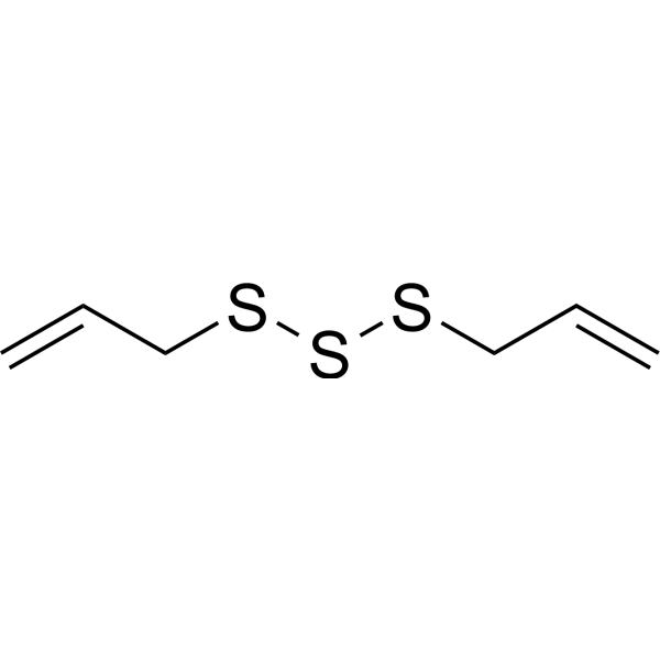
Diallyl TrisulfideAnticancer Agent -
HY-N0315
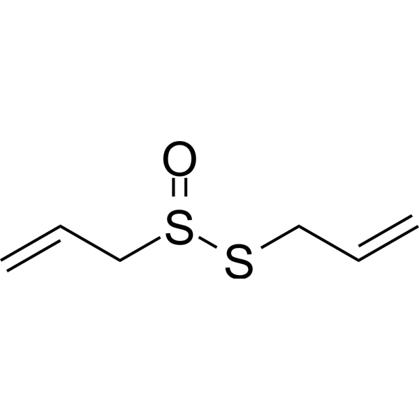
AllicinBacterial Inhibitor -
HY-128447
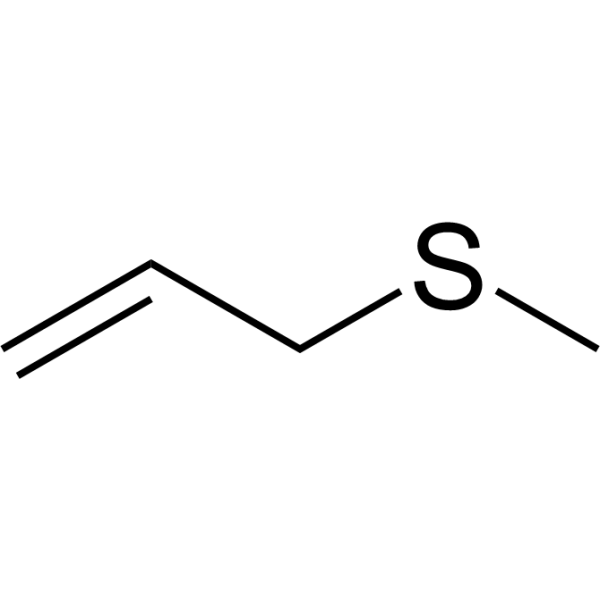
Allyl methyl sulfideOrganic Sulfide -

HY-W015635R
Diallyl disulfide (Standard)Standard of Diallyl disulfide -
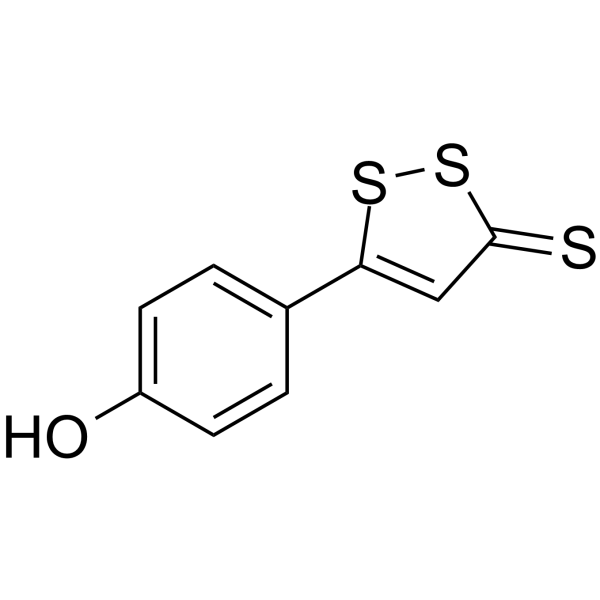
HY-109582
ADT-OHAnti-cancer Agent -
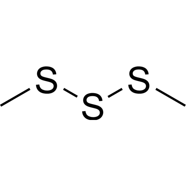
HY-128454
Dimethyl trisulfideCyanide Scavenger -
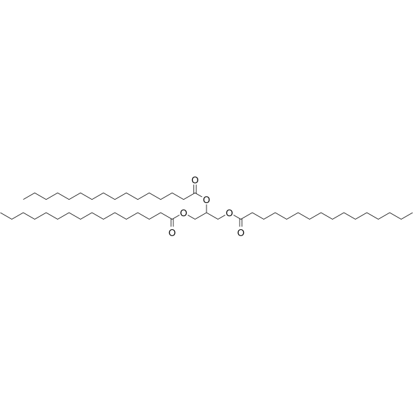
HY-W013061
Propane-1,2,3-triyl tripalmitateGlycerol Tristearate Lymphatic Transport Promoter -
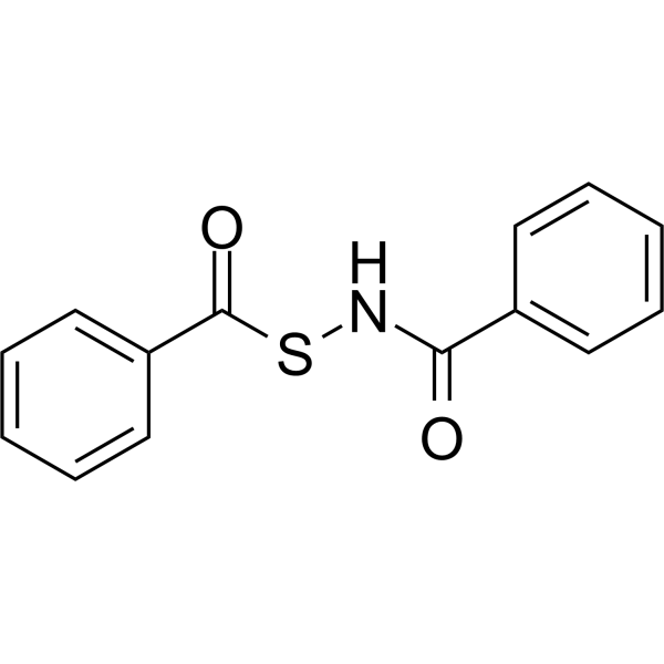
HY-130347
H2S Donor 5aH2S Donor -

HY-W196803
Dipropyl disulfideEndogenous Metabolites -
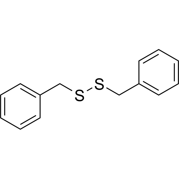
HY-W009516
Dibenzyl disulfide -
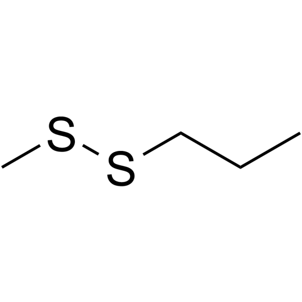
HY-N7436
Methyl propyl disulfideSulfide
Publications Citing Use of MedChemExpress (MCE) Diallyl disulfide
More

Biological Activity
Description
Cellular Effect
|
Cell Line

|
Type | Value | Description | References |
|---|---|---|---|---|
| A549 | IC50 |
100 μM
Compound: Diallyl disulfide
|
Antiproliferative activity against human A549 cells
Antiproliferative activity against human A549 cells
|
[PMID: 30245394] |
| Caco-2 | IC50 |
100 μM
Compound: Diallyl disulfide
|
Antiproliferative activity against human Caco2 cells
Antiproliferative activity against human Caco2 cells
|
[PMID: 30245394] |
| CHO | EC50 |
7.55 μM
Compound: 3
|
Agonist activity at human TRPA1 channel expressed in CHO cells assessed as increase in intracellular calcium levels
Agonist activity at human TRPA1 channel expressed in CHO cells assessed as increase in intracellular calcium levels
|
[PMID: 20356305] |
| DLD-1 | IC50 |
100 μM
Compound: Diallyl disulfide
|
Antiproliferative activity against human DLD1 cells
Antiproliferative activity against human DLD1 cells
|
[PMID: 30245394] |
| HCT-15 | IC50 |
100 μM
Compound: Diallyl disulfide
|
Antiproliferative activity against human HCT15 cells
Antiproliferative activity against human HCT15 cells
|
[PMID: 30245394] |
| MDCK | IC50 |
1.82 mM
Compound: 2
|
Cytotoxicity against MDCK cells assessed as decrease in cell viability after 16 hrs by tetrazolium salt WST-1 assay
Cytotoxicity against MDCK cells assessed as decrease in cell viability after 16 hrs by tetrazolium salt WST-1 assay
|
[PMID: 23163425] |
| SK-MEL-2 | IC50 |
100 μM
Compound: Diallyl disulfide
|
Antiproliferative activity against human SK-MEL-2 cells
Antiproliferative activity against human SK-MEL-2 cells
|
[PMID: 30245394] |
In Vitro
Diallyl disulfide (30-240 μM; 12-48 h) suppresses HT-29 cell growth[2].
Diallyl disulfide (30-120 μM; 24 h) induces cell cycle arrest in the G2/M-phase in HT-29 cells in a concentration-independent manner[2].
Diallyl disulfide (120 μM) induces the mRNA levels of ARPC3, SPINT2, SOD1, p21 and TACSTD2[2].
Diallyl disulfide (10-120 μM; 24 h) can protect Caco-2 and HepG2 cells from the cytotoxicity induced by ethyl carbamate. Compared with the group exposed to ethyl carbamate alone, it can significantly increase the cell survival rate[5].
Diallyl disulfide (10-40 μM; 24 h) can reduce the ROS production induced by carbamate exposure for 24 hours in Caco-2 and HepG2 cells[5].
Diallyl disulfide (10-40 μM; 24 h) can protect Caco-2 and HepG2 cells from the apoptosis and necrosis induced by carbamate. Compared with the group exposed to carbamate alone, it can significantly reduce the proportion of apoptotic and necrotic cells[5].
MedChemExpress (MCE) has not independently confirmed the accuracy of these methods. They are for reference only. Further protocols information, click here.
-
Cell Line:HT-29 cells
-
Concentration:30, 60, 120 and 240 μM
-
Incubation Time:12, 24, 48 h
-
Result:Suppressed HT-29 cell growth by 5.3, 23.1, 45.6 and 68.3% with 30, 60, 120 and 240 µM for 24 h, respectively.
-
Cell Line:HT-29 cells
-
Concentration:30, 60, 120 μM
-
Incubation Time:24 h
-
Result:The percentage of cells in the G2/M-phase was found to be 23.6, 25.9, 39.7 and 61.0% with 30, 60, 120 and 240 µM, respectively.
-
Cell Line:human colorectal adenocarcinoma (Caco-2) and hepatocarcinoma (HepG2) cells
-
Concentration:10, 20 and 40 μM
-
Incubation Time:24 h
-
Result:Reduced ethyl carbamate-induced apoptosis (increased by 20% vs negative control) and necrosis (increased by 5% vs negative control) in Caco-2 cells by approximately 70% vs ethyl carbamate alone. Reduced ethyl carbamate-induced apoptosis and necrosis (increased by 10% vs negative control) in HepG2 cells compared to ethyl carbamate alone.
Did not induce apoptosis or necrosis in either cell line vs solvent control when used alone.
In Vivo
Diallyl disulfide (30-90 mg/kg; i.g.; three times/week for the last 30 days) dose-dependently reduces cigarette smoke (CS) -induced leukocyte infiltration into the airways and improves lung histology in C57BL/6 mice were exposed to CS for 60 consecutive days[4].
MedChemExpress (MCE) has not independently confirmed the accuracy of these methods. They are for reference only.
-
Animal Model:Male C57BL6/J mice (6-8 weeks)[3]
-
Dosage:40, 80 mg/kg
-
Administration:IP; pre-treatment 1 h before a single LPS injection (100 μg/kg)
-
Result:Prevented LPS-induced increases in immobility time in the TST and FST in mice.
Did not affect the locomotor activity in mice with or without LPS treatment.


Chemical Information
-
CAS No. 2179-57-9
-
Appearance Liquid (Density: 1.010 g/cm3)
-
Molecular Weight 146.27
-
Formula C6H10S2
-
Color Colorless to light yellow
-
SMILES
C=CCSSCC=C
-
Structure Classification
-
Initial Source
-
Shipping
Room temperature in continental US; may vary elsewhere.
-
Storage
4°C, stored under nitrogen
* In solvent : -80°C, 6 months; -20°C, 1 month (stored under nitrogen)


Publications (1)
-
Journal Impact Factor
-
Most Recent


Solvent & Solubility
In Vitro:
DMSO : 100 mg/mL (683.67 mM; Need ultrasonic; Hygroscopic DMSO has a significant impact on the solubility of product, please use newly opened DMSO)
Please refer to the solubility information to select the appropriate solvent. Once prepared, please aliquot and store the solution to prevent product inactivation from repeated freeze-thaw cycles.
Storage method and period of stock solution: -80°C, 6 months; -20°C, 1 month (stored under nitrogen). When stored at -80°C, please use it within 6 months. When stored at -20°C, please use it within 1 month.
Please refer to the solubility information to select the appropriate solvent. Once prepared, please aliquot and store the solution to prevent product inactivation from repeated freeze-thaw cycles.
Storage method and period of stock solution: -80°C, 6 months; -20°C, 1 month (stored under nitrogen). When stored at -80°C, please use it within 6 months. When stored at -20°C, please use it within 1 month.
Concentration (start) × Volume (start) = Concentration (final) × Volume (final)
In Vivo:
Select the appropriate dissolution method based on your experimental animal and administration route.
- For the following dissolution methods, please ensure to first prepare a clear stock solution using an In Vitro approach and then sequentially add co-solvents:
- To ensure reliable experimental results, the clarified stock solution can be appropriately stored based on storage conditions. As for the working solution for In Vivo experiments, it is recommended to prepare freshly and use it on the same day.
- The percentages shown for the solvents indicate their volumetric ratio in the final prepared solution. If precipitation or phase separation occurs during preparation, heat and/or sonication can be used to aid dissolution.
Add each solvent one by one: 10% DMSO 40% PEG300 5% Tween-80 45% Saline
Solubility: ≥ 2.5 mg/mL (17.09 mM); Clear solution
This protocol yields a clear solution of ≥ 2.5 mg/mL (saturation unknown).
Taking 1 mL working solution as an example, add 100 μL DMSO stock solution (25.0 mg/mL) to 400 μL PEG300, and mix evenly; then add 50 μL Tween-80 and mix evenly; then add 450 μL Saline to adjust the volume to 1 mL.
Preparation of Saline: Dissolve 0.9 g sodium chloride in ddH₂O and dilute to 100 mL to obtain a clear Saline solution.
Add each solvent one by one: 10% DMSO 90% (20% SBE-β-CD in Saline)
Solubility: ≥ 2.5 mg/mL (17.09 mM); Clear solution
This protocol yields a clear solution of ≥ 2.5 mg/mL (saturation unknown).
Taking 1 mL working solution as an example, add 100 μL DMSO stock solution (25.0 mg/mL) to 900 μL 20% SBE-β-CD in Saline, and mix evenly.
Preparation of 20% SBE-β-CD in Saline (4°C, storage for one week): 2 g SBE-β-CD powder is dissolved in 10 mL Saline, completely dissolve until clear.
In Vivo Dissolution Calculator
Please enter the basic information of animal experiments:
-
-
-
-
Recommended: Prepare an additional quantity of animals to account for potential losses during experiments.
Please enter your animal formula composition:
-
%DMSO +
Recommended: Keep the proportion of DMSO in working solution below 2% if your animal is weak.
-
%+
-
+%Tween-80 + +
-
%Saline +
The co-solvents required include: DMSO, . All of co-solvents are available by MedChemExpress (MCE). , Tween 80. All of co-solvents are available by MedChemExpress (MCE).
Working solution concentration: 0.22 mg/mL
Method for preparing stock solution: mg drug dissolved in μL DMSO. Stock solution concentration: mg/mL. * In solvent : -80°C, 6 months; -20°C, 1 month (stored under nitrogen)
1. Take μL DMSO stock solution;
2. Add μL .
μL , mix evenly;
3. Then add μL Tween 80, mix evenly;
4. Then add μL
Please ensure that the stock solution in the first step is dissolved to a clear state, and add co-solvents in sequence. You can use ultrasonic heating (ultrasonic cleaner, recommended frequency 20-40 kHz), vortexing, etc. to assist dissolution.

Protocols
-
Apoptosis
Apoptosis, also called programmed cell death, is generally characterized by distinct morphological characteristics.
-
TUNEL staining for apoptotic DNA fragmentation
TUNEL staining detects DNA strand breaks by using terminal deoxynucleotidyl transferase to add labeled nucleotides to exposed 3′-OH DNA termini, generating either microscopic staining in fixed cells or tissue sections, or fluorescence/cytometric signal in cell suspensions. TUNEL positivity reflects DNA fragmentation but should not be interpreted alone as definitive apoptosis, because TUNEL can also label necrotic, autolytic, mechanically damaged, or DNA-repair-associated DNA breaks.
-
Cell Cytotoxicity Assay
Cytotoxicity assays are usually based on the assessment of cell membrane damage, which can also be indirectly detected by measuring cell viability. Detection methods include MTT assay, CKK-8 assay, LDH assay and ATP assay, etc.
-
Mammalian live/dead viability and cytotoxicity staining
Live/dead viability and cytotoxicity staining assays are based on the simultaneous detection of intracellular esterase activity in metabolically active (viable) cells and membrane integrity loss in non-viable cells. In commonly used dual-staining approaches, membrane-permeant fluorogenic substrates are converted by intracellular esterases into fluorescent products in live cells, while impermeant DNA-binding dyes selectively enter cells with compromised plasma membranes and label nucleic acids in dead or dying cells, enabling discrimination between viable and non-viable populations by fluorescence microscopy or flow cytometry.
-
Annexin V plus membrane-impermeant dye apoptosis staining
Annexin V-based apoptosis assays rely on the detection of phosphatidylserine (PS) externalization from the inner leaflet of the plasma membrane to the outer leaflet, an early biochemical hallmark of apoptosis. Fluorescently labeled Annexin V binds PS in a calcium-dependent manner, enabling identification of early apoptotic cells by flow cytometry or fluorescence microscopy. When combined with a membrane-impermeant DNA-binding dye (e. g. , propidium iodide), this approach allows discrimination between viable (Annexin V−/dye−), early apoptotic (Annexin V+/dye−), and late apoptotic or necrotic (Annexin V+/dye+) cell populations by assessing membrane integrity and PS exposure.
-
Protocol for Tail Suspension Test (TST)
The Tail Suspension Test is a mouse behavioral assay in which an animal is suspended by the tail in an inescapable position, causing alternating escape-directed activity and immobility; the main readout is immobility time, and antidepressant-like treatments generally reduce immobility compared with vehicle controls. The assay detects behavioral response to acute inescapable stress rather than a molecular event; immobility is interpreted as passive stress-coping behavior, while reduced immobility is used as a predictive screen for antidepressant-like activity, with important limitations related to strain, locomotor activity, and tail-climbing behavior.
-
Apoptosis Solutions
Apoptosis is a regulated, generally non-lytic cell-death pathway that removes unwanted, damaged, infected, or abnormal cells through coordinated morphological changes, caspase activation, DNA fragmentation, and membrane remodeling. The intrinsic apoptosis pathway is controlled mainly by mitochondrial outer membrane permeabilization, BCL-2 family proteins, cytochrome c release, apoptosome formation, caspase-9 activation, and downstream executioner caspase-3/7 activation. The extrinsic apoptosis pathway is initiated by death receptors such as Fas, TNFR, and TRAIL receptors, which recruit adaptor proteins and activate caspase-8 before engaging executioner caspases or mitochondrial amplification through BID cleavage. Apoptosis is linked to many phenotypes, including cancer cell killing, tissue homeostasis, immune regulation, neurodegeneration, infection response, and treatment-induced cytotoxicity; unresolved questions include how apoptosis interacts with necroptosis, pyroptosis, ferroptos
-
Research Protocol for Inflammation-related Diseases
The NLRP3 inflammasome is a cytosolic innate immune signaling platform that integrates priming signals and danger-signal activation to promote caspase-1 activation, maturation of IL-1β and IL-18, and gasdermin D-mediated pyroptotic cell death. The core experimental logic is to determine whether inflammatory disease phenotypes are driven by increased NLRP3 expression, ASC-containing inflammasome assembly, caspase-1 cleavage, GSDMD cleavage, and extracellular release of IL-1β/IL-18 rather than by nonspecific cell injury alone. The pathway is strongly linked to inflammation-related disease phenotypes because monosodium urate crystals activate NALP3/NLRP3 inflammasome signaling in gout-like crystal inflammation, cholesterol crystals activate NLRP3 inflammasomes in atherogenesis models, and DSS-induced intestinal inflammation has been reported to involve NLRP3 inflammasome activity. However, experimental colitis studies also show context-dependent protective effects of NLRP3 inflammasome co
-
Protocol for Forced Swim Test (FST)
The Forced Swim Test is a rodent behavioral assay in which a mouse or rat is placed in an inescapable cylinder of water, and the main readout is the time spent immobile versus active escape-related behaviors such as swimming or climbing. Reduced immobility after treatment has historically been interpreted as antidepressant-like activity, but the assay should be interpreted as a behavioral response to acute inescapable stress rather than a complete model of human depression.
-
Protocol for Sucrose Preference Test (SPT)
The Sucrose Preference Test is a rodent two-bottle choice assay used to estimate reward-related behavior by measuring preference for a sweet sucrose solution over water. Reduced sucrose preference is commonly interpreted as an anhedonia-like phenotype in stress-based depression models, but it can also be affected by thirst, hunger, body weight, learning, motivation, and general fluid intake.
-
Cell Viability Determination by MTT Colorimetric Assay
The following protocol uses the MTT colorimetric assay as a classic literature-established method for assessing cell viability/metabolic activity in cultured mammalian cells. MTT[3-(4,5-dimethylthiazol-2-yl)-2,5-diphenyltetrazolium bromide] is reduced by metabolically active cells to a colored formazan product; the amount of formazan is quantified spectrophotometrically and provides an indirect measure of metabolically active viable cells. Importantly, MTT reduction reflects cellular oxidoreductase/metabolic activity rather than an absolute direct count of living cells, so changes in cellular metabolism can alter the signal independently of cell number.


Purity & Documentation
-
Data Sheet (276 KB)
-
SDS (481 KB)
- English - EN (481 KB)
- Français - FR (481 KB)
- Deutsch - DE (481 KB)
- Norwegian - NO (481 KB)
- Español - ES (481 KB)
- Swedish - SV (481 KB)
- Italian - IT (481 KB)
- Korean - KR (481 KB)
- Portuguese - PT (481 KB)
-
Handling Instructions (2659 KB)
References
[1]. Gupta N, et al. Garlic and garlic-derived compounds inhibit human squalene monooxygenase. J Nutr. 2001;131(6):1662-1667. [Content Brief]
[2]. You-Sheng Huang, et al. Diallyl disulfide inhibits the proliferation of HT-29 human colon cancer cells by inducing differentially expressed genes. Mol Med Rep. 2011 May-Jun;4(3):553-9. [Content Brief]
[3]. Xiaoyou Wei, et al. Acute Diallyl Disulfide Administration Prevents and Reveres Lipopolysaccharide-Induced Depression-Like Behaviors in Mice via Regulating Neuroinflammation and Oxido-Nitrosative Stress. Inflammation. 2021 Aug;44(4):1381-1395. [Content Brief]
[4]. Aline de Oliveira Pontes Cardoso, et al. Diallyl disulfide prevents cigarette smoke-induced emphysema in mice. Pulm Pharmacol Ther. 2021 Aug:69:102053. [Content Brief]
[5]. Sanchez CA, et al. Diallyl Disulfide Reduces Ethyl Carbamate-Induced Cytotoxicity and Apoptosis in Intestinal and Hepatic Cells. Chem Res Toxicol. 2025 Apr 21;38(4):623-634. [Content Brief]


Complete Stock Solution Preparation Table
Please refer to the solubility information to select the appropriate solvent. Once prepared, please aliquot and store the solution to prevent product inactivation from repeated freeze-thaw cycles.
Storage method and period of stock solution: -80°C, 6 months; -20°C, 1 month (stored under nitrogen). When stored at -80°C, please use it within 6 months. When stored at -20°C, please use it within 1 month.
| Optional Solvent | Concentration Solvent Mass | 1 mg | 5 mg | 10 mg | 25 mg |
|---|---|---|---|---|---|
| DMSO | 1 mM | 6.8367 mL | 34.1834 mL | 68.3667 mL | 170.9168 mL |
| 5 mM | 1.3673 mL | 6.8367 mL | 13.6733 mL | 34.1834 mL | |
| 10 mM | 0.6837 mL | 3.4183 mL | 6.8367 mL | 17.0917 mL | |
| 15 mM | 0.4558 mL | 2.2789 mL | 4.5578 mL | 11.3945 mL | |
| 20 mM | 0.3418 mL | 1.7092 mL | 3.4183 mL | 8.5458 mL | |
| 25 mM | 0.2735 mL | 1.3673 mL | 2.7347 mL | 6.8367 mL | |
| 30 mM | 0.2279 mL | 1.1394 mL | 2.2789 mL | 5.6972 mL | |
| 40 mM | 0.1709 mL | 0.8546 mL | 1.7092 mL | 4.2729 mL | |
| 50 mM | 0.1367 mL | 0.6837 mL | 1.3673 mL | 3.4183 mL | |
| 60 mM | 0.1139 mL | 0.5697 mL | 1.1394 mL | 2.8486 mL | |
| 80 mM | 0.0855 mL | 0.4273 mL | 0.8546 mL | 2.1365 mL | |
| 100 mM | 0.0684 mL | 0.3418 mL | 0.6837 mL | 1.7092 mL |


Diallyl disulfide Related Classifications
HY-W015635 Related Classifications
 Powered by Bioz