



DIBA-Cy5
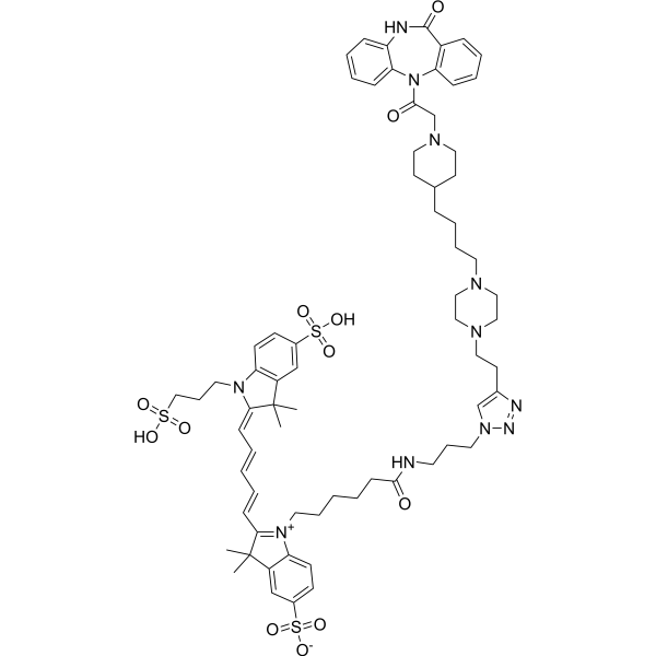
DIBA-Cy5 is a fluorescent DIBA antagonist made up be DIBA-alkyne binding Cyanine5 fluorophores (Cy5) and polyethylene glycol (PEG) biomolecules. DIBA-Cy5 can serve as a fluorescent ligand, suitable for probe attachment through click chemistry. DIBA-Cy5 exerts a high binding affinity to type-2 mAChR (M2R) with the Kd value of 1.80 nM, can directly stain M2R receptors in the sinoatrial node of a mouse heart.
For research use only. We do not sell to patients.
- Formula: C69H89N11O12S3
- Molecular Weight:1360.71
-
Storage:
Please store the product under the recommended conditions in the Certificate of Analysis.

To place orders, for customer services and technical support, please contact: MedChemExpress USA
Tel: 609-228-6898 E-mail: [email protected] [email protected]


-
 Biological Activity
-
Chemical Information
- Protocols
- Purity & Documentation
- References
-
 Help & FAQs




Biological Activity
Description
IC50 & Target
[1]|
mAChR2 1.80 nM (Kd) |
mAChR1 104.5 nM (Kd) |
In Vitro
DIBA-Cy5 (0.01 nM-1 mM; 5 h) shows selective binding property for type-2 mAChR (M2R) over M1R, with Kd of 1.08 nM and 104.5 nM, respectively[1].
DIBA-Cy5 (10 nM; 3 h) exerts the competitive binding mode with orthosteric antagonist Atropine (HY-B1205)/allosteric modulator LY2119620 (HY-15885), indicating a dualsteric binding mode of the DIBA-type antagonist to M2R[1].
DIBA-Cy5 (50 nM; 16 h) results direct staining to M2R receptors in the sinoatrial node of a mouse heart. DIBA-Cy5 can be used binding assays that screen compounds for M2R as the receptor target[1].
MedChemExpress (MCE) has not independently confirmed the accuracy of these methods. They are for reference only. Further protocols information, click here.
Emission (Em)
668
Excitation (Ex)
647


Chemical Information
-
Molecular Weight 1360.71
-
Formula C69H89N11O12S3
-
SMILES
CC1(C2=CC(S(=O)([O-])=O)=CC=C2[N+](CCCCCC(NCCCN3C=C(N=N3)CCN4CCN(CC4)CCCCC5CCN(CC5)CC(N6C7=CC=CC=C7NC(C8=C6C=CC=C8)=O)=O)=O)=C1/C=C/C=C/C=C9N(C%10=C(C/9(C)C)C=C(C=C%10)S(=O)(O)=O)CCCS(=O)(O)=O)C
-
Shipping
Room temperature in continental US; may vary elsewhere.
-
Storage
Please store the product under the recommended conditions in the Certificate of Analysis.

Protocols
-
EdU Incorporation Assay (Click Chemistry-Based DNA Synthesis Measurement)
The EdU incorporation assay measures DNA synthesis by adding the thymidine analog 5-ethynyl-2′-deoxyuridine to cells or tissues, where it is incorporated into newly synthesized DNA during S phase. Incorporated EdU is detected by copper-catalyzed azide-alkyne cycloaddition, in which a fluorescent azide covalently reacts with the ethynyl group on EdU, allowing S-phase cells to be detected by fluorescence microscopy, flow cytometry, or high-content imaging. EdU detection does not require DNA denaturation or anti-BrdU antibody access, which preserves sample structure and improves compatibility with immunostaining and multiparameter cytometry compared with BrdU-based detection. EdU can be cytotoxic in a cell-type- and exposure-dependent manner, so pulse duration, concentration, and continuous-labeling designs should be validated for each cell type.


Purity & Documentation
References

Calculators
Concentration (start) × Volume (start) = Concentration (final) × Volume (final)


DIBA-Cy5 Related Classifications
HY-151801 Related Classifications
 Powered by Bioz