



Difluorinated Curcumin

Based on 1 Customer Validation
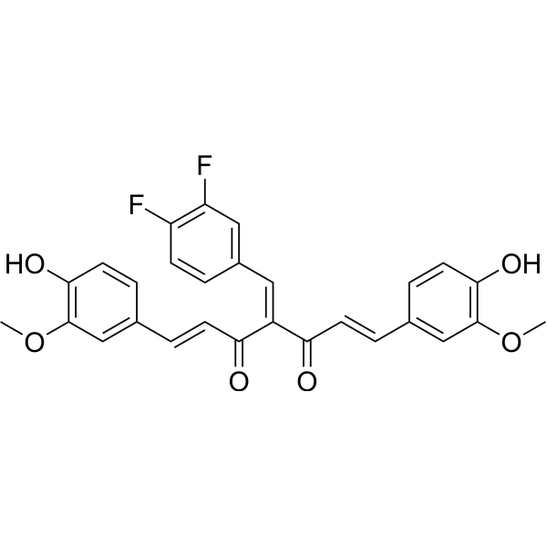
Difluorinated Curcumin (3,4-Difluorobenzylidene curcumin) is a fluorinated curcumin analog with high bioavailability and anticancer activity. Difluorinated Curcumin inhibits the self-renewal ability of tumor stem/stem-like cells, clonogenicity, invasiveness and angiogenesis of tumor cells. Difluorinated Curcumin also increases cell sensitivity to chemotherapy.
For research use only. We do not sell to patients.
- Purity : 99.56%
- CAS No.: 1170354-22-9
- Formula: C28H22F2O6
- Molecular Weight:492.47
-
Storage:Powder -20°C, 3 years ; In solvent -80°C, 6 months , -20°C, 1 month

To place orders, for customer services and technical support, please contact: MedChemExpress USA
Tel: 609-228-6898 E-mail: [email protected] [email protected]


-
 Biological Activity
-
Chemical Information
- Protocols
- Purity & Documentation
- References
-
 Help & FAQs




Biological Activity
Description


Chemical Information
-
CAS No. 1170354-22-9
-
Appearance Solid
-
Molecular Weight 492.47
-
Formula C28H22F2O6
-
Color Light yellow to yellow
-
SMILES
FC(C=C1/C=C(C(/C=C/C2=CC(OC)=C(C=C2)O)=O)/C(/C=C/C3=CC(OC)=C(C=C3)O)=O)=C(C=C1)F
-
Synonyms
3,4-Difluorobenzylidene curcumin
-
Shipping
Room temperature in continental US; may vary elsewhere.
-
Storage
Powder -20°C 3 years In solvent -80°C 6 months -20°C 1 month

Protocols
-
Human pluripotent stem cell endothelial-cell differentiation
Human pluripotent stem cell endothelial differentiation is based on stepwise developmental patterning: early activation of WNT/GSK3β inhibition promotes mesodermal or vascular progenitor entry, followed by endothelial specification using VEGF-related signaling, BMP4, FGF2, Notch modulation, or cAMP depending on the published protocol. Endothelial differentiation is read out by acquisition of CD31, CD34, VE-cadherin/CD144, KDR/VEGFR2, vWF, Tie2, NOS3, acetylated LDL uptake, tube/network formation, barrier function, and in vivo vessel-forming capacity where tested.
-
Vascular/Branching Fractal Analysis
Vascular/branching fractal analysis quantifies the geometric complexity of vessel trees or vascular networks from segmented 2D images, commonly by converting vessels into binary and/or skeletonized maps and estimating fractal dimension using box-counting or related approaches. Fractal dimension is interpreted as an image-derived readout of vascular branching complexity, space filling, or density, and has been applied to retinal photographs, fluorescein angiography, OCT angiography, capillary perfusion maps, and in vitro Matrigel angiogenesis networks. The assay readout is generated from vessel-positive pixels after image preprocessing, vessel segmentation, binarization, and optional skeletonization; reported outputs include fractal dimension, vessel density, branchpoint density, endpoint density, vessel length density, tortuosity, and generation-based branching metrics when VESGEN-style analysis is used. The biological interpretation is limited to quantitative vascular patterning and s
-
Protocol for Pharmacokinetic Study
Pharmacokinetic studies quantify how an organism handles a drug over time through absorption, distribution, metabolism, and excretion, and the core experimental readout is the concentration-time profile of parent drug and, when relevant, metabolites in biological matrices such as plasma, whole blood, urine, bile, or tissue. Pharmacokinetic analysis links dose, route, exposure, clearance, half-life, distribution, bioavailability, and systemic exposure to drug efficacy and toxicity hypotheses rather than measuring a signaling pathway directly. The literature links pharmacokinetics to drug-development phenotypes by showing that drug metabolism and pharmacokinetics influence compound progression, exposure-response interpretation, safety margins, dosing strategy, and failure risk during discovery and development. DMPK science contributes to compound optimization by integrating physicochemical properties, in vitro metabolism, transporter behavior, in vivo exposure, and pharmacodynamic contex
-
Endothelial Tube Formation Assay
Endothelial tube formation assay evaluates the ability of endothelial cells to attach, migrate, align, and organize into capillary-like networks when cultured on gelled basement membrane extract or Matrigel; the readout is the morphology and quantity of tube-like networks, which reflects an in vitro endothelial morphogenesis step related to angiogenesis. Basement membrane extract/Matrigel provides laminin-rich extracellular matrix cues that support endothelial differentiation into capillary-like structures, but it can contain biologically active growth factors, so growth-factor-reduced matrix is preferred when testing defined angiogenic stimulators or inhibitors.
-
hPSC maintenance and expansion
This protocol maintains and expands human pluripotent stem cells under feeder-free, chemically defined conditions using E8 medium and vitronectin-coated culture surfaces; the readout is sustained adherent colony growth with undifferentiated morphology and retained pluripotency-marker expression during serial passaging. E8-based hPSC culture relies on defined soluble factors and matrix-dependent adhesion rather than feeder cells; vitronectin supports hPSC attachment through integrin-mediated interactions, and EDTA passaging dissociates colonies as small aggregates without enzymatic digestion, centrifugation, or routine ROCK-inhibitor treatment.
-
Endothelial Cell Migration/Angiogenic Sprouting Assay
Endothelial cell migration and angiogenic sprouting assays are in vitro (and partially ex vivo-adapted) functional models that quantify the ability of endothelial cells to undergo coordinated migration, extracellular matrix invasion, and multicellular organization into capillary-like sprouts in response to pro-angiogenic stimuli such as VEGF, bFGF, or conditioned microenvironments. These assays are used to model early angiogenic events including tip-cell formation, directional migration, and lumen-like sprout extension, which collectively reflect angiogenic activation and vascular morphogenesis processes observed in vivo.


Purity & Documentation
-
Data Sheet (266 KB)
-
SDS (251 KB)
- English - EN (251 KB)
- Français - FR (251 KB)
- Deutsch - DE (251 KB)
- Norwegian - NO (251 KB)
- Español - ES (251 KB)
- Swedish - SV (251 KB)
- Italian - IT (251 KB)
- Korean - KR (251 KB)
- Portuguese - PT (251 KB)
-
Handling Instructions (2659 KB)
References

Calculators
Concentration (start) × Volume (start) = Concentration (final) × Volume (final)


Difluorinated Curcumin Related Classifications
HY-139211 Related Classifications
 Powered by Bioz