



Dornase alfa
Based on 1 publication(s) in Google Scholar
Dornase alfa (rhDNase) is a recombinant human deoxyribonuclease I (rhDNase) that can specifically degrade extracellular DNA. Dornase alfa catalyzes the cleavage of DNA released by neutrophils in respiratory mucus, reduces sputum viscosity, thereby improving mucus clearance efficiency, reducing airway obstruction and alleviating inflammatory responses. Dornase alfa can be used to improve lung function (such as FEV_1) in cystic fibrosis (CF), reduce the risk of acute pulmonary exacerbations, and has good in vivo tolerability. Dornase alfa acts locally on the respiratory tract through aerosol inhalation, specifically improving the high viscosity of mucus caused by DNA accumulation and related respiratory symptoms.
Para uso exclusivo en investigación. No vendemos a pacientes.
- No. CAS: 143831-71-4
-
Almacenamiento:
Please store the product under the recommended conditions in the Certificate of Analysis.


To place orders, for customer services and technical support, please contact: MedChemExpress USA
Tel: 609-228-6898 E-mail: [email protected] [email protected]


-
 Actividad biológica
-
Chemical Information
- Protocolo
- Pureza y Documentación
- Referencias
-
 Help & FAQs


Publications Citing Use of MedChemExpress (MCE) Dornase alfa
MoreVer todos los productos específicos de isoformas DNA/RNA Synthesis
More

Actividad biológica
Descripciòn
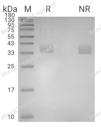
MCE Validation Data
In Vitro
In vitro enzyme activity assays of Dornase alfa (2.5 mg/2.5 mL; nebulized) showed that after treatment with different types of nebulizers (spray type, static type, and vibration type), there was no significant loss of enzyme activity, and the protein structure remained intact without aggregation or degradation[3].
MedChemExpress (MCE) has not independently confirmed the accuracy of these methods. They are for reference only. Further protocols information, click here.
In Vivo
Dornase alfa can reduce the number of tumor metastases in a mouse B16 melanoma model and change the cfDNA pattern in serum[4].
MedChemExpress (MCE) has not independently confirmed the accuracy of these methods. They are for reference only.
-
Animal Model:Female BALB/c mice (7-8 weeks old), healthy murine model[3]
-
Dosage:2.5 mg/2.5 mL
-
Administration:Nebulization via four nebulizers (jet nebulizers: PARI BOY SX-red/blue nozzles; static mesh: NE-U150; vibrating mesh: NE-SM1), single administration for 1 minute after 30 seconds of pre-nebulization
-
Result:The PARI BOY SX-red nozzle jet nebulizer delivered the highest amount of dornase alfa to the lungs, correlating with its aerosol particle size (Dv(50)=2.5-3.0 μm).
Ensayo clínico
| NCT Number | Sponsor | Condition | Start Date |
Phase

|
|---|---|---|---|---|
| NCT01329991 | Plexxikon| | 2011-05 | PHASE1 |
Specific Activity
1000 IU/mg protein
Unit Definition
One Dnase unit results in an increase in absorbance at 260nm of 0.001/minute at 25°C when acting upon highly polymerized solution of DNA at pH-5.


Chemical Information
-
No. CAS 143831-71-4
-
Appearance Liquid
-
Color Colorless to light yellow
-
SMILES
[Dornase alfa]
-
Synonyms
rhDNase
-
Envío
Room temperature in continental US; may vary elsewhere.
-
Almacenamiento
Please store the product under the recommended conditions in the Certificate of Analysis.


Publications (1)
-
Journal Impact Factor
-
Most Recent
-
Carbohydr Polym
Exploring the antibiofilm potential of chitosan nanoparticles by functional modification with chloroquine and deoxyribonuclease. [Abstract]2025 Jan 1:347:122726. PMID: 39486956

Protocolo
-
Fibrosis/Collagen Morphometry
Fibrosis and collagen morphometry is based on the quantitative visualization of fibrillar collagen deposition in tissue sections using histochemical stains such as Sirius Red (Picrosirius Red) or Masson's trichrome, followed by image-based or polarization-enhanced analysis to estimate collagen proportional area as a surrogate of extracellular matrix accumulation during fibrotic remodeling. Sirius Red combined with polarized light microscopy enhances detection of collagen fibers due to birefringence properties, enabling more specific visualization of collagen type I and III fibrils compared to conventional bright-field histology, while whole-section or region-restricted digital morphometry reduces field-selection bias in fibrosis assessment. Alternative quantitative approaches include second harmonic generation (SHG) and two-photon excited fluorescence microscopy, which enable label-free detection of fibrillar collagen and have been validated against histological staining and biochemica
-
Collagen: Sirius Red Staining
Sirius Red or picrosirius red staining is a histochemical method for visualizing collagen-rich extracellular matrix in tissue sections, and collagen fibers are detected as red-stained structures under bright-field microscopy with enhanced birefringence under polarized light. Picrosirius red is useful for assessing total collagen organization, distribution, and fibrosis burden, but polarized color should not be interpreted as a definitive collagen type I versus type III readout because color is affected by fiber orientation, thickness, and packing.
-
Connective Tissue: Masson's Trichrome/Collagen Trichrome Staining
Masson’s Trichrome (collagen/trichrome staining) is a histological technique that differentially stains tissue compartments using sequential acidic dyes to distinguish collagen from muscle and cytoplasmic components based on dye affinity and tissue permeability differences, enabling visualization of fibrosis and connective tissue architecture in histological sections. The classical formulation typically uses Weigert's iron hematoxylin for nuclear staining, Biebrich scarlet-acid fuchsin for cytoplasm and muscle, and aniline blue (or light green variants) for collagen, producing a characteristic blue/green collagen signal contrasted against red cytoplasm and dark nuclei. The staining principle relies on selective displacement of smaller dye molecules by larger anionic dyes in collagen-rich regions under controlled acidified conditions, which enhances collagen-specific dye retention. This property makes the method widely used for fibrosis assessment in organs such as heart, liver, lung, a
-
Protocol for Pharmacokinetic Study
Pharmacokinetic studies quantify how an organism handles a drug over time through absorption, distribution, metabolism, and excretion, and the core experimental readout is the concentration-time profile of parent drug and, when relevant, metabolites in biological matrices such as plasma, whole blood, urine, bile, or tissue. Pharmacokinetic analysis links dose, route, exposure, clearance, half-life, distribution, bioavailability, and systemic exposure to drug efficacy and toxicity hypotheses rather than measuring a signaling pathway directly. The literature links pharmacokinetics to drug-development phenotypes by showing that drug metabolism and pharmacokinetics influence compound progression, exposure-response interpretation, safety margins, dosing strategy, and failure risk during discovery and development. DMPK science contributes to compound optimization by integrating physicochemical properties, in vitro metabolism, transporter behavior, in vivo exposure, and pharmacodynamic contex


Pureza y Documentación
-
Ficha de datos (271 KB)
-
SDS (251 KB)
- English - EN (251 KB)
- Français - FR (251 KB)
- Deutsch - DE (251 KB)
- Norwegian - NO (251 KB)
- Español - ES (251 KB)
- Swedish - SV (251 KB)
- Italian - IT (251 KB)
- Korean - KR (251 KB)
- Portuguese - PT (251 KB)
-
Instrucciones de manejo (2659 KB)
Referencias
[1]. Connie Yang, et al. Dornase alfa for cystic fibrosis. Cochrane Database Syst Rev. 2021 Mar 18;3(3):CD001127. [Content Brief]
[2]. Konstan MW, et al. Effect of dornase alfa on inflammation and lung function: potential role in the early treatment of cystic fibrosis. J Cyst Fibros. 2012 Mar;11(2):78-83. [Content Brief]
[3]. Chang KH, et al. Aerosol Delivery of Dornase Alfa Generated by Jet and Mesh Nebulizers. Pharmaceutics. 2020 Jul 31;12(8):721. [Content Brief]

Calculators
Concentration (start) × Volume (start) = Concentration (final) × Volume (final)


Dornase alfa Related Classifications
HY-108858 Related Classifications
 Powered by Bioz