



LL-37 amide
Based on 2 publication(s) in Google Scholar
LL-37 amide is a selective agonist of formyl peptide receptor-like FPRL1, effectively inhibiting periodontal pathogens (ED99=8.5-8.7 μg/mL). LL-37 amide exerts its bactericidal effect by activating FPRL1-mediated immune cell chemotaxis and disrupting bacterial cell membrane integrity. It can also regulate inflammatory responses (inhibiting the release of factors such as TNF-α) and promote angiogenesis. Amidation modification reduces its sensitivity to serum inhibition and improves its stability. LL-37 amide possesses key activities in bactericidal action, immunomodulation, and wound healing, and is mainly used in research on infection-related diseases such as periodontal disease and deep tissue injuries (pressure ulcers), and wound healing.
For research use only. We do not sell to patients.
- Purity : 99.77%
- CAS No.: 597562-32-8
- Formula: C205H341N61O52
- Molecular Weight:4492.28
-
Storage:
Sealed storage, away from moisture.
Powder -80°C, 2 years , -20°C, 1 year* In solvent : -80°C, 6 months; -20°C, 1 month (sealed storage, away from moisture)

To place orders, for customer services and technical support, please contact: MedChemExpress USA
Tel: 609-228-6898 E-mail: [email protected] [email protected]


-
 Biological Activity
-
Chemical Information
-
Solvent & Solubility
- Protocols
- Purity & Documentation
- References
-
 Help & FAQs


Publications Citing Use of MedChemExpress (MCE) LL-37 amide
More

Biological Activity
Description
In Vitro
LL-37 amide is the carboxyl-terminal amidated form of LL-37. Compared to natural LL-37, it exhibits significantly improved stability and enhanced resistance to enzymatic degradation[1].
Antibacterial activity: It shows good antibacterial effects against both Gram-positive bacteria (such as *Bacillus megaterium*) and Gram-negative bacteria (such as *Escherichia coli* D21). The MIC against D21 is approximately 5 μM, comparable to that of natural LL-37[1].
Cytotoxicity: The toxicity to eukaryotic cells is concentration-dependent, with a safe concentration range of 0.1-10 μM. Toxicity increases significantly at concentrations exceeding 10 μM[1].
LL-37 amide (8.5-8.7 μg/mL; 1 h) exhibits bactericidal activity against *Actinobacillus actinomycetemcomitans* FDC-Y4 and *Porphyromonas gingivalis* ATCC 33123, with ED99 values ??of 8.7 μg/mL and 8.5 μg/mL, respectively[3].
MedChemExpress (MCE) has not independently confirmed the accuracy of these methods. They are for reference only.
In Vivo
MedChemExpress (MCE) has not independently confirmed the accuracy of these methods. They are for reference only.
- Research Protocol for Infectious Diseases
- Human pluripotent stem cell endothelial-cell differentiation
- Vascular/Branching Fractal Analysis
- Cell migration
- Zigmond Chamber Chemotaxis Assay
- Under-Agarose Cell Migration Assay
- Transwell/Boyden Chamber Migration Assay
- Endothelial Tube Formation Assay
- Dunn/Insall Chamber Chemotaxis Assay
- Cell-Exclusion Zone Migration Assay
- Endothelial Cell Migration/Angiogenic Sprouting Assay


Chemical Information
-
CAS No. 597562-32-8
-
Appearance Solid
-
Molecular Weight 4492.28
-
Formula C205H341N61O52
-
Color White to off-white
-
Sequence
Leu-Leu-Gly-Asp-Phe-Phe-Arg-Lys-Ser-Lys-Glu-Lys-Ile-Gly-Lys-Glu-Phe-Lys-Arg-Ile-Val-Gln-Arg-Ile-Lys-Asp-Phe-Leu-Arg-Asn-Leu-Val-Pro-Arg-Thr-Glu-Ser-NH2
-
Sequence Shortening
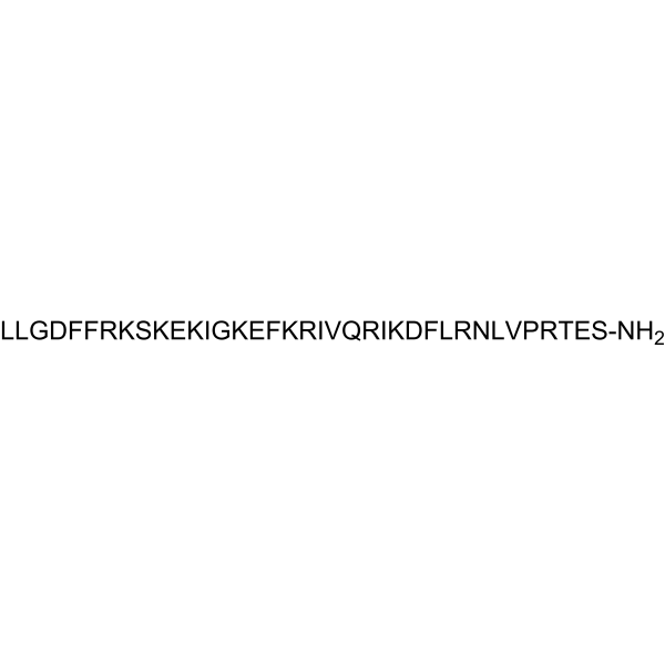
LLGDFFRKSKEKIGKEFKRIVQRIKDFLRNLVPRTES-NH2
-
Shipping
Room temperature in continental US; may vary elsewhere.
-
Storage
Sealed storage, away from moisture
Powder -80°C 2 years -20°C 1 year * In solvent : -80°C, 6 months; -20°C, 1 month (sealed storage, away from moisture)


Publications (2)
-
Journal Impact Factor
-
Most Recent
-
Eur J Pharmacol
Systemic administration of Neochamaejasmin B inhibits mast cell activation to reduce inflammation in a rosacea mouse model by targeting MRGPRX2. [Abstract]2025 Sep 15:1003:177988. PMID: 40706973 -


Solvent & Solubility
In Vitro:
H2O : ≥ 100 mg/mL (22.26 mM)
* "≥" means soluble, but saturation unknown.
Please refer to the solubility information to select the appropriate solvent. Once prepared, please aliquot and store the solution to prevent product inactivation from repeated freeze-thaw cycles.
Storage method and period of stock solution: -80°C, 6 months; -20°C, 1 month (sealed storage, away from moisture). When stored at -80°C, please use it within 6 months. When stored at -20°C, please use it within 1 month.
* Note: If you choose water as the stock solution, please dilute it to the working solution, then filter and sterilize it with a 0.22 μm filter before use.
Please refer to the solubility information to select the appropriate solvent. Once prepared, please aliquot and store the solution to prevent product inactivation from repeated freeze-thaw cycles.
Storage method and period of stock solution: -80°C, 6 months; -20°C, 1 month (sealed storage, away from moisture). When stored at -80°C, please use it within 6 months. When stored at -20°C, please use it within 1 month.
* Note: If you choose water as the stock solution, please dilute it to the working solution, then filter and sterilize it with a 0.22 μm filter before use.
Concentration (start) × Volume (start) = Concentration (final) × Volume (final)


Purity & Documentation
-
Data Sheet (273 KB)
-
SDS (252 KB)
- English - EN (252 KB)
- Français - FR (252 KB)
- Deutsch - DE (252 KB)
- Norwegian - NO (252 KB)
- Español - ES (252 KB)
- Swedish - SV (252 KB)
- Italian - IT (252 KB)
- Korean - KR (252 KB)
- Portuguese - PT (252 KB)
-
Handling Instructions (2659 KB)
References
[1]. Johansson J, et al. Conformation-dependent antibacterial activity of the naturally occurring human peptide LL-37. J Biol Chem. 1998 Feb 6;273(6):3718-24. [Content Brief]
[2]. De Yang, et al. LL-37, the neutrophil granule- and epithelial cell-derived cathelicidin, utilizes formyl peptide receptor-like 1 (FPRL1) as a receptor to chemoattract human peripheral blood neutrophils, monocytes, and T cells. J Exp Med. 2000 Oct 2;192(7):1069-74. [Content Brief]
[3]. Tanaka D, et al. Sensitivity of Actinobacillus actinomycetemcomitans and Capnocytophaga spp. to the bactericidal action of LL-37: a cathelicidin found in human leukocytes and epithelium. Oral Microbiol Immunol. 2000 Aug;15(4):226-31. [Content Brief]
[4]. Yang X, et al. Chitosan hydrogel encapsulated with LL-37 peptide promotes deep tissue injury healing in a mouse model. Mil Med Res. 2020 Apr 22;7(1):20. [Content Brief]


Complete Stock Solution Preparation Table
Please refer to the solubility information to select the appropriate solvent. Once prepared, please aliquot and store the solution to prevent product inactivation from repeated freeze-thaw cycles.
Storage method and period of stock solution: -80°C, 6 months; -20°C, 1 month (sealed storage, away from moisture). When stored at -80°C, please use it within 6 months. When stored at -20°C, please use it within 1 month.
| Optional Solvent | Concentration Solvent Mass | 1 mg | 5 mg | 10 mg | 25 mg |
|---|---|---|---|---|---|
| H2O | 1 mM | 0.2226 mL | 1.1130 mL | 2.2260 mL | 5.5651 mL |
| 5 mM | 0.0445 mL | 0.2226 mL | 0.4452 mL | 1.1130 mL | |
| 10 mM | 0.0223 mL | 0.1113 mL | 0.2226 mL | 0.5565 mL | |
| 15 mM | 0.0148 mL | 0.0742 mL | 0.1484 mL | 0.3710 mL | |
| 20 mM | 0.0111 mL | 0.0557 mL | 0.1113 mL | 0.2783 mL |
* Note: If you choose water as the stock solution, please dilute it to the working solution, then filter and sterilize it with a 0.22 μm filter before use.


LL-37 amide Related Classifications
HY-P4744 Related Classifications
 Powered by Bioz