



TPEN
Based on 29 publication(s) in Google Scholar
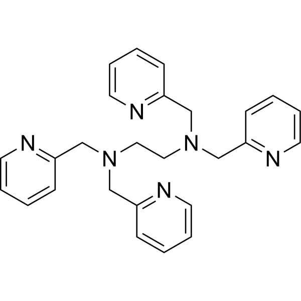
TPEN (TPEDA) is a specific cell-permeable heavy metal chelator. TPEN has a higher affinity for Zn2+, but a lower affinity for Mg2+ and Ca2+. TPEN induces DNA damage and increases intracellular ROS production. TPEN also inhibits cell proliferation and induces apoptosis.
Nos produits utilisent uniquement pour la recherche. Nous ne vendons pas aux patients.
- Pureté : 99.91%
- CAS No.: 16858-02-9
- Formule: C26H28N6
- Masse moléculaire:424.55
-
Stockage:Powder -20°C, 3 years , 4°C, 2 years ; In solvent -80°C, 2 years , -20°C, 1 year

To place orders, for customer services and technical support, please contact: MedChemExpress USA
Tel: 609-228-6898 E-mail: [email protected] [email protected]


-
 Activité biologique
-
Chemical Information
-
Solvant et solubilité
- Protocole
- Pureté et documentation
- Références
-
 Help & FAQs


-
HY-119287
TSQCellular Zinc Fluorescent Sensor -
HY-100545
BAPTA-AMCa2+ Chelator -
HY-B0988
Deferoxamine mesylateIron Chelator -

HY-100558
Bafilomycin A1V-ATPase Inhibitor -

HY-B0215
AcetylcysteineMucolytic Agent -
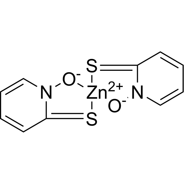
HY-B0572
Zinc PyrithioneAntimicrobial Agent -
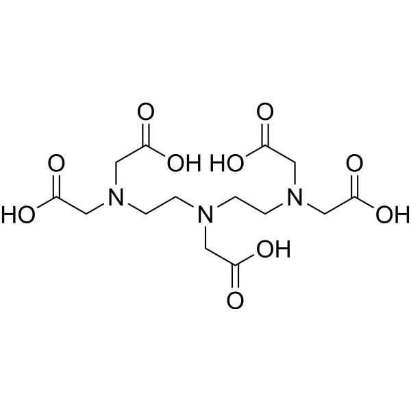
HY-B1335
Pentetic acidMagnetic Adsorbent -

HY-B1820
Zinc sulphateTyrosinase Inhibitor, Glutathione Reductase Inhibitor, Dopachrome Tautomerase Cofactor, Melanocortin Receptor 1 and 4 Modulator, Eumelanogenesis Inhibitor, Hair Hypopigmentation Inducer, Hair Cycle Modulator, Antioxidant, Healing Accelerator, Tensile Strength Enhancer, Anti-inflammatory Agent -

HY-Y0320
Dimethyl sulfoxide, meets analytical specification of Ch.P.Aprotic Solvent -
HY-50895
GefitinibEGFR Inhibitor -
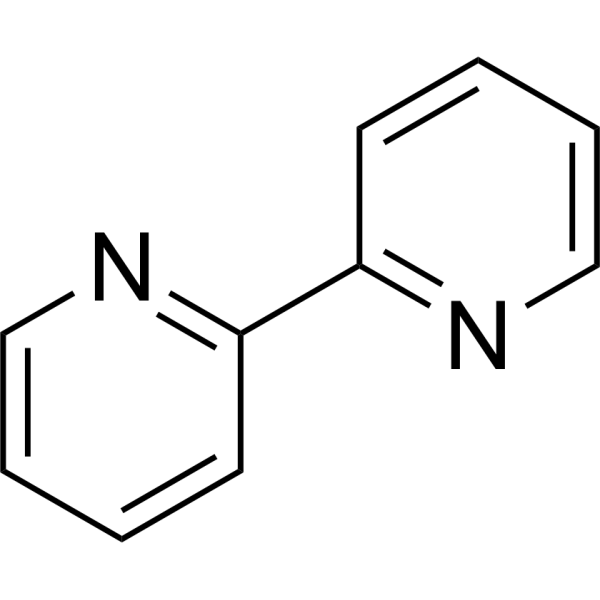
HY-D0020
2,2'-BipyridineMetal Chelating Ligand -
HY-B1625
DeferoxamineIron Chelator
Publications Citing Use of MedChemExpress (MCE) TPEN
More- Gut. 2026 Jan 13:gutjnl-2025-337019. [Abstract]
- Nat Commun. 2026 Jun 9;17(1):5072. [Abstract]
- J Nanobiotechnology. 2023 Sep 4;21(1):316. [Abstract]
- Adv Sci (Weinh). 2026 Aug 5:e76941.
- Adv Sci (Weinh). 2025 Sep;12(33):e08280. [Abstract]
- Small. 2025 Jul 20:e02783. [Abstract]
- J Hazard Mater. 2024 Jul 5:472:134466. [Abstract]
- Acta Biomater. 2026 Mar:213:666-677. [Abstract]
- Food Chem. 2024 Jun 1:442:138386. [Abstract]
- Int J Biol Macromol. 2026 May 27:152746. [Abstract]
- Br J Pharmacol. 2021 Jan;178(2):346-362. [Abstract]
- CNS Neurosci Ther. 2020 Oct;26(10):1058-1068. [Abstract]
- Food Funct. 2021 Sep 20;12(18):8626-8634. [Abstract]
- Int Immunopharmacol. 2026 Jan 1;168(Pt 1):115768. [Abstract]
- Int J Mol Sci. 2025 May 22;26(11):4978. [Abstract]
- Front Pharmacol. 2022 Feb 23;13:816133. [Abstract]
- Front Pharmacol. 2021 Jul 14:12:684538. [Abstract]
- BMC Microbiol. 2020 Jun 16;20(1):165. [Abstract]
- Cell Calcium. 2026 Mar:134:103125. [Abstract]
- PLoS Pathog. 2024 Aug 22;20(8):e1012444. [Abstract]
- FASEB J. 2026 Jun 15;40(11):e72023. [Abstract]
- BMC Cancer. 2025 May 28;25(1):956. [Abstract]
- Mol Immunol. 2021 Sep:137:155-162. [Abstract]
- Cancer Med. 2019 May;8(5):2462-2473. [Abstract]
- Infect Drug Resist. 2020 Aug 18:13:2883-2890. [Abstract]
- Physiol Rep. 2026 Jun;14(11):e70975. [Abstract]
- bioRxiv. 2026 Jun 16.
- bioRxiv. 2026 Apr 29.
- Heliyon. 2024 Jul 6;10(14):e33994. [Abstract]
 Customer Validation & Images

-
WB
-
Flow Cytometry
-
Cell Imaging/Staining

-
Cell Migration/Invasion Assay
-
WB


Activité biologique
Description
Cellular Effect
|
Cell Line

|
Type | Value | Description | References |
|---|---|---|---|---|
| Fibroblast | IC50 |
6.7 μM
Compound: TPEN
|
Cytotoxicity against human Fibroblast assessed as reduction in cell viability incubated for 48 hrs by cell titer-glo assay
Cytotoxicity against human Fibroblast assessed as reduction in cell viability incubated for 48 hrs by cell titer-glo assay
|
[PMID: 31935092] |
| PC-12 | EC50 |
38 μM
Compound: 1, TPEN
|
Induction of apoptosis in rat PC12 cells after 24 hrs by trypan blue exclusion assay
Induction of apoptosis in rat PC12 cells after 24 hrs by trypan blue exclusion assay
|
[PMID: 23859779] |
In Vitro
Heavy metal chelator TPEN attenuates fura-2 fluorescence changes induced by cadmium, mercury and methylmercury. TPEN, a cell-permeable chelator for heavy metal cations with a low affinity for Ca2+. In cells stimulated with 10 or 30 μM cadmium chloride, the addition of TPEN at 3 hr after exposure significantly decreases the elevated fura-2 fluorescence ratio to the basal levels within 10 min (119.6±2.4% or 109±1.5% decrease in ΔRatio (F340/F380) induced by 10 or 30 μM cadmium chloride, respectively), suggesting that a cadmium chloride-induced increase in the fura-2 fluorescence ratio is dependent on an increase in intracellular heavy metal cations but not intracellular Ca2+[1].
TPEN is a metal chelator, which targets colon cancer cells through redox cycling of copper. TPEN reduces cell viability in a dose- and time-dependent manner. TPEN-induced cell death is also dependent on the redox cycling of copper since the copper chelator neocuproine inhibited DNA damage and reduced pChk1, γ-H2AX, and ATM protein expression. Cell death by low TPEN concentrations, involved ATM/ATR signaling in all 3 cell lines, since pre-incubation with specific inhibitors of ATM and DNA-PK led to the recovery of cells from TPEN-induced DNA damage[2].
MedChemExpress (MCE) has not independently confirmed the accuracy of these methods. They are for reference only.
- Annexin V plus membrane-impermeant dye apoptosis staining
- ROS/oxidative-stress fluorescent staining
- BrdU Incorporation Assay
- Somatic Cell Culture
- CFSE Dye Dilution Proliferation Assay
- Dye-dilution cell tracking and proliferation staining
- CCK-8/WST-8 Cell Proliferation Assay
- Protocol for Cell Cycle
- Ca2+ Staining Technique
- Cell Counting-Based Growth Curve Assay
- Colony Formation (Clonogenic) Assay
- Cell Viability Determination by MTT Colorimetric Assay
- EdU Incorporation Assay (Click Chemistry-Based DNA Synthesis Measurement)
- Ki-67 Immunostaining Proliferation Assay
- Detection of Apoptosis
- TUNEL staining for apoptotic DNA fragmentation
- PCNA Immunodetection Proliferation Assay
- Protocol for Cell Counting and Cell Density Analysis
- Apoptosis Solutions
- MTT Cell Proliferation Assay
- Genotoxicity/Mutagenicity Study


Chemical Information
-
CAS No. 16858-02-9
-
Appearance Solid
-
Masse moléculaire 424.55
-
Formule C26H28N6
-
Color Light yellow to brown
-
SMILES
N(CC1=NC=CC=C1)(CC2=NC=CC=C2)CCN(CC3=NC=CC=C3)CC4=NC=CC=C4
-
Synonyms
TPEDA
-
Livraison
Room temperature in continental US; may vary elsewhere.
-
Stockage
Powder -20°C 3 years 4°C 2 years In solvent -80°C 2 years -20°C 1 year


Publications (29)
-
Journal Impact Factor
-
Most Recent
-
Gut
Zinc-dependent RNA-binding protein controls hepatocyte senescence and recovery from alcohol-related liver failure. [Abstract]2026 Jan 13:gutjnl-2025-337019. PMID: 41534893 -
Nat Commun
CLPX acquires an iron-sulfur cluster to sustain mitochondrial proteostasis in cancer cells. [Abstract]2026 Jun 9;17(1):5072. PMID: 42265110 -
J Nanobiotechnology
Metal-organic framework materials promote neural differentiation of dental pulp stem cells in spinal cord injury. [Abstract]2023 Sep 4;21(1):316. PMID: 37667307
TPEN purchased from MedChemExpress. Usage Cited in: J Nanobiotechnology. 2023 Sep 4;21(1):316. [Abstract]
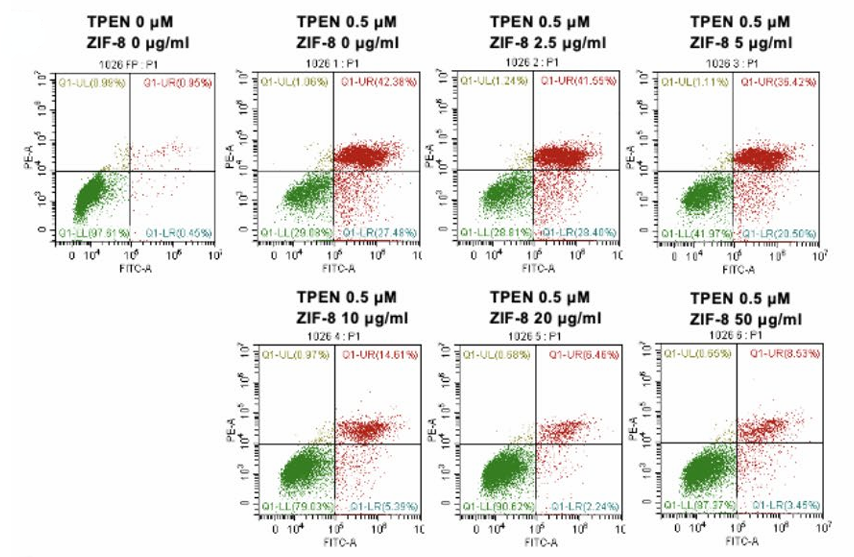
DPSCs were treated with TPEN (0 and 0.5 µM) combined with ZIF-8 at a concentration gradient (0, 2.5, 5, 10, 20, and 50 µg/ml). After 24 h of treatment, flow cytometry was used to detect FITC and PE.
TPEN purchased from MedChemExpress. Usage Cited in: J Nanobiotechnology. 2023 Sep 4;21(1):316. [Abstract]
Fluorescent images of calcein-labeled DPSCs cells treated with TPEN (0, 0.25, 0.5, 1, 1.5, and 2 µM) with ZIF-8 (20 µg/ml) in B27 medium for 6 days.
TPEN purchased from MedChemExpress. Usage Cited in: J Nanobiotechnology. 2023 Sep 4;21(1):316. [Abstract]
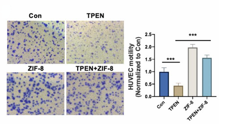
DPSCs were treated with TPEN (0 and 0.5 µM) combined with ZIF-8 at a concentration gradient (0 and 20 µg/ml), and the medium was co-cultured with HUVECs. Then, cells were stained with crystal violet stain for Transwell experiments. The number of transferred cells was quantified.
-
-
Adv Sci (Weinh)
2025 Sep;12(33):e08280. PMID: 40642900
TPEN purchased from MedChemExpress. Usage Cited in: Adv Sci (Weinh). 2025 Sep;12(33):e08280. [Abstract]
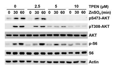
HCT116 cells were treated with TPEN (0, 2.5, 5, and 10 μM) for 1 h, starved in PBS for 1 h, and then supplemented with ZnSO4 for the indicated time. The levels of pS473-AKT, pT308-AKT, p-S6, and the indicated protein were detected by WB.
-
Small
2025 Jul 20:e02783. PMID: 40685755 -
J Hazard Mater
The involvement of SigmaR1K142 degradation mediated by ERAD in neural senescence linked with CdCl2 exposure. [Abstract]2024 Jul 5:472:134466. PMID: 38718507 -
Acta Biomater
A metal-free cascade nanoreactor reprograms the cystine-glutathione axis for disulfidptosis-pyroptosis therapy. [Abstract]2026 Mar:213:666-677. PMID: 41662918 -
Food Chem
Green synthesis of Zn2+ nanocarriers from Auricularia auricula fermentation broth with excellent antioxidant activity. [Abstract]2024 Jun 1:442:138386. PMID: 38219568 -
Int J Biol Macromol
SLC39A1 promotes pancreatic cancer metastasis by elevating intracellular zinc and activating the Src/FAK signaling pathway. [Abstract]2026 May 27:152746. PMID: 42208832 -
Br J Pharmacol
Small intestinal glucose and sodium absorption through calcium-induced calcium release and store-operated Ca2+ entry mechanisms. [Abstract]2021 Jan;178(2):346-362. PMID: 33080043 -
CNS Neurosci Ther
Laminin degradation by matrix metalloproteinase 9 promotes ketamine-induced neuronal apoptosis in the early developing rat retina. [Abstract]2020 Oct;26(10):1058-1068. PMID: 32562453
TPEN purchased from MedChemExpress. Usage Cited in: CNS Neurosci Ther. 2020 Oct;26(10):1058-1068. [Abstract]
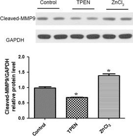
The Western blot results show that compared to the control group, the expression of cleaved MMP9 is increased by ZnCl2 treatment and decreased by 100 μM TPEN.
TPEN purchased from MedChemExpress. Usage Cited in: CNS Neurosci Ther. 2020 Oct;26(10):1058-1068. [Abstract]
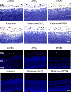
The immunohistochemistry assay reveales that exposure to 100 μM TPEN significantly decreases the extent of neuronal apoptosis in the GCL of the rat retina.
-
Food Funct
Zinc delivery system constructed from food-borne nanoparticles derived from Undaria pinnatifida. [Abstract]2021 Sep 20;12(18):8626-8634. PMID: 34346455 -
Int Immunopharmacol
Colistin-induced acute kidney injury via zinc Dyshomeostasis-triggered GSDMD-executed Pyroptosis. [Abstract]2026 Jan 1;168(Pt 1):115768. PMID: 41176907 -
Int J Mol Sci
Fisetin Attenuates Zinc Overload-Induced Hepatotoxicity in Mice via Autophagy-Dependent Nrf2 Activation. [Abstract]2025 May 22;26(11):4978. PMID: 40507790 -
Front Pharmacol
Ca2+-Permeable Channels/Ca2+ Signaling in the Regulation of Ileal Na+/Gln Co-Transport in Mice. [Abstract]2022 Feb 23;13:816133. PMID: 35281933 -
Front Pharmacol
Role of Serosal TRPV4-Constituted SOCE Mechanism in Secretagogues-Stimulated Intestinal Epithelial Anion Secretion. [Abstract]2021 Jul 14:12:684538. PMID: 34335254 -
BMC Microbiol
The effects of clioquinol in morphogenesis, cell membrane and ion homeostasis in Candida albicans. [Abstract]2020 Jun 16;20(1):165. PMID: 32546212 -
Cell Calcium
MONNA alleviates MPTP-induced Parkinson's disease in zebrafish by activating TFEB dependently on ER Calcium. [Abstract]2026 Mar:134:103125. PMID: 41637953 -
PLoS Pathog
Genome-wide CRISPR/Cas9 screen identifies SLC39A9 and PIK3C3 as crucial entry factors for Ebola virus infection. [Abstract]2024 Aug 22;20(8):e1012444. PMID: 39173055 -
FASEB J
Low Zinc Ameliorated the Decrease in Intestinal Copper Level Induced by Low Dietary Copper: Involvement of SUMOylated-Mtf-1 and Mtf-1/Atp7a Pathway. [Abstract]2026 Jun 15;40(11):e72023. PMID: 42257536 -
BMC Cancer
TRPM2 channels mediate ROS-induced actin remodeling and cell migration of prostate cancer cells. [Abstract]2025 May 28;25(1):956. PMID: 40437388 -
Mol Immunol
Naringenin inhibits pro‑inflammatory cytokine production in macrophages through inducing MT1G to suppress the activation of NF‑κB. [Abstract]2021 Sep:137:155-162. PMID: 34252709 -
Cancer Med
Zinc cooperates with p53 to inhibit the activity of mitochondrial aconitase through reactive oxygen species accumulation. [Abstract]2019 May;8(5):2462-2473. PMID: 30972978 -
Infect Drug Resist
Zinc Chelator N,N,N',N'-Tetrakis(2-Pyridylmethyl)Ethylenediamine Reduces the Resistance of Mycobacterium abscessus to Imipenem. [Abstract]2020 Aug 18:13:2883-2890. PMID: 32903882 -
Physiol Rep
Monosodium glutamate-mediated Ca2+-dependent intestinal epithelial ion transports in health and IBS-D in male mice. [Abstract]2026 Jun;14(11):e70975. PMID: 42281391 -
-
-
Heliyon
N,N,N',N'-Tetrakis(2-pyridylmethyl)ethylenediamine induces endothelium-dependent hyperpolarization-mediated vasorelaxation via store-operated calcium entry mechanism in healthy and intestinal inflammatory mice. [Abstract]2024 Jul 6;10(14):e33994. PMID: 39108891


Solvant et solubilité
In Vitro:
DMSO : 20 mg/mL (47.11 mM; Need ultrasonic; Hygroscopic DMSO has a significant impact on the solubility of product, please use newly opened DMSO)
Please refer to the solubility information to select the appropriate solvent. Once prepared, please aliquot and store the solution to prevent product inactivation from repeated freeze-thaw cycles.
Storage method and period of stock solution: -80°C, 2 years; -20°C, 1 year. When stored at -80°C, please use it within 2 years. When stored at -20°C, please use it within 1 year.
Please refer to the solubility information to select the appropriate solvent. Once prepared, please aliquot and store the solution to prevent product inactivation from repeated freeze-thaw cycles.
Storage method and period of stock solution: -80°C, 2 years; -20°C, 1 year. When stored at -80°C, please use it within 2 years. When stored at -20°C, please use it within 1 year.
Concentration (start) × Volume (start) = Concentration (final) × Volume (final)
In Vivo:
Select the appropriate dissolution method based on your experimental animal and administration route.
- For the following dissolution methods, please ensure to first prepare a clear stock solution using an In Vitro approach and then sequentially add co-solvents:
- To ensure reliable experimental results, the clarified stock solution can be appropriately stored based on storage conditions. As for the working solution for In Vivo experiments, it is recommended to prepare freshly and use it on the same day.
- The percentages shown for the solvents indicate their volumetric ratio in the final prepared solution. If precipitation or phase separation occurs during preparation, heat and/or sonication can be used to aid dissolution.
Add each solvent one by one: 10% DMSO 40% PEG300 5% Tween-80 45% Saline
Solubility: ≥ 2 mg/mL (4.71 mM); Clear solution
This protocol yields a clear solution of ≥ 2 mg/mL (saturation unknown).
Taking 1 mL working solution as an example, add 100 μL DMSO stock solution (20.0 mg/mL) to 400 μL PEG300, and mix evenly; then add 50 μL Tween-80 and mix evenly; then add 450 μL Saline to adjust the volume to 1 mL.
Preparation of Saline: Dissolve 0.9 g sodium chloride in ddH₂O and dilute to 100 mL to obtain a clear Saline solution.
Add each solvent one by one: 10% DMSO 90% (20% SBE-β-CD in Saline)
Solubility: ≥ 2 mg/mL (4.71 mM); Clear solution
This protocol yields a clear solution of ≥ 2 mg/mL (saturation unknown).
Taking 1 mL working solution as an example, add 100 μL DMSO stock solution (20.0 mg/mL) to 900 μL 20% SBE-β-CD in Saline, and mix evenly.
Preparation of 20% SBE-β-CD in Saline (4°C, storage for one week): 2 g SBE-β-CD powder is dissolved in 10 mL Saline, completely dissolve until clear.
In Vivo Dissolution Calculator
Please enter the basic information of animal experiments:
-
-
-
-
Recommended: Prepare an additional quantity of animals to account for potential losses during experiments.
Please enter your animal formula composition:
-
%DMSO +
Recommended: Keep the proportion of DMSO in working solution below 2% if your animal is weak.
-
%+
-
+%Tween-80 + +
-
%Saline +
The co-solvents required include: DMSO, . All of co-solvents are available by MedChemExpress (MCE). , Tween 80. All of co-solvents are available by MedChemExpress (MCE).
Working solution concentration: 0.22 mg/mL
Method for preparing stock solution: mg drug dissolved in μL DMSO. Stock solution concentration: mg/mL.
1. Take μL DMSO stock solution;
2. Add μL .
μL , mix evenly;
3. Then add μL Tween 80, mix evenly;
4. Then add μL
Please ensure that the stock solution in the first step is dissolved to a clear state, and add co-solvents in sequence. You can use ultrasonic heating (ultrasonic cleaner, recommended frequency 20-40 kHz), vortexing, etc. to assist dissolution.


Pureté et documentation
-
Fiche technique (281 KB)
-
SDS (396 KB)
- English - EN (396 KB)
- Français - FR (396 KB)
- Deutsch - DE (396 KB)
- Norwegian - NO (396 KB)
- Español - ES (396 KB)
- Swedish - SV (396 KB)
- Italian - IT (396 KB)
- Korean - KR (396 KB)
- Portuguese - PT (396 KB)
-
Instruction de manipulation (2659 KB)
Références
[1]. Ohkubo M, et al. Heavy metal chelator TPEN attenuates fura-2 fluorescence changes induced by cadmium, mercury and methylmercury. J Vet Med Sci. 2016 Jun 1;78(5):761-7. [Content Brief]
[2]. Rahal ON, et al. Chk1 and DNA-PK mediate TPEN-induced DNA damage in a ROS dependent manner in human colon cancer cells. Cancer Biol Ther. 2016 Nov;17(11):1139-1148. [Content Brief]
[3]. E Aizenman, et al. Induction of neuronal apoptosis by thiol oxidation: putative role of intracellular zinc release. J Neurochem. 2000 Nov;75(5):1878-88. [Content Brief]


Complete Stock Solution Preparation Table
Please refer to the solubility information to select the appropriate solvent. Once prepared, please aliquot and store the solution to prevent product inactivation from repeated freeze-thaw cycles.
Storage method and period of stock solution: -80°C, 2 years; -20°C, 1 year. When stored at -80°C, please use it within 2 years. When stored at -20°C, please use it within 1 year.
| Optional Solvent | Concentration Solvent Mass | 1 mg | 5 mg | 10 mg | 25 mg |
|---|---|---|---|---|---|
| DMSO | 1 mM | 2.3554 mL | 11.7772 mL | 23.5544 mL | 58.8859 mL |
| 5 mM | 0.4711 mL | 2.3554 mL | 4.7109 mL | 11.7772 mL | |
| 10 mM | 0.2355 mL | 1.1777 mL | 2.3554 mL | 5.8886 mL | |
| 15 mM | 0.1570 mL | 0.7851 mL | 1.5703 mL | 3.9257 mL | |
| 20 mM | 0.1178 mL | 0.5889 mL | 1.1777 mL | 2.9443 mL | |
| 25 mM | 0.0942 mL | 0.4711 mL | 0.9422 mL | 2.3554 mL | |
| 30 mM | 0.0785 mL | 0.3926 mL | 0.7851 mL | 1.9629 mL | |
| 40 mM | 0.0589 mL | 0.2944 mL | 0.5889 mL | 1.4721 mL |


TPEN Related Classifications
HY-100202 Related Classifications
 Powered by Bioz