



RA306

Based on 1 Customer Validation
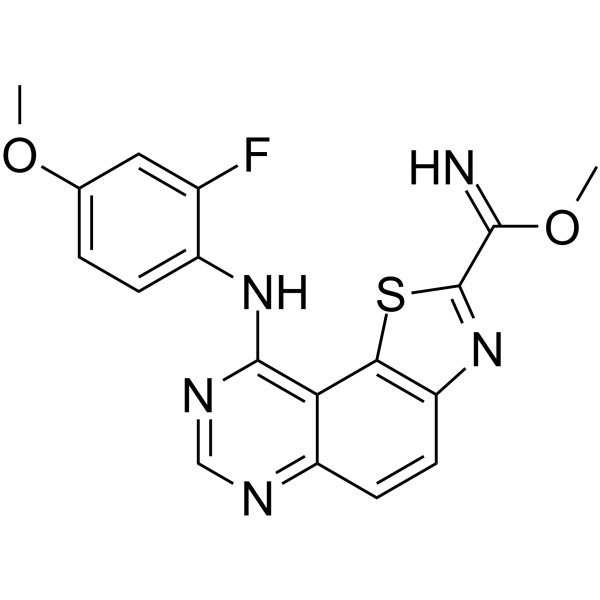
RA306 is an orally active CaMKIIδ/γ inhibitor with IC50 values of 15, 25, 61, and 420 nM against CaMKIIδ, γ, α, and β isoforms, respectively. RA306 reduces the serine/threonine phosphorylation level of PEAK1, inhibits PEAK1-mediated cancer cell migration and invasion, and attenuates the growth and metastasis of cancer xenografts. RA306 can be used in research related to triple-negative breast cancer and dilated cardiomyopathy.
Para uso exclusivo en investigación. No vendemos a pacientes.
- Pureza : 99.54%
- Fòrmula: C29H41N5O5S
- Peso molecular:571.73
-
Almacenamiento:Powder -20°C, 3 years , 4°C, 2 years ; In solvent -80°C, 6 months , -20°C, 1 month


To place orders, for customer services and technical support, please contact: MedChemExpress USA
Tel: 609-228-6898 E-mail: [email protected] [email protected]


-
 Actividad biológica
-
Chemical Information
-
Solvente y solubilidad
- Protocolo
- Pureza y Documentación
- Referencias
-
 Help & FAQs




Actividad biológica
Descripciòn
IC50 & Target
[2]|
CaMK IIδ 15 nM (IC50) |
CaMK IIγ 25 nM (IC50) |
CaMK IIα 61 nM (IC50) |
CaMK IIβ 420 nM (IC50) |
In Vitro
RA306 (1 μM; 24 h) significantly inhibits serine/threonine phosphorylation of PEAK1 in MDA-MB-231_EcoR cells stably expressing Flag-tagged PEAK1[1].
RA306 (120 h) reduces the viability of MDA-MB-231_HM cells in a dose-dependent manner in both high-adhesion and ultra-low-adhesion plates[1].
RA306 blocks PEAK1-mediated migration of MDA-MB-231 and MDA-MB-468 cells in transwell migration assays[1].
RA306 blocks PEAK1-mediated invasion of MDA-MB-231 cells in Matrigel chamber invasion assays[1].
MedChemExpress (MCE) has not independently confirmed the accuracy of these methods. They are for reference only.
-
Cell Line:MDA-MB-231_EcoR cells stably expressing Flag-tagged PEAK1
-
Concentration:1 μM
-
Incubation Time:24 h
-
Result:Significantly inhibited serine/threonine phosphorylation of PEAK1, with relative phosphorylation levels reduced compared to untreated controls.
Parmacokinetics
In Vivo
RA306 (30 mg/kg; p.o.; twice daily; 14 days) significantly improves the ejection fraction and cardiac output of α-actin transgenic (TG) mice with dilated cardiomyopathy, via a mechanism of reducing myocardial CaMKII activity by inhibiting PLN threonine-17 phosphorylation[2].
RA306 (30 mg/kg; p.o.; once daily for two months) significantly improves the ejection fraction and cardiac output of α-actin TG mice with advanced dilated cardiomyopathy, and exerts no effect on the detected cardiac remodeling parameters[2].
MedChemExpress (MCE) has not independently confirmed the accuracy of these methods. They are for reference only.
-
Animal Model:BALB/c athymic nude (6-week-old, female)[1]
-
Dosage:30 mg/kg
-
Administration:p.o.; once daily; 28 days
-
Result:Attenuated primary tumour growth, with a reduction in total bioluminescent imaging signal comparable to that observed with PEAK1 gene knockout.
Blocked lung metastasis from primary tumours to a similar extent as PEAK1 gene knockout, with a significant reduction in the number of mice exhibiting obvious metastatic growth.
-
Animal Model:alpha-actin transgenic (TG) mice (8-10 months old, C57BL/6 background, dilated cardiomyopathy model)[2]
-
Dosage:30 mg/kg
-
Administration:p.o.; twice daily; 14 days
-
Result:Increased ejection fraction from baseline 34.2% to 44.8%.
Increased cardiac output from baseline 13.22 mL/min to 15.60 mL/min.
Caused no notable effect on heart rate.
Significantly inhibited cardiac phospholamban (PLN) phosphorylation at threonine-17.
Caused no significant increase in PLN phosphorylation at serine-16.
-
Animal Model:alpha-actin transgenic (TG) mice (8 months old, C57BL/6 background, dilated cardiomyopathy model)[2]
-
Dosage:30 mg/kg
-
Administration:p.o.; once daily; 2 months
-
Result:Significantly increased ejection fraction and cardiac output from baseline at both 2 weeks and 2 months post-treatment initiation.
Caused no notable effect on heart rate or left ventricular internal diameter.
Resulted in minimal to mild focal/multifocal myocardial fibrosis.
Caused no significant difference in heart weight-to-body weight ratio compared to vehicle-treated TG mice.
Showed no signs of myocardial inflammation or cardiomyocyte apoptosis.
- Western Blot
- Scratch/Wound-Healing Migration Assay
- Cell migration
- Neural Crest/Neuronal Cell Migration Explant Assay
- Breast Cancer Modeling
- Kinase activity and phosphorylation assays
- Zigmond Chamber Chemotaxis Assay
- Under-Agarose Cell Migration Assay
- Transwell/Boyden Chamber Migration Assay
- 3D Collagen/Hydrogel Matrix Migration Assay
- Cell invasion
- Protocol for Kinase activity and phosphorylation assays
- Cell-Exclusion Zone Migration Assay
- Dunn/Insall Chamber Chemotaxis Assay
- Orthotopic Cell-Line Xenograft


Chemical Information
-
Appearance Solid
-
Peso molecular 571.73
-
Fòrmula C29H41N5O5S
-
Color Light yellow to yellow
-
SMILES
COC1=CC(N(CC2)CCC2N3C[C@H](CO)OCC3)=CC=C1NC4=NC=C5C(C(OC(C)C)=C(C(C)(O)C)S5)=N4
-
Envío
Room temperature in continental US; may vary elsewhere.
-
Almacenamiento
Powder -20°C 3 years 4°C 2 years In solvent -80°C 6 months -20°C 1 month


Solvente y solubilidad
In Vitro:
DMSO : 100 mg/mL (174.91 mM; Need ultrasonic; Hygroscopic DMSO has a significant impact on the solubility of product, please use newly opened DMSO)
Please refer to the solubility information to select the appropriate solvent. Once prepared, please aliquot and store the solution to prevent product inactivation from repeated freeze-thaw cycles.
Storage method and period of stock solution: -80°C, 6 months; -20°C, 1 month. When stored at -80°C, please use it within 6 months. When stored at -20°C, please use it within 1 month.
Please refer to the solubility information to select the appropriate solvent. Once prepared, please aliquot and store the solution to prevent product inactivation from repeated freeze-thaw cycles.
Storage method and period of stock solution: -80°C, 6 months; -20°C, 1 month. When stored at -80°C, please use it within 6 months. When stored at -20°C, please use it within 1 month.
Concentration (start) × Volume (start) = Concentration (final) × Volume (final)
In Vivo:
Select the appropriate dissolution method based on your experimental animal and administration route.
- For the following dissolution methods, please ensure to first prepare a clear stock solution using an In Vitro approach and then sequentially add co-solvents:
- To ensure reliable experimental results, the clarified stock solution can be appropriately stored based on storage conditions. As for the working solution for In Vivo experiments, it is recommended to prepare freshly and use it on the same day.
- The percentages shown for the solvents indicate their volumetric ratio in the final prepared solution. If precipitation or phase separation occurs during preparation, heat and/or sonication can be used to aid dissolution.
Add each solvent one by one: 10% DMSO 40% PEG300 5% Tween-80 45% Saline
Solubility: ≥ 2.5 mg/mL (4.37 mM); Clear solution
This protocol yields a clear solution of ≥ 2.5 mg/mL (saturation unknown).
Taking 1 mL working solution as an example, add 100 μL DMSO stock solution (25.0 mg/mL) to 400 μL PEG300, and mix evenly; then add 50 μL Tween-80 and mix evenly; then add 450 μL Saline to adjust the volume to 1 mL.
Preparation of Saline: Dissolve 0.9 g sodium chloride in ddH₂O and dilute to 100 mL to obtain a clear Saline solution.
Add each solvent one by one: 10% DMSO 90% (20% SBE-β-CD in Saline)
Solubility: ≥ 2.5 mg/mL (4.37 mM); Clear solution
This protocol yields a clear solution of ≥ 2.5 mg/mL (saturation unknown).
Taking 1 mL working solution as an example, add 100 μL DMSO stock solution (25.0 mg/mL) to 900 μL 20% SBE-β-CD in Saline, and mix evenly.
Preparation of 20% SBE-β-CD in Saline (4°C, storage for one week): 2 g SBE-β-CD powder is dissolved in 10 mL Saline, completely dissolve until clear.
In Vivo Dissolution Calculator
Please enter the basic information of animal experiments:
-
-
-
-
Recommended: Prepare an additional quantity of animals to account for potential losses during experiments.
Please enter your animal formula composition:
-
%DMSO +
Recommended: Keep the proportion of DMSO in working solution below 2% if your animal is weak.
-
%+
-
+%Tween-80 + +
-
%Saline +
The co-solvents required include: DMSO, . All of co-solvents are available by MedChemExpress (MCE). , Tween 80. All of co-solvents are available by MedChemExpress (MCE).
Working solution concentration: 0.22 mg/mL
Method for preparing stock solution: mg drug dissolved in μL DMSO. Stock solution concentration: mg/mL.
1. Take μL DMSO stock solution;
2. Add μL .
μL , mix evenly;
3. Then add μL Tween 80, mix evenly;
4. Then add μL
Please ensure that the stock solution in the first step is dissolved to a clear state, and add co-solvents in sequence. You can use ultrasonic heating (ultrasonic cleaner, recommended frequency 20-40 kHz), vortexing, etc. to assist dissolution.


Pureza y Documentación
-
Ficha de datos (278 KB)
-
SDS (251 KB)
- English - EN (251 KB)
- Français - FR (251 KB)
- Deutsch - DE (251 KB)
- Norwegian - NO (251 KB)
- Español - ES (251 KB)
- Swedish - SV (251 KB)
- Italian - IT (251 KB)
- Korean - KR (251 KB)
- Portuguese - PT (251 KB)
-
Instrucciones de manejo (2659 KB)
Referencias
[1]. Yang X, et al. Activation of CAMK2 by pseudokinase PEAK1 represents a targetable pathway in triple negative breast cancer. Nat Commun. 2025;16(1):1871. Published 2025 Feb 22. [Content Brief]
[2]. Beauverger P, et al. Reversion of cardiac dysfunction by a novel orally available calcium/calmodulin-dependent protein kinase II inhibitor, RA306, in a genetic model of dilated cardiomyopathy. Cardiovasc Res. 2020;116(2):329-338. [Content Brief]


Complete Stock Solution Preparation Table
Please refer to the solubility information to select the appropriate solvent. Once prepared, please aliquot and store the solution to prevent product inactivation from repeated freeze-thaw cycles.
Storage method and period of stock solution: -80°C, 6 months; -20°C, 1 month. When stored at -80°C, please use it within 6 months. When stored at -20°C, please use it within 1 month.
| Optional Solvent | Concentration Solvent Mass | 1 mg | 5 mg | 10 mg | 25 mg |
|---|---|---|---|---|---|
| DMSO | 1 mM | 1.7491 mL | 8.7454 mL | 17.4908 mL | 43.7269 mL |
| 5 mM | 0.3498 mL | 1.7491 mL | 3.4982 mL | 8.7454 mL | |
| 10 mM | 0.1749 mL | 0.8745 mL | 1.7491 mL | 4.3727 mL | |
| 15 mM | 0.1166 mL | 0.5830 mL | 1.1661 mL | 2.9151 mL | |
| 20 mM | 0.0875 mL | 0.4373 mL | 0.8745 mL | 2.1863 mL | |
| 25 mM | 0.0700 mL | 0.3498 mL | 0.6996 mL | 1.7491 mL | |
| 30 mM | 0.0583 mL | 0.2915 mL | 0.5830 mL | 1.4576 mL | |
| 40 mM | 0.0437 mL | 0.2186 mL | 0.4373 mL | 1.0932 mL | |
| 50 mM | 0.0350 mL | 0.1749 mL | 0.3498 mL | 0.8745 mL | |
| 60 mM | 0.0292 mL | 0.1458 mL | 0.2915 mL | 0.7288 mL | |
| 80 mM | 0.0219 mL | 0.1093 mL | 0.2186 mL | 0.5466 mL | |
| 100 mM | 0.0175 mL | 0.0875 mL | 0.1749 mL | 0.4373 mL |


RA306 Related Classifications
HY-173117 Related Classifications
 Powered by Bioz