



Glucobrassicin potassium

Based on 1 Customer Validation
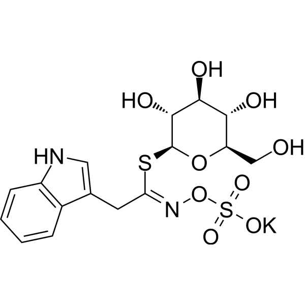
Glucobrassicin potassium is an indole-based anticancer agent and plant growth-regulating hormone. Glucobrassicin potassium exerts its biological activity by disrupting the integrity of microtubule networks in both plant and mammalian cells. At high concentrations, Glucobrassicin potassium inhibits seed germination and root growth; it can also specifically induce apoptosis in mammalian cancer cells and interfere with the intercellular transmission of viruses that rely on microtubules. In plants, Glucobrassicin potassium can be catalyzed by myrosinase to release growth-regulating substances, exhibiting a concentration-dependent growth-regulating effect.
For research use only. We do not sell to patients.
- Purity : 95.01%
- CAS No.: 143231-38-3
- Formula: C16H19KN2O9S2
- Molecular Weight:486.56
-
Storage:
-20°C, sealed storage, away from moisture and light
* In solvent : -80°C, 6 months; -20°C, 1 month (sealed storage, away from moisture and light)

To place orders, for customer services and technical support, please contact: MedChemExpress USA
Tel: 609-228-6898 E-mail: [email protected] [email protected]


-
 Biological Activity
-
Chemical Information
-
Solvent & Solubility
- Protocols
- Purity & Documentation
- References
-
 Help & FAQs


-
HY-N0583
HydrocortisoneGlucocorticoid Receptor Agonist -
HY-N0593
Deoxycholic acidBile Acid Receptor Activator -
HY-13417
AICARAMPK Activator -
HY-N0170
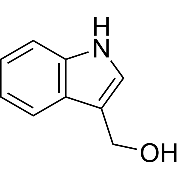
Indole-3-carbinolEndogenous Metabolite -
HY-13433
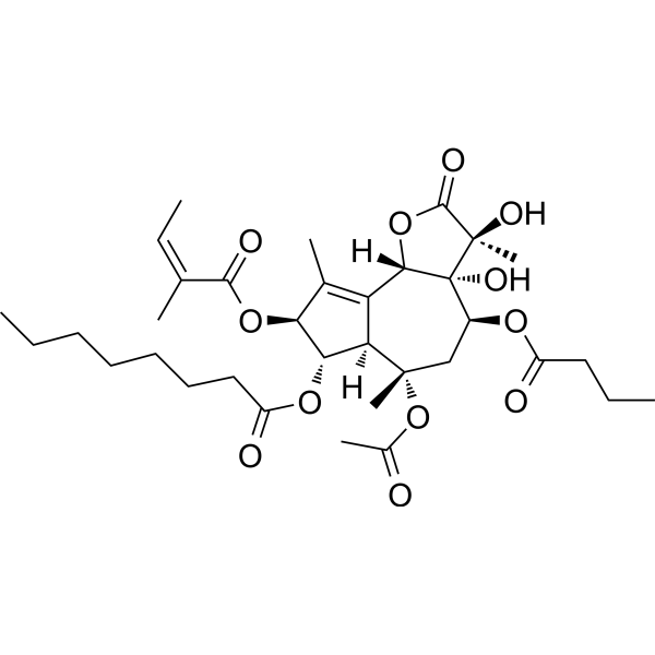
ThapsigarginER Stress Inducer -
HY-15760
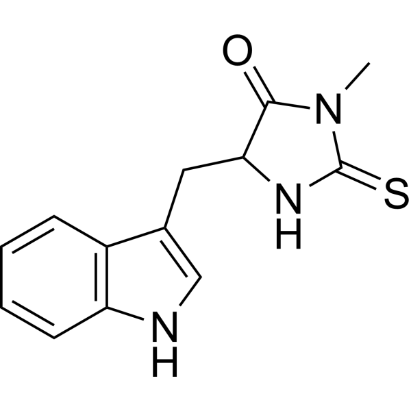
Necrostatin-1RIPK1 Inhibitor -
HY-100545
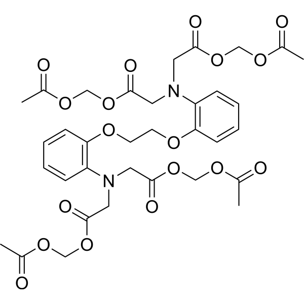
BAPTA-AMCa2+ Chelator -
HY-B1272
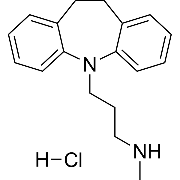
Desipramine hydrochlorideSerotonin Transporter Inhibitor
All Phytohormone Isoforms
More

Biological Activity
Description
In Vitro
Glucobrassicin potassium (50 μM; 30-180 min) induces time-dependent elimination of microtubules in tobacco BY-2 TuA3-GFP cells, with nearly complete loss of microtubule integrity observed after 180 min of treatment[1].
Glucobrassicin potassium (50 μM) drastically reduces the mitotic index and disrupts all stages of mitotic microtubule organization in Nicotiana tabacum BY-2 TuA3-GFP cells[1].
Glucobrassicin potassium (50 μM; 30-90 min) induces rapid and sustained elimination of microtubules in HeLa GFP-α-tubulin/H2B-mCherry cells[1].
Glucobrassicin potassium (25 μM; 24 h) induces pronounced apoptosis in HeLa GFP-α-tubulin/H2B-mCherry cells after 24 h of treatment[1].
MedChemExpress (MCE) has not independently confirmed the accuracy of these methods. They are for reference only. Further protocols information, click here.
In Vivo
MedChemExpress (MCE) has not independently confirmed the accuracy of these methods. They are for reference only.


Chemical Information
-
CAS No. 143231-38-3
-
Appearance Solid
-
Molecular Weight 486.56
-
Formula C16H19KN2O9S2
-
Color Pale purple to light pink
-
SMILES
OC[C@@H]1[C@@H](O)[C@H](O)[C@@H](O)[C@H](S/C(CC2=CNC3=C2C=CC=C3)=N\OS(=O)(O[K])=O)O1
-
Shipping
Room temperature in continental US; may vary elsewhere.
-
Storage
-20°C, sealed storage, away from moisture and light
* In solvent : -80°C, 6 months; -20°C, 1 month (sealed storage, away from moisture and light)


Solvent & Solubility
In Vitro:
H2O : 125 mg/mL (256.91 mM; Need ultrasonic)
Please refer to the solubility information to select the appropriate solvent. Once prepared, please aliquot and store the solution to prevent product inactivation from repeated freeze-thaw cycles.
Storage method and period of stock solution: -80°C, 6 months; -20°C, 1 month (sealed storage, away from moisture and light). When stored at -80°C, please use it within 6 months. When stored at -20°C, please use it within 1 month.
* Note: If you choose water as the stock solution, please dilute it to the working solution, then filter and sterilize it with a 0.22 μm filter before use.
Please refer to the solubility information to select the appropriate solvent. Once prepared, please aliquot and store the solution to prevent product inactivation from repeated freeze-thaw cycles.
Storage method and period of stock solution: -80°C, 6 months; -20°C, 1 month (sealed storage, away from moisture and light). When stored at -80°C, please use it within 6 months. When stored at -20°C, please use it within 1 month.
* Note: If you choose water as the stock solution, please dilute it to the working solution, then filter and sterilize it with a 0.22 μm filter before use.
Concentration (start) × Volume (start) = Concentration (final) × Volume (final)

Protocols
-
Apoptosis
Apoptosis, also called programmed cell death, is generally characterized by distinct morphological characteristics.
-
TUNEL staining for apoptotic DNA fragmentation
TUNEL staining detects DNA strand breaks by using terminal deoxynucleotidyl transferase to add labeled nucleotides to exposed 3′-OH DNA termini, generating either microscopic staining in fixed cells or tissue sections, or fluorescence/cytometric signal in cell suspensions. TUNEL positivity reflects DNA fragmentation but should not be interpreted alone as definitive apoptosis, because TUNEL can also label necrotic, autolytic, mechanically damaged, or DNA-repair-associated DNA breaks.
-
Annexin V plus membrane-impermeant dye apoptosis staining
Annexin V-based apoptosis assays rely on the detection of phosphatidylserine (PS) externalization from the inner leaflet of the plasma membrane to the outer leaflet, an early biochemical hallmark of apoptosis. Fluorescently labeled Annexin V binds PS in a calcium-dependent manner, enabling identification of early apoptotic cells by flow cytometry or fluorescence microscopy. When combined with a membrane-impermeant DNA-binding dye (e. g. , propidium iodide), this approach allows discrimination between viable (Annexin V−/dye−), early apoptotic (Annexin V+/dye−), and late apoptotic or necrotic (Annexin V+/dye+) cell populations by assessing membrane integrity and PS exposure.
-
Research Protocol for Endocrine Diseases
Endocrine diseases often arise from disrupted hormone production, hormone signaling, or target-tissue responsiveness; for diabetes-focused endocrine disease models, insulin signaling regulates glucose uptake, hepatic glucose output, lipid metabolism, and β-cell compensation. Type 2 diabetes develops through interacting defects in insulin resistance, β-cell dysfunction, adipose inflammation, hepatic glucose overproduction, altered incretin signaling, and ectopic lipid metabolism. A major unresolved question is whether endocrine dysfunction is driven primarily by target-tissue insulin resistance, intrinsic β-cell failure, immune/inflammatory stress, or combined multi-organ failure that differs by disease stage.
-
Apoptosis Solutions
Apoptosis is a regulated, generally non-lytic cell-death pathway that removes unwanted, damaged, infected, or abnormal cells through coordinated morphological changes, caspase activation, DNA fragmentation, and membrane remodeling. The intrinsic apoptosis pathway is controlled mainly by mitochondrial outer membrane permeabilization, BCL-2 family proteins, cytochrome c release, apoptosome formation, caspase-9 activation, and downstream executioner caspase-3/7 activation. The extrinsic apoptosis pathway is initiated by death receptors such as Fas, TNFR, and TRAIL receptors, which recruit adaptor proteins and activate caspase-8 before engaging executioner caspases or mitochondrial amplification through BID cleavage. Apoptosis is linked to many phenotypes, including cancer cell killing, tissue homeostasis, immune regulation, neurodegeneration, infection response, and treatment-induced cytotoxicity; unresolved questions include how apoptosis interacts with necroptosis, pyroptosis, ferroptos


Purity & Documentation
-
Data Sheet (279 KB)
-
SDS (393 KB)
- English - EN (393 KB)
- Français - FR (393 KB)
- Deutsch - DE (393 KB)
- Norwegian - NO (393 KB)
- Español - ES (393 KB)
- Swedish - SV (393 KB)
- Italian - IT (393 KB)
- Korean - KR (393 KB)
- Portuguese - PT (393 KB)
-
Handling Instructions (2659 KB)
References


Complete Stock Solution Preparation Table
Please refer to the solubility information to select the appropriate solvent. Once prepared, please aliquot and store the solution to prevent product inactivation from repeated freeze-thaw cycles.
Storage method and period of stock solution: -80°C, 6 months; -20°C, 1 month (sealed storage, away from moisture and light). When stored at -80°C, please use it within 6 months. When stored at -20°C, please use it within 1 month.
| Optional Solvent | Concentration Solvent Mass | 1 mg | 5 mg | 10 mg | 25 mg |
|---|---|---|---|---|---|
| H2O | 1 mM | 2.0552 mL | 10.2762 mL | 20.5524 mL | 51.3811 mL |
| 5 mM | 0.4110 mL | 2.0552 mL | 4.1105 mL | 10.2762 mL | |
| 10 mM | 0.2055 mL | 1.0276 mL | 2.0552 mL | 5.1381 mL | |
| 15 mM | 0.1370 mL | 0.6851 mL | 1.3702 mL | 3.4254 mL | |
| 20 mM | 0.1028 mL | 0.5138 mL | 1.0276 mL | 2.5691 mL | |
| 25 mM | 0.0822 mL | 0.4110 mL | 0.8221 mL | 2.0552 mL | |
| 30 mM | 0.0685 mL | 0.3425 mL | 0.6851 mL | 1.7127 mL | |
| 40 mM | 0.0514 mL | 0.2569 mL | 0.5138 mL | 1.2845 mL | |
| 50 mM | 0.0411 mL | 0.2055 mL | 0.4110 mL | 1.0276 mL | |
| 60 mM | 0.0343 mL | 0.1713 mL | 0.3425 mL | 0.8564 mL | |
| 80 mM | 0.0257 mL | 0.1285 mL | 0.2569 mL | 0.6423 mL | |
| 100 mM | 0.0206 mL | 0.1028 mL | 0.2055 mL | 0.5138 mL |
* Note: If you choose water as the stock solution, please dilute it to the working solution, then filter and sterilize it with a 0.22 μm filter before use.


Glucobrassicin potassium Related Classifications
HY-N7659A Related Classifications
 Powered by Bioz