



GSK-3685032
Based on 15 publication(s) in Google Scholar
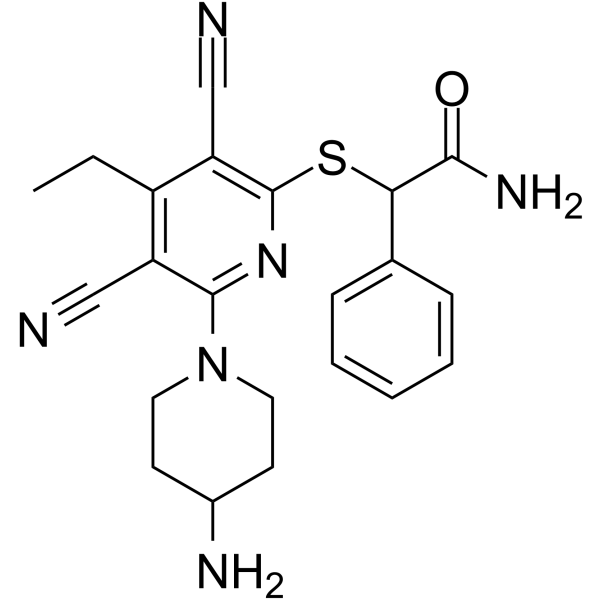
GSK-3685032 is a non-time-dependent, noncovalently, first-in-class reversible DNMT1-selective inhibitor, with an IC50 of 0.036 μM. GSK-3685032 induces robust loss of DNA methylation, transcriptional activation, and cancer cell growth inhibition.
For research use only. We do not sell to patients.
- Purity : 99.41%
- CAS No.: 2170137-61-6
- Formula: C22H24N6OS
- Molecular Weight:420.53
-
Storage:Powder -20°C, 3 years , 4°C, 2 years ; In solvent -80°C, 2 years , -20°C, 1 year

To place orders, for customer services and technical support, please contact: MedChemExpress USA
Tel: 609-228-6898 E-mail: [email protected] [email protected]


-
 Biological Activity
-
Chemical Information
-
Solvent & Solubility
- Protocols
- Purity & Documentation
- References
-
 Help & FAQs


-
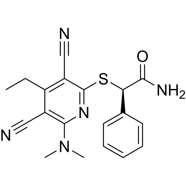
HY-135146
GSK-3484862DNMT1 Inhibitor -
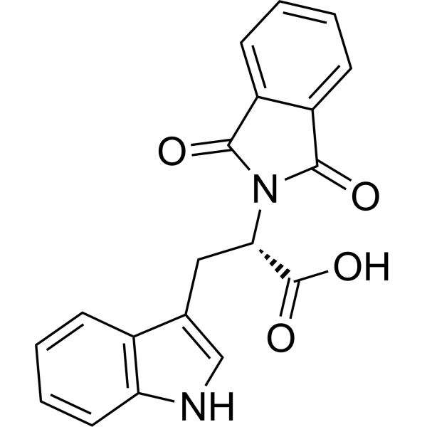
HY-13642
RG108DNMT Inhibitor -
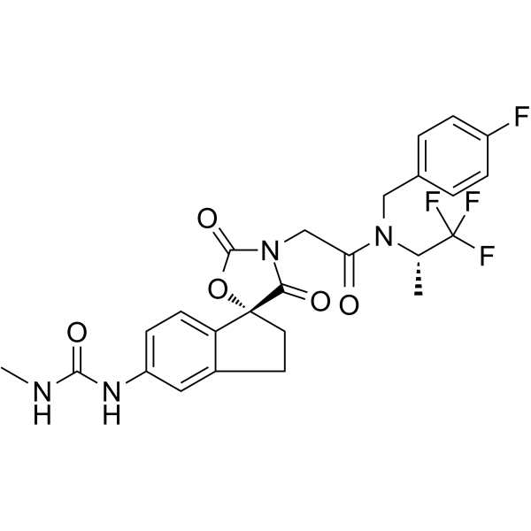
HY-107455
A-485p300/CBP Inhibitor -
HY-A0004
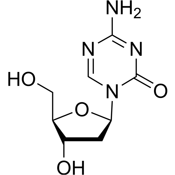
DecitabineDNMT Inhibitor -
HY-141644
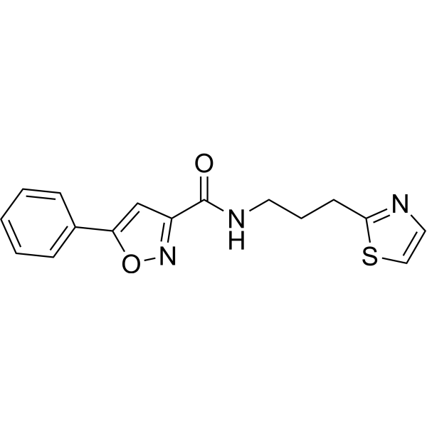
PY-60YAP Activator -
HY-10586
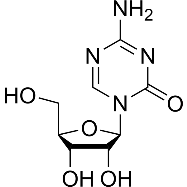
5-AzacytidineDNMT Inhibitor -
HY-50855
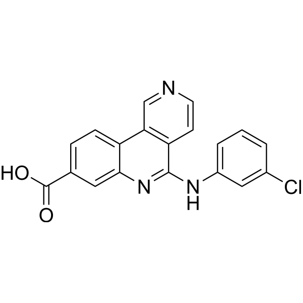
SilmitasertibCasein Kinase 2 Inhibitor -
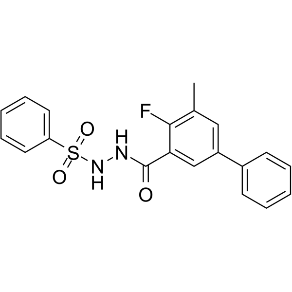
HY-102060
WM-8014MOZ Inhibitor -
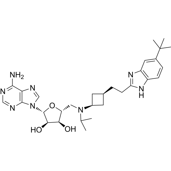
HY-15593
PinometostatDOT1L Inhibitor -
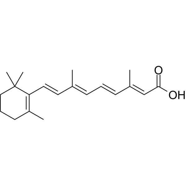
HY-14649
Retinoic acidRAR Agonist -
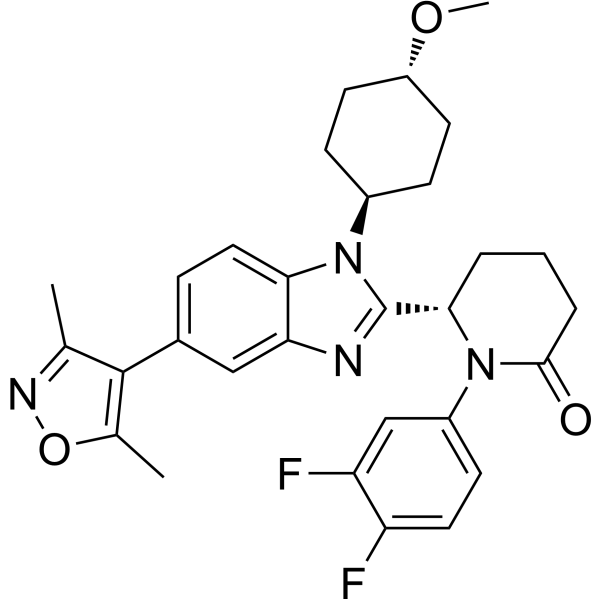
HY-111784
Inobrodibp300/CBP Inhibitor -
HY-15424
5-IodotubercidinAdenosine Kinase Inhibitor
Publications Citing Use of MedChemExpress (MCE) GSK-3685032
More- Cell. 2026 Jun 11;189(12):3541-3552.e18. [Abstract]
- Sci Immunol. 2025 Mar 7;10(105):eado9933. [Abstract]
- J Crohns Colitis. 2026 Feb 5;20(2):jjag003. [Abstract]
- NPJ Breast Cancer. 2023 Aug 11;9(1):66. [Abstract]
- Stem Cell Res Ther. 2025 Jul 18;16(1):389. [Abstract]
- J Med Chem. 2024 Sep 12;67(17):15098-15117. [Abstract]
- J Cell Biol. 2024 Apr 1;223(4):e202307026. [Abstract]
- Clin Epigenetics. 2025 Jul 3;17(1):114. [Abstract]
- Res Sq. 2026 May 4.
- University of Rochester. 2025.
- bioRxiv. 2025 Jul 31:2025.07.28.666975. [Abstract]
- Patent. US20250152731A1.
- bioRxiv. 2024 November 06.
- bioRxiv. 2024 Apr 3:2024.04.03.587980. [Abstract]
- bioRxiv. 2023 May 9.
 Customer Validation & Images

-
Cell Proliferation/Viability Assay
-
IF
-
Cell Proliferation/Viability Assay
-
Flow Cytometry
-
WB
All DNA Methyltransferase Isoforms
More

Biological Activity
Description
IC50 & Target
DNMT1[1]
Cellular Effect
|
Cell Line

|
Type | Value | Description | References |
|---|---|---|---|---|
| HL-60 | IC50 |
0.32 μM
Compound: GSK3685032 ; GSK5032
|
Inhibition of cell viability in human HL-60 cells incubated for 4 days in presence of SAHA by CCK8 assay
Inhibition of cell viability in human HL-60 cells incubated for 4 days in presence of SAHA by CCK8 assay
|
[PMID: 39264152] |
| Kasumi 1 | IC50 |
0.38 μM
Compound: GSK3685032 ; GSK5032
|
Inhibition of cell viability in human Kasumi 1 cells incubated for 4 days in presence of SAHA by CCK8 assay
Inhibition of cell viability in human Kasumi 1 cells incubated for 4 days in presence of SAHA by CCK8 assay
|
[PMID: 39264152] |
| MV4-11 | IC50 |
>5 μM
Compound: GSK3685032 ; GSK5032
|
Antiproliferative activity against human MV4-11 cells incubated for 96 hrs by CCK8 assay
Antiproliferative activity against human MV4-11 cells incubated for 96 hrs by CCK8 assay
|
[PMID: 39264152] |
| MV4-11 | IC50 |
0.4 μM
Compound: GSK3685032 ; GSK5032
|
Inhibition of cell viability in human MV4-11 cells incubated for 4 days in presence of SAHA by CCK8 assay
Inhibition of cell viability in human MV4-11 cells incubated for 4 days in presence of SAHA by CCK8 assay
|
[PMID: 39264152] |
In Vitro
GSK-3685032 (6 days) has cell growth inhibition of majority cancer cell lines, with a median growth IC50 value of 0.64 μM[1].
GSK-3685032 (0.1-1000 nM, 1-6 days) exhibits growth inhibition after 3 days, with decreasing growth IC50 throughout a 6 d time course[1].
GSK3685032 (10-10000 nM, day 4) dose-dependently increases the immune-related gene transcription[1].
GSK3685032 (3.2-10,000 nM, 2 days) inhibits DNMT1 protein expression[1].
GSK3685032 induces DNA hypomethylation and gene activation[1].
MedChemExpress (MCE) has not independently confirmed the accuracy of these methods. They are for reference only. Further protocols information, click here.
-
Cell Line:15 leukemia, 29 lymphoma and 7 multiple myeloma cell lines, e.g., EOL-1, Ki-JK, MM.IR cells.
-
Concentration:0.01-100 μM
-
Incubation Time:6 days
-
Result:Showed cell growth inhibition of majority cancer cell lines, with a median growth IC50 value of 0.64 μM.
-
Cell Line:MV4-11 cells
-
Concentration:0.1-1000 nM
-
Incubation Time:1-6 days
-
Result:Exhibited growth inhibition after 3 days, with decreasing growth IC50 throughout a 6 d time course.
-
Cell Line:MV4-11 cells
-
Concentration:10-10000 nM
-
Incubation Time:4 days
-
Result:Dose-dependent increased of CXCL11, IFI27, HLA-DQA1 and MAGEA4 following treatment of MV4-11 cells.
-
Cell Line:GDM-1 cells
-
Concentration:3.2-10,000 nM
-
Incubation Time:2 days
-
Result:Inhibited DNMT1 protein expression
In Vivo
Summary of mouse pharmacokinetic parameters for GSK-3685032[1]
| Dose,Route | Cmax (ng/mL) |
AUC0-8hr (h*ng/mL) |
DNAUC (h*kg*ng/mL/mg) |
Clearance (mL/min/kg) |
Volumedss (L/kg) |
T1/2 (h) |
| 2 mg/kg,IV | 5103 | 2418 | 1209 | 13 | 1.3 | 1.8 |
| 2 mg/kg,SC | 252 | 921 | 461 | NA | NA | 2.8 |
| 2 mg/kg,SC | 5473 | 15400 | 513 | NA | NA | ND |
MedChemExpress (MCE) has not independently confirmed the accuracy of these methods. They are for reference only.
-
Animal Model:MV4-11 xenograft models (female CD1-Foxn1
mice, 12 weeks of age) or SKM-1 xenograft models (NOD. CB17-Prkdc 1NCrCrl mice, 8-11 weeks of age)[1] -
Dosage:1, 5, 15, 30, 45 mg/kg (10% captisol adjusted to pH 4.5-5 with 1 M acetic acid, stored for up to 1 week at 4 °C)
-
Administration:Subcutaneous injection, twice daily for 4 weeks
-
Result:Revealed statistically significant dose-dependent tumor growth inhibition with clear regression at ≥30 mg/kg.


Chemical Information
-
CAS No. 2170137-61-6
-
Appearance Solid
-
Molecular Weight 420.53
-
Formula C22H24N6OS
-
Color White to off-white
-
SMILES
N#CC1=C(SC(C2=CC=CC=C2)C(N)=O)N=C(N3CCC(CC3)N)C(C#N)=C1CC
-
Shipping
Room temperature in continental US; may vary elsewhere.
-
Storage
Powder -20°C 3 years 4°C 2 years In solvent -80°C 2 years -20°C 1 year


Publications (15)
-
Journal Impact Factor
-
Most Recent
-
Cell
2026 Jun 11;189(12):3541-3552.e18. PMID: 41999746 -
Sci Immunol
2025 Mar 7;10(105):eado9933. PMID: 40053603 -
J Crohns Colitis
Intermittent ketogenic diet promotes Treg differentiation and alleviates Crohn's disease via the β-hydroxybutyrate driven AHCY-DNMT1-Foxp3 axis. [Abstract]2026 Feb 5;20(2):jjag003. PMID: 41527294 -
NPJ Breast Cancer
Epigenetically upregulating TROP2 and SLFN11 enhances therapeutic efficacy of TROP2 antibody drug conjugate sacitizumab govitecan. [Abstract]2023 Aug 11;9(1):66. PMID: 37567892
GSK-3685032 purchased from MedChemExpress. Usage Cited in: NPJ Breast Cancer. 2023 Aug 11;9(1):66. [Abstract]
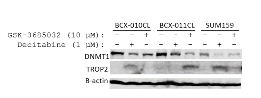
Breast cancer cell lines were treated with GSK-3685032 at 10 μM and decitabine at 1 µM for 3 days. DNMT1 and TROP2 levels were examined by immunoblotting.
-
Stem Cell Res Ther
Highly efficient XIST reactivation in female hPSC by transient dual inhibition of TP53 and DNA methylation during Cas9 mediated genome editing. [Abstract]2025 Jul 18;16(1):389. PMID: 40682144
GSK-3685032 purchased from MedChemExpress. Usage Cited in: Stem Cell Res Ther. 2025 Jul 18;16(1):389. [Abstract]
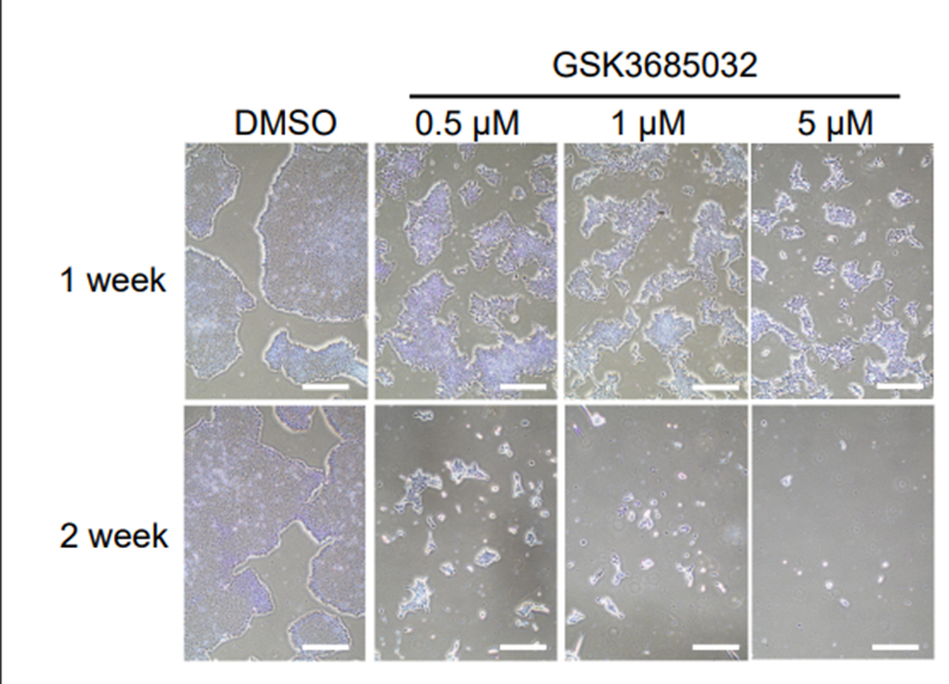
GSK3685032 (0.5, 1, 5 μM) treatment resulted in dose-dependent growth inhibition, with most cells dying after two weeks.
GSK-3685032 purchased from MedChemExpress. Usage Cited in: Stem Cell Res Ther. 2025 Jul 18;16(1):389. [Abstract]
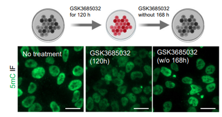
DNAme levels were signifcantly reduced upon GSK3685032 (1 μM, 120 h) treatmen by immunofuorescence (IF) analysis.
-
J Med Chem
Discovery of a Novel Benzimidazole Derivative Targeting Histone Deacetylase to Induce Ferroptosis and Trigger Immunogenic Cell Death. [Abstract]2024 Sep 12;67(17):15098-15117. PMID: 39145486 -
J Cell Biol
Tunable DNMT1 degradation reveals DNMT1/DNMT3B synergy in DNA methylation and genome organization. [Abstract]2024 Apr 1;223(4):e202307026. PMID: 38376465 -
Clin Epigenetics
Exposing the DNA methylation-responsive compartment of the leukaemic genome in T-ALL cell lines support its potential as a novel therapeutic target in T-ALL. [Abstract]2025 Jul 3;17(1):114. PMID: 40611304
GSK-3685032 purchased from MedChemExpress. Usage Cited in: Clin Epigenetics. 2025 Jul 3;17(1):114. [Abstract]
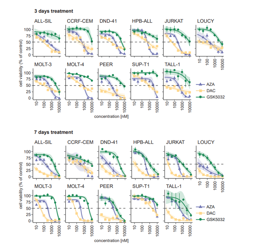
Cell viability as percentage of untreated control for eleven T-ALL cells treated with GSK-3685032 (10, 100, 1000, 10000 nM) for 3 and 7 days.
GSK-3685032 purchased from MedChemExpress. Usage Cited in: Clin Epigenetics. 2025 Jul 3;17(1):114. [Abstract]
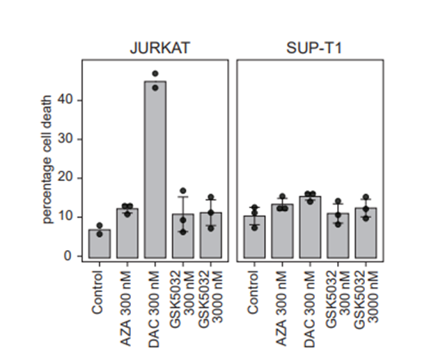
Percentage of dead cells (apoptotic and necrotic) after treatment of JURKAT and SUP-T1 cells with indicated concentrations of GSK5032 (300 nM) for 3 days based on flow cytometry analysis.
-
-
-
bioRxiv
2025 Jul 31:2025.07.28.666975. PMID: 40766615 -
-
-
bioRxiv
2024 Apr 3:2024.04.03.587980. PMID: 38617249 -


Solvent & Solubility
In Vitro:
DMSO : 25 mg/mL (59.45 mM; Need ultrasonic; Hygroscopic DMSO has a significant impact on the solubility of product, please use newly opened DMSO)
Please refer to the solubility information to select the appropriate solvent. Once prepared, please aliquot and store the solution to prevent product inactivation from repeated freeze-thaw cycles.
Storage method and period of stock solution: -80°C, 2 years; -20°C, 1 year. When stored at -80°C, please use it within 2 years. When stored at -20°C, please use it within 1 year.
Please refer to the solubility information to select the appropriate solvent. Once prepared, please aliquot and store the solution to prevent product inactivation from repeated freeze-thaw cycles.
Storage method and period of stock solution: -80°C, 2 years; -20°C, 1 year. When stored at -80°C, please use it within 2 years. When stored at -20°C, please use it within 1 year.
Concentration (start) × Volume (start) = Concentration (final) × Volume (final)
In Vivo:
Select the appropriate dissolution method based on your experimental animal and administration route.
- For the following dissolution methods, please ensure to first prepare a clear stock solution using an In Vitro approach and then sequentially add co-solvents:
- To ensure reliable experimental results, the clarified stock solution can be appropriately stored based on storage conditions. As for the working solution for In Vivo experiments, it is recommended to prepare freshly and use it on the same day.
- The percentages shown for the solvents indicate their volumetric ratio in the final prepared solution. If precipitation or phase separation occurs during preparation, heat and/or sonication can be used to aid dissolution.
Add each solvent one by one: 10% DMSO 40% PEG300 5% Tween-80 45% Saline
Solubility: ≥ 2.5 mg/mL (5.94 mM); Clear solution
This protocol yields a clear solution of ≥ 2.5 mg/mL (saturation unknown).
Taking 1 mL working solution as an example, add 100 μL DMSO stock solution (25.0 mg/mL) to 400 μL PEG300, and mix evenly; then add 50 μL Tween-80 and mix evenly; then add 450 μL Saline to adjust the volume to 1 mL.
Preparation of Saline: Dissolve 0.9 g sodium chloride in ddH₂O and dilute to 100 mL to obtain a clear Saline solution.
In Vivo Dissolution Calculator
Please enter the basic information of animal experiments:
-
-
-
-
Recommended: Prepare an additional quantity of animals to account for potential losses during experiments.
Please enter your animal formula composition:
-
%DMSO +
Recommended: Keep the proportion of DMSO in working solution below 2% if your animal is weak.
-
%+
-
+%Tween-80 + +
-
%Saline +
The co-solvents required include: DMSO, . All of co-solvents are available by MedChemExpress (MCE). , Tween 80. All of co-solvents are available by MedChemExpress (MCE).
Working solution concentration: 0.22 mg/mL
Method for preparing stock solution: mg drug dissolved in μL DMSO. Stock solution concentration: mg/mL.
1. Take μL DMSO stock solution;
2. Add μL .
μL , mix evenly;
3. Then add μL Tween 80, mix evenly;
4. Then add μL
Please ensure that the stock solution in the first step is dissolved to a clear state, and add co-solvents in sequence. You can use ultrasonic heating (ultrasonic cleaner, recommended frequency 20-40 kHz), vortexing, etc. to assist dissolution.

Protocols
-
Cell Cytotoxicity Assay
Cytotoxicity assays are usually based on the assessment of cell membrane damage, which can also be indirectly detected by measuring cell viability. Detection methods include MTT assay, CKK-8 assay, LDH assay and ATP assay, etc.
-
Research Protocol for Epigenomic Data Analysis
Epigenomic data analysis identifies genome-wide regulatory features that influence gene expression, chromatin state, and phenotype without changing the underlying DNA sequence. In this strategy, the core regulatory layer includes chromatin accessibility, transcription-factor or histone-mark occupancy, DNA methylation, and chromatin-state patterns; these features are measured by sequencing-based assays and interpreted as regulatory elements, promoters, enhancers, repressive domains, methylated cytosines, or candidate phenotype-associated chromatin programs. The literature links epigenomic features to phenotype by showing that functional genomic elements can be mapped across human cell types and tissues, and that integrated epigenomic maps reveal cell-type-specific regulatory programs. ENCODE integrated transcription, chromatin accessibility, transcription-factor occupancy, and histone modification data to annotate functional elements in the human genome, while the Roadmap Epigenomics Co
-
Protocol for Cell Counting and Cell Density Analysis
Cell counting and cell-density analysis estimate the number of cells in a known volume or field area. Manual hemocytometer counting uses a chamber of defined geometry to convert counted cells into cells/mL, while automated counters and image-analysis workflows detect cell objects from optical, brightfield, fluorescence, impedance, or digital-image features. Trypan blue viability counting is based on dye exclusion: viable cells with intact membranes exclude dye, while non-viable cells with compromised membranes stain blue. The readout is total cell density, viable-cell density, dead-cell density, and percent viability. Cell density can also be estimated from microscopy images by counting objects per image area, from flow cytometry using calibrated volume or reference particles, or from in situ microscopy in bioreactors after calibration against reference methods such as hemocytometer or flow cytometry.
-
Somatic Cell Culture
A method of simulating the in vivo environment in vitro to maintain the cell growth, differentation and main functions.
-
CCK-8/WST-8 Cell Proliferation Assay
The CCK-8/WST-8 assay is based on the reduction of the water-soluble tetrazolium salt WST-8 to a water-soluble formazan product by cellular dehydrogenases in metabolically active cells, where the generated formazan amount is proportional to the number of living cells and is quantified by measuring absorbance in the visible range, providing a colorimetric readout for cell viability and proliferation assessment. This class of tetrazolium-based assays improves upon earlier MTT-based systems by producing a water-soluble formazan, eliminating the need for organic solubilization steps and enabling direct spectrophotometric measurement in culture medium.
-
Cell Counting-Based Growth Curve Assay
Cell counting-based growth curve assays quantify cell proliferation by directly measuring changes in viable cell number over time using manual or automated counting methods such as hemocytometer-based counting or instrument-assisted cell enumeration, enabling construction of growth curves that reflect population expansion dynamics in response to culture conditions. A widely used approach is trypan blue exclusion with hemocytometer counting, where membrane-compromised (non-viable) cells take up the dye, allowing discrimination between viable and non-viable cells while simultaneously enabling total cell number quantification. Repeated sampling across time points allows estimation of proliferation rate, growth phases, and comparative growth kinetics between experimental conditions.
-
MTT Cell Proliferation Assay
The MTT assay is a colorimetric endpoint assay for estimating viable cell number, cell growth, cytotoxicity, or cell activation in cultured mammalian cells. Living cells reduce the yellow tetrazolium salt MTT into purple/blue formazan, while dead cells do not generate the same signal; the resulting color can be quantified with a multiwell spectrophotometer. MTT reduction is commonly interpreted as a readout of metabolic activity that often correlates with viable cell number, but it should not be treated as a direct cell-counting method unless the assay is optimized for the cell type and experimental condition. Studies show that MTT reduction can involve mitochondrial and non-mitochondrial reducing systems, and formazan may accumulate in intracellular lipid droplets rather than simply marking mitochondria.
-
Cell Viability Determination by MTT Colorimetric Assay
The following protocol uses the MTT colorimetric assay as a classic literature-established method for assessing cell viability/metabolic activity in cultured mammalian cells. MTT[3-(4,5-dimethylthiazol-2-yl)-2,5-diphenyltetrazolium bromide] is reduced by metabolically active cells to a colored formazan product; the amount of formazan is quantified spectrophotometrically and provides an indirect measure of metabolically active viable cells. Importantly, MTT reduction reflects cellular oxidoreductase/metabolic activity rather than an absolute direct count of living cells, so changes in cellular metabolism can alter the signal independently of cell number.


Purity & Documentation
-
Data Sheet (283 KB)
-
SDS (251 KB)
- English - EN (251 KB)
- Français - FR (251 KB)
- Deutsch - DE (251 KB)
- Norwegian - NO (251 KB)
- Español - ES (251 KB)
- Swedish - SV (251 KB)
- Italian - IT (251 KB)
- Korean - KR (251 KB)
- Portuguese - PT (251 KB)
-
Handling Instructions (2659 KB)
References


Complete Stock Solution Preparation Table
Please refer to the solubility information to select the appropriate solvent. Once prepared, please aliquot and store the solution to prevent product inactivation from repeated freeze-thaw cycles.
Storage method and period of stock solution: -80°C, 2 years; -20°C, 1 year. When stored at -80°C, please use it within 2 years. When stored at -20°C, please use it within 1 year.
| Optional Solvent | Concentration Solvent Mass | 1 mg | 5 mg | 10 mg | 25 mg |
|---|---|---|---|---|---|
| DMSO | 1 mM | 2.3780 mL | 11.8898 mL | 23.7795 mL | 59.4488 mL |
| 5 mM | 0.4756 mL | 2.3780 mL | 4.7559 mL | 11.8898 mL | |
| 10 mM | 0.2378 mL | 1.1890 mL | 2.3780 mL | 5.9449 mL | |
| 15 mM | 0.1585 mL | 0.7927 mL | 1.5853 mL | 3.9633 mL | |
| 20 mM | 0.1189 mL | 0.5945 mL | 1.1890 mL | 2.9724 mL | |
| 25 mM | 0.0951 mL | 0.4756 mL | 0.9512 mL | 2.3780 mL | |
| 30 mM | 0.0793 mL | 0.3963 mL | 0.7927 mL | 1.9816 mL | |
| 40 mM | 0.0594 mL | 0.2972 mL | 0.5945 mL | 1.4862 mL | |
| 50 mM | 0.0476 mL | 0.2378 mL | 0.4756 mL | 1.1890 mL |


GSK-3685032 Related Classifications
HY-139664 Related Classifications
 Powered by Bioz