



ATP-Red 1
Based on 13 publication(s) in Google Scholar
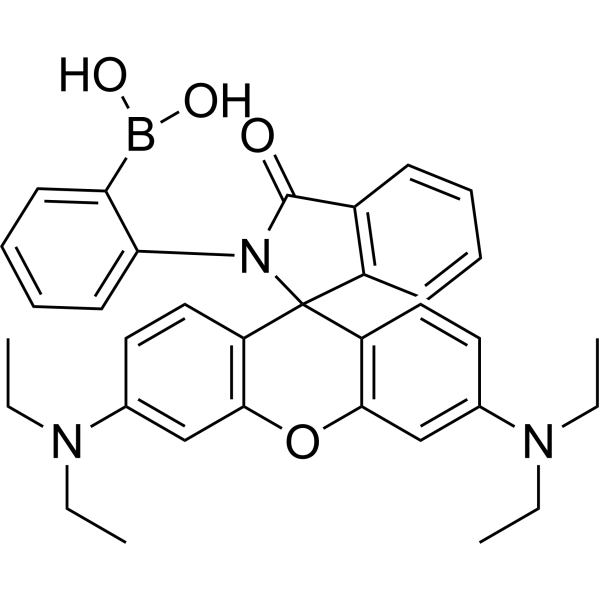
ATP-Red 1 is a multisite-binding switchable fluorescent probe, and can selectively and rapidly responds to intracellular concentrations of ATP in living cells (Ex/Em = 510/590 nm).
For research use only. We do not sell to patients.
- Purity : 98.0%
- CAS No.: 1847485-97-5
- Formula: C34H36BN3O4
- Molecular Weight:561.48
-
Storage:
4°C, protect from light
* In solvent : -80°C, 6 months; -20°C, 1 month (protect from light)

To place orders, for customer services and technical support, please contact: MedChemExpress USA
Tel: 609-228-6898 E-mail: [email protected] [email protected]


-
 Biological Activity
-
Chemical Information
-
Solvent & Solubility
- Purity & Documentation
- References
-
 Help & FAQs


-
HY-D0940
H2DCFDAROS-Sensitive Probe -

HY-15371
ForskolinAdenylate Cyclase Activator -
HY-16592
Brefeldin AProtein Transport Inhibitor -

HY-N6782
OligomycinH+-ATP-Synthase Inhibitor -

HY-10219
RapamycinmTOR Inhibitor -
HY-13757A
TamoxifenEstrogen Receptor Modulator -
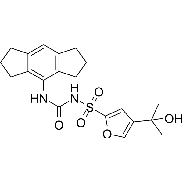
HY-12815
MCC950NLRP3 Inhibitor -
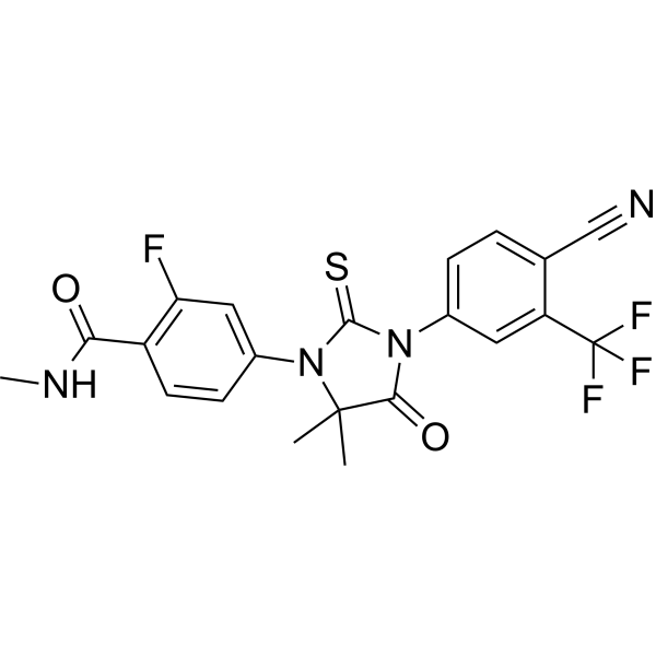
HY-70002
EnzalutamideAndrogen Receptor Antagonist -
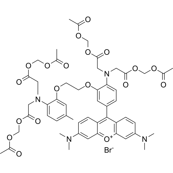
HY-D0989
Rhod-2 AMCa(2+) Indicator -

HY-100558
Bafilomycin A1V-ATPase Inhibitor -
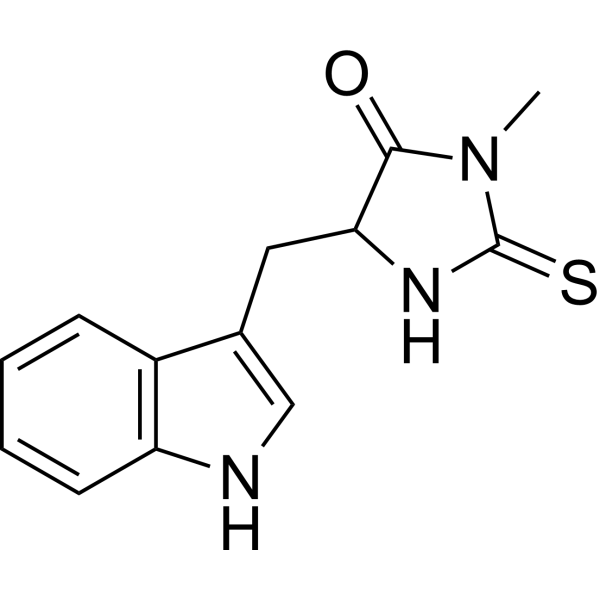
HY-15760
Necrostatin-1RIPK1 Inhibitor -
HY-10432
A 83-01ALK4/5/7 Inhibitor
Publications Citing Use of MedChemExpress (MCE) ATP-Red 1
More- Science. 2026 Apr 9;392(6794):eadt2178. [Abstract]
- Free Radic Biol Med. 2026 Sep:253:785-802. [Abstract]
- Br J Pharmacol. 2025 Aug;182(16):3923-3951. [Abstract]
- Postharvest Biol Technol. 2026 Jun 2;241:114495.
- Cells. 2022 Dec 23;12(1):68. [Abstract]
- Int J Mol Sci. 2024 Dec 24;26(1):44. [Abstract]
- Eur J Med Res. 2025 Oct 17;30(1):991. [Abstract]
- FASEB J. 2025 Jul 31;39(14):e70846. [Abstract]
- Drug Dev Res. 2025 Sep;86(6):e70150. [Abstract]
- Naunyn Schmiedebergs Arch Pharmacol. 2025 Jul;398(7):8899-8908. [Abstract]
- Mechanobiol Med. 2026 Jul 13.
- Tissue Cell. 2025 Apr:93:102739. [Abstract]
- Res Sq. 2026 May 20.
 Customer Validation & Images

-
Cell Imaging/Staining
-
Cell Imaging/Staining
-
Cell Imaging/Staining
-
Cell Imaging/Staining
-
Cell Imaging/Staining


Biological Activity
Description
In Vitro
Guide (The following is our recommended protocol. This protocol is only a guide and should be modified according to your specific needs).
1. Solution preparation
1.1 Preparation of stock solution
Solvent: DMSO
Concentration: 10 mM is recommended.
Storage: Store at -20°C or -80°C in dark after aliquoting. Avoid repeated freezing and thawing.
1.2 Preparation of working solution
Dilute to 1-10 μM with PBS or serum-free cell culture medium (optimized according to the experiment).
Note: The working solution should be prepared and used immediately. Keep it away from light.
2. Cell staining (suspended cells)
2.1 Collect cells by centrifugation and wash twice with PBS for 5 minutes each time. The cell density is 1×10⁶/mL.
2.2 Add ATP-Red 1 at a final concentration of 5-10 μM.
2.3 Incubate cells at room temperature or 37°C for 15 minutes (or up to 30 minutes for live cell imaging).
2.4 Centrifuge at 400 g for 3-4 minutes and discard the supernatant.
2.5 Add PBS to wash the cells twice, 5 minutes each time.
2.6 Resuspend the cells of serum-free medium or PBS and observe using a fluorescence microscope or flow cytometer.
3. Cell staining (adherent cells)
3.1 Culture the adherent cells on a sterile coverslip.
3.2 Remove the coverslip from the culture medium and remove the excess culture medium.
3.3 Add ATP-Red 1 at a final concentration of 5-10 μM.
3.4 Incubate cells at room temperature or 37°C for 15 minutes (or up to 30 minutes for live cell imaging).
3.5 Aspirate the dye working solution, wash 2-3 times with culture medium, 5 minutes each time, and observe using a fluorescence microscope (Ex/Em = 510/590 nm) or flow cytometer.
Note: If flow cytometry is required, the cells need to be digested with trypsin and resuspended before staining.
MedChemExpress (MCE) has not independently confirmed the accuracy of these methods. They are for reference only.
Emission (Em)
590
Excitation (Ex)
510


Chemical Information
-
CAS No. 1847485-97-5
-
Appearance Solid
-
Molecular Weight 561.48
-
Formula C34H36BN3O4
-
Color Pale purple to purple
-
SMILES
O=C1N(C2=CC=CC=C2B(O)O)C3(C4=C(OC5=C3C=CC(N(CC)CC)=C5)C=C(N(CC)CC)C=C4)C6=C1C=CC=C6
-
Shipping
Room temperature in continental US; may vary elsewhere.
-
Storage
4°C, protect from light
* In solvent : -80°C, 6 months; -20°C, 1 month (protect from light)


Publications (13)
-
Journal Impact Factor
-
Most Recent
-
Science
2026 Apr 9;392(6794):eadt2178. PMID: 41610195 -
Free Radic Biol Med
Bushen Tiaojing decoction ameliorates meiotic impairment to improve oocyte quality during aging through blocking the iron overload-induced p53/CDK1/ESPL1 pathway. [Abstract]2026 Sep:253:785-802. PMID: 42214643 -
Br J Pharmacol
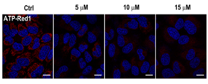
A mitochondria-targeting and G-quadruplex structure-binding ligand inducing calcium overload and ferroptosis in human cancer cells. [Abstract]2025 Aug;182(16):3923-3951. PMID: 40344208
ATP-Red 1 purchased from MedChemExpress. Usage Cited in: Br J Pharmacol. 2025 Aug;182(16):3923-3951. [Abstract]
ATP production in HeLa cells after BYB (0–15 μM) treatment for 24 h. The ATP was evaluated with ATP-Red 1 (red, λex = 561 nm; 2 μM; 30 min).
-
-
Cells
Low Expression of Phosphodiesterase 2 (PDE2A) Promotes the Progression by Regulating Mitochondrial Morphology and ATP Content and Predicts Poor Prognosis in Hepatocellular Carcinoma. [Abstract]2022 Dec 23;12(1):68. PMID: 36611861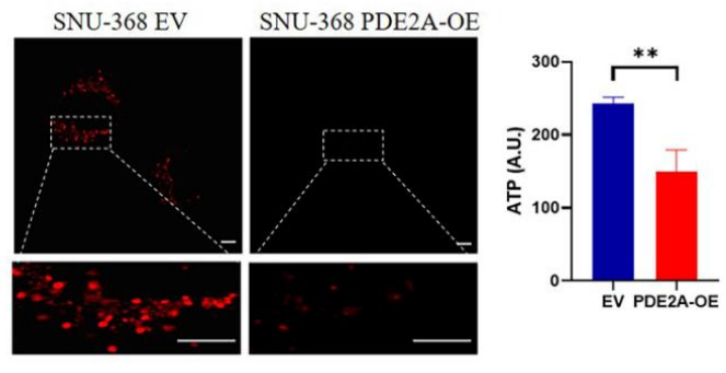
ATP-Red 1 purchased from MedChemExpress. Usage Cited in: Cells. 2022 Dec 23;12(1):68. [Abstract]
ATP-Red1 detection of ATP content in SNU-368 under PDE2A-overexpressed treatment and EV as control.
-
Int J Mol Sci
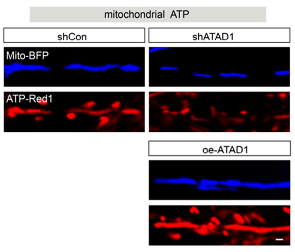
ATAD1 Regulates Neuronal Development and Synapse Formation Through Tuning Mitochondrial Function. [Abstract]2024 Dec 24;26(1):44. PMID: 39795902
ATP-Red 1 purchased from MedChemExpress. Usage Cited in: Int J Mol Sci. 2024 Dec 24;26(1):44. [Abstract]
Representative images of ATP-Red 1 staining (red, labeling mitochondrial ATP; 5 μM; 30 min) in shCon-expressing-control,shATAD1-expressing and oe-ATAD1-expressing neuron.
-
Eur J Med Res
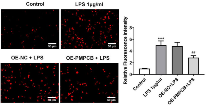
Identification of diagnostic biomarkers and mitochondrial metabolic characteristics in sepsis-associated acute kidney injury. [Abstract]2025 Oct 17;30(1):991. PMID: 41107920
ATP-Red 1 purchased from MedChemExpress. Usage Cited in: Eur J Med Res. 2025 Oct 17;30(1):991. [Abstract]
ATP production level as measured by fluorescence intensity in HK-2 cells by ATP-Red 1.
-
FASEB J
Reverse Effects of Nicotinamide Mononucleotide Supplementation on Declining Quality of Oocytes With Polycystic Ovary Syndrome. [Abstract]2025 Jul 31;39(14):e70846. PMID: 40678956 -
Drug Dev Res
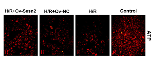
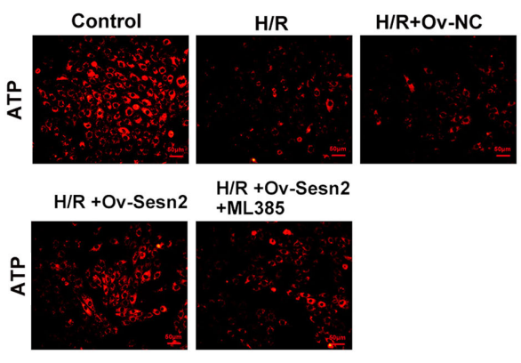
Sestrin2 Regulates Mitochondrial Function and Autophagy via Nrf2/SIRT3 Signaling to Ameliorate Hypoxia/Reoxygenation-Induced Rat's Cardiomyocyte Injury. [Abstract]2025 Sep;86(6):e70150. PMID: 40878255
ATP-Red 1 purchased from MedChemExpress. Usage Cited in: Drug Dev Res. 2025 Sep;86(6):e70150. [Abstract]
ATP‐Red 1 probe (30 min) was used to measure ATP content in H9C2 cells. Sesn2 overexpression increased ATP levels and alleviated the decrease in ATP caused by H/R.
ATP-Red 1 purchased from MedChemExpress. Usage Cited in: Drug Dev Res. 2025 Sep;86(6):e70150. [Abstract]
ATP‐Red 1 probe (30 min) was used to measure ATP content in H9C2 cells. ML385 was found to reduce ATP content.
-
Naunyn Schmiedebergs Arch Pharmacol
Ginkgolide B regulates apoptosis, oxidative stress, and mitochondrial dysfunction in MPP+-induced SK-N-SH cells by targeting HDAC4/JNK pathway. [Abstract]2025 Jul;398(7):8899-8908. PMID: 39878815 -
-
Tissue Cell
PCSK9 exacerbates sevoflurane-induced neuroinflammatory response and apoptosis by up-regulating cGAS-STING signal. [Abstract]2025 Apr:93:102739. PMID: 39818066 -


Solvent & Solubility
In Vitro:
DMSO : 50 mg/mL (89.05 mM; Need ultrasonic; Hygroscopic DMSO has a significant impact on the solubility of product, please use newly opened DMSO)
Please refer to the solubility information to select the appropriate solvent. Once prepared, please aliquot and store the solution to prevent product inactivation from repeated freeze-thaw cycles.
Storage method and period of stock solution: -80°C, 6 months; -20°C, 1 month (protect from light). When stored at -80°C, please use it within 6 months. When stored at -20°C, please use it within 1 month.
Please refer to the solubility information to select the appropriate solvent. Once prepared, please aliquot and store the solution to prevent product inactivation from repeated freeze-thaw cycles.
Storage method and period of stock solution: -80°C, 6 months; -20°C, 1 month (protect from light). When stored at -80°C, please use it within 6 months. When stored at -20°C, please use it within 1 month.
Concentration (start) × Volume (start) = Concentration (final) × Volume (final)


Purity & Documentation
-
Data Sheet (285 KB)
-
SDS (252 KB)
- English - EN (252 KB)
- Français - FR (252 KB)
- Deutsch - DE (252 KB)
- Norwegian - NO (252 KB)
- Español - ES (252 KB)
- Swedish - SV (252 KB)
- Italian - IT (252 KB)
- Korean - KR (252 KB)
- Portuguese - PT (252 KB)
-
Handling Instructions (2659 KB)
References


Complete Stock Solution Preparation Table
Please refer to the solubility information to select the appropriate solvent. Once prepared, please aliquot and store the solution to prevent product inactivation from repeated freeze-thaw cycles.
Storage method and period of stock solution: -80°C, 6 months; -20°C, 1 month (protect from light). When stored at -80°C, please use it within 6 months. When stored at -20°C, please use it within 1 month.
| Optional Solvent | Concentration Solvent Mass | 1 mg | 5 mg | 10 mg | 25 mg |
|---|---|---|---|---|---|
| DMSO | 1 mM | 1.7810 mL | 8.9050 mL | 17.8101 mL | 44.5252 mL |
| 5 mM | 0.3562 mL | 1.7810 mL | 3.5620 mL | 8.9050 mL | |
| 10 mM | 0.1781 mL | 0.8905 mL | 1.7810 mL | 4.4525 mL | |
| 15 mM | 0.1187 mL | 0.5937 mL | 1.1873 mL | 2.9683 mL | |
| 20 mM | 0.0891 mL | 0.4453 mL | 0.8905 mL | 2.2263 mL | |
| 25 mM | 0.0712 mL | 0.3562 mL | 0.7124 mL | 1.7810 mL | |
| 30 mM | 0.0594 mL | 0.2968 mL | 0.5937 mL | 1.4842 mL | |
| 40 mM | 0.0445 mL | 0.2226 mL | 0.4453 mL | 1.1131 mL | |
| 50 mM | 0.0356 mL | 0.1781 mL | 0.3562 mL | 0.8905 mL | |
| 60 mM | 0.0297 mL | 0.1484 mL | 0.2968 mL | 0.7421 mL | |
| 80 mM | 0.0223 mL | 0.1113 mL | 0.2226 mL | 0.5566 mL |


ATP-Red 1 Related Classifications
HY-U00451 Related Classifications
 Powered by Bioz