



Flunisolide
Based on 1 publication(s) in Google Scholar
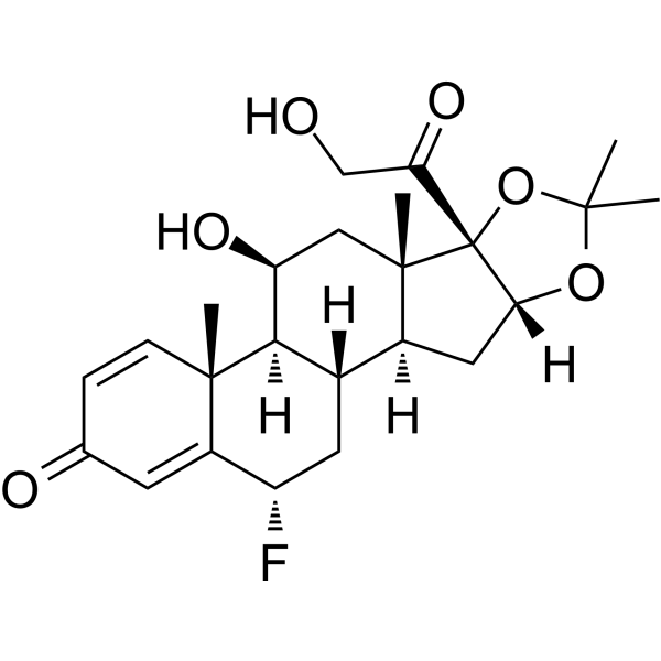
Flunisolide is a corticosteroid, which is an orally active glucocorticoid receptor activator with anti-inflammatory activity. Flunisolide can induce eosinophil apoptosis, and is used for the research of asthma or rhinitis, and inflammation.
For research use only. We do not sell to patients.
- Purity : 99.87%
- CAS No.: 3385-03-3
- Formula: C24H31FO6
- Molecular Weight:434.50
-
Storage:Powder -20°C, 3 years , 4°C, 2 years ; In solvent -80°C, 2 years , -20°C, 1 year

To place orders, for customer services and technical support, please contact: MedChemExpress USA
Tel: 609-228-6898 E-mail: [email protected] [email protected]


-
 Biological Activity
-
Chemical Information
-
Solvent & Solubility
- Protocols
- Purity & Documentation
- References
-
 Help & FAQs


-
HY-13580
BudesonideGlucocorticoid Receptor Agonist -
HY-13693
Mometasone furoateGlucocorticoid Receptor Agonist -
HY-B0636
Triamcinolone acetonideAnti-Inflammatory Agent -
HY-B0154
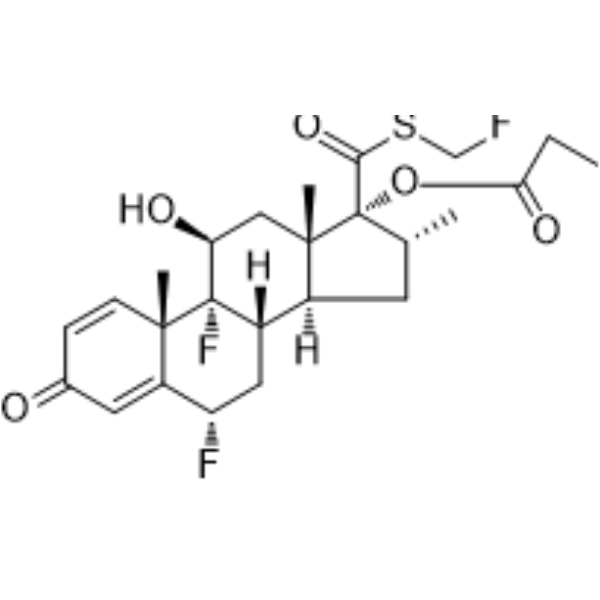
Fluticasone propionateGlucocorticoid Receptor Agonist -
HY-B0328
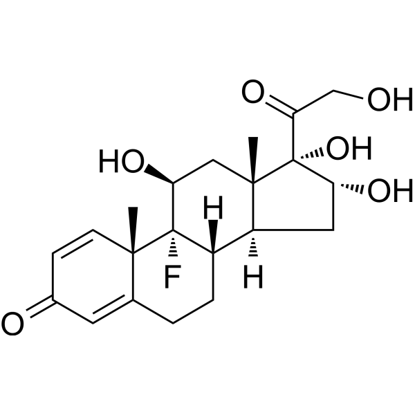
TriamcinoloneSHRs Agonist -
HY-13570
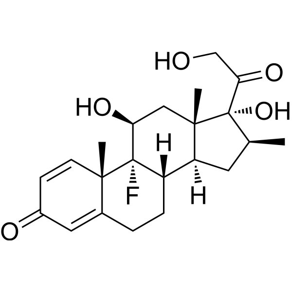
BetamethasoneGlucocorticoid Receptor Agonist -

HY-13571AR
Beclometasone dipropionate (Standard)Glucocorticoid Receptor Agonist -
HY-13600
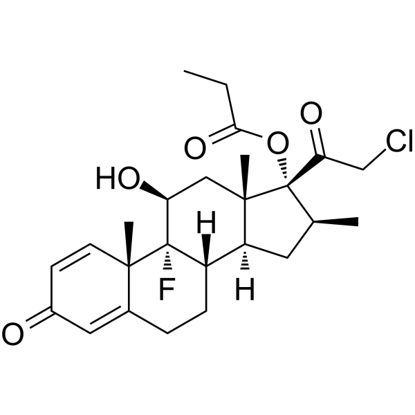
Clobetasol propionateCYP3A5 Inhibitor -
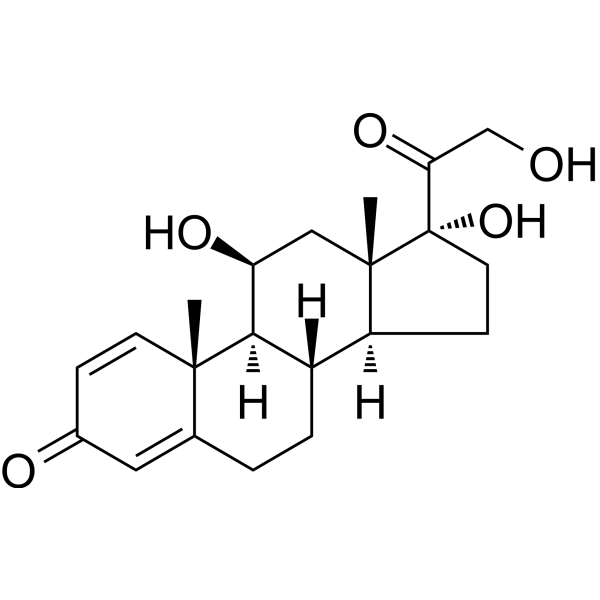
HY-17463
PrednisoloneGlucocorticoid -
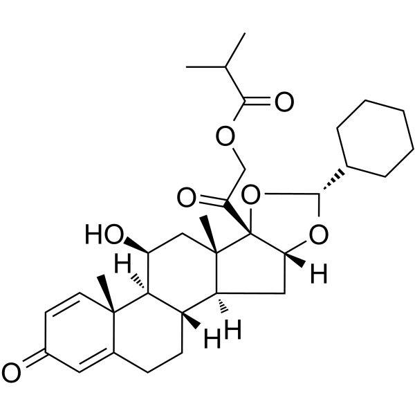
HY-B0625
CiclesonideGlucocorticoid -
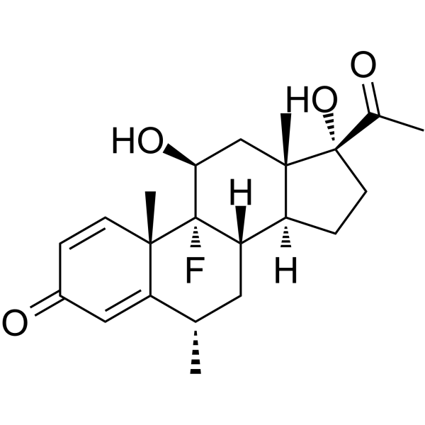
HY-B1893
FluorometholoneGlucocorticoid Receptor Agonist -
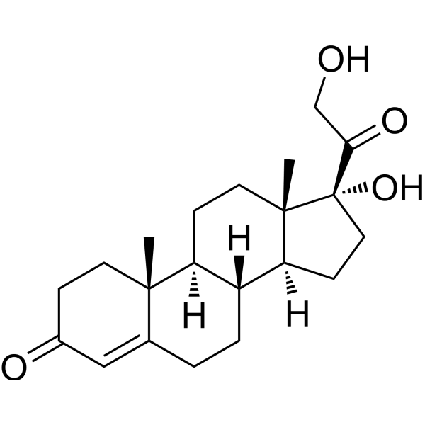
HY-77839
CortodoxoneGlucocorticoid Steroid Hormone
Publications Citing Use of MedChemExpress (MCE) Flunisolide
More

Biological Activity
Description
In Vitro
Flunisolide (0.1-10 μM, 1 h) inhibits lung fibroblast (Isolated from lung) activation[1]. Flunisolide (10 μM, 24 h) reduces MMP-9, TIMP-1, TGF-β and fibronectin release by sputum cells (isolated from mild to moderate asthmatics), and induces sputum eosinophil apoptosis[2]. Flunisolide (0.1-10 μM μM, 24 h) effectively inhibits ICAM-1 expression and GM-CSF and IL-5 release induced by TNF-alpha in BEAS-2B cells[3]. Flunisolide (115 μM, 0-3 h) can be transported in a polarized way in the apical (ap) to basolateral (bl) direction in Calu-3 cells and is demonstrated to be ATP-dependent[4].
MedChemExpress (MCE) has not independently confirmed the accuracy of these methods. They are for reference only. Further protocols information, click here.
-
Cell Line:Eosinophil
-
Concentration:10 μM
-
Incubation Time:24 h
-
Result:Induced sputum eosinophil apoptosis.
In Vivo
MedChemExpress (MCE) has not independently confirmed the accuracy of these methods. They are for reference only.
-
Animal Model:Male Swiss Webster mice (instilled, intranasally, with crystalline silica, 10 mg/50 µL, particle size 0.5-10 µm)[1]
-
Dosage:0.3-10 µg/mouse, daily, from days 21–27
-
Administration:Intranasal administration
-
Result:Reduced both granulomatous response, collagen deposition, concerning granuloma formation caused by silica particles. Reduced the number of F4/80 and α-SMA positive cells.


Chemical Information
-
CAS No. 3385-03-3
-
Appearance Solid
-
Molecular Weight 434.50
-
Formula C24H31FO6
-
Color White to off-white
-
SMILES
C[C@@]12[C@@]3(C(CO)=O)[C@@](OC(C)(O3)C)([H])C[C@@]1([H])[C@]4([H])C[C@H](F)C5=CC(C=C[C@]5(C)[C@@]4([H])[C@@H](O)C2)=O
-
Shipping
Room temperature in continental US; may vary elsewhere.
-
Storage
Powder -20°C 3 years 4°C 2 years In solvent -80°C 2 years -20°C 1 year


Publications (1)
-
Journal Impact Factor
-
Most Recent
-
Drug Test Anal
High-throughput liquid chromatography tandem mass spectrometry assay as initial testing procedure for analysis of total urinary fraction. [Abstract]2021 Feb;13(2):283-298. PMID: 32852861


Solvent & Solubility
In Vitro:
DMSO : 100 mg/mL (230.15 mM; Need ultrasonic; Hygroscopic DMSO has a significant impact on the solubility of product, please use newly opened DMSO)
Please refer to the solubility information to select the appropriate solvent. Once prepared, please aliquot and store the solution to prevent product inactivation from repeated freeze-thaw cycles.
Storage method and period of stock solution: -80°C, 2 years; -20°C, 1 year. When stored at -80°C, please use it within 2 years. When stored at -20°C, please use it within 1 year.
Please refer to the solubility information to select the appropriate solvent. Once prepared, please aliquot and store the solution to prevent product inactivation from repeated freeze-thaw cycles.
Storage method and period of stock solution: -80°C, 2 years; -20°C, 1 year. When stored at -80°C, please use it within 2 years. When stored at -20°C, please use it within 1 year.
Concentration (start) × Volume (start) = Concentration (final) × Volume (final)
In Vivo:
Select the appropriate dissolution method based on your experimental animal and administration route.
- For the following dissolution methods, please ensure to first prepare a clear stock solution using an In Vitro approach and then sequentially add co-solvents:
- To ensure reliable experimental results, the clarified stock solution can be appropriately stored based on storage conditions. As for the working solution for In Vivo experiments, it is recommended to prepare freshly and use it on the same day.
- The percentages shown for the solvents indicate their volumetric ratio in the final prepared solution. If precipitation or phase separation occurs during preparation, heat and/or sonication can be used to aid dissolution.
Add each solvent one by one: 10% DMSO 40% PEG300 5% Tween-80 45% Saline
Solubility: ≥ 2.08 mg/mL (4.79 mM); Clear solution
This protocol yields a clear solution of ≥ 2.08 mg/mL (saturation unknown).
Taking 1 mL working solution as an example, add 100 μL DMSO stock solution (20.8 mg/mL) to 400 μL PEG300, and mix evenly; then add 50 μL Tween-80 and mix evenly; then add 450 μL Saline to adjust the volume to 1 mL.
Preparation of Saline: Dissolve 0.9 g sodium chloride in ddH₂O and dilute to 100 mL to obtain a clear Saline solution.
Add each solvent one by one: 10% DMSO 90% (20% SBE-β-CD in Saline)
Solubility: ≥ 2.08 mg/mL (4.79 mM); Clear solution
This protocol yields a clear solution of ≥ 2.08 mg/mL (saturation unknown).
Taking 1 mL working solution as an example, add 100 μL DMSO stock solution (20.8 mg/mL) to 900 μL 20% SBE-β-CD in Saline, and mix evenly.
Preparation of 20% SBE-β-CD in Saline (4°C, storage for one week): 2 g SBE-β-CD powder is dissolved in 10 mL Saline, completely dissolve until clear.
In Vivo Dissolution Calculator
Please enter the basic information of animal experiments:
-
-
-
-
Recommended: Prepare an additional quantity of animals to account for potential losses during experiments.
Please enter your animal formula composition:
-
%DMSO +
Recommended: Keep the proportion of DMSO in working solution below 2% if your animal is weak.
-
%+
-
+%Tween-80 + +
-
%Saline +
The co-solvents required include: DMSO, . All of co-solvents are available by MedChemExpress (MCE). , Tween 80. All of co-solvents are available by MedChemExpress (MCE).
Working solution concentration: 0.22 mg/mL
Method for preparing stock solution: mg drug dissolved in μL DMSO. Stock solution concentration: mg/mL.
1. Take μL DMSO stock solution;
2. Add μL .
μL , mix evenly;
3. Then add μL Tween 80, mix evenly;
4. Then add μL
Please ensure that the stock solution in the first step is dissolved to a clear state, and add co-solvents in sequence. You can use ultrasonic heating (ultrasonic cleaner, recommended frequency 20-40 kHz), vortexing, etc. to assist dissolution.

Protocols
-
Apoptosis
Apoptosis, also called programmed cell death, is generally characterized by distinct morphological characteristics.
-
TUNEL staining for apoptotic DNA fragmentation
TUNEL staining detects DNA strand breaks by using terminal deoxynucleotidyl transferase to add labeled nucleotides to exposed 3′-OH DNA termini, generating either microscopic staining in fixed cells or tissue sections, or fluorescence/cytometric signal in cell suspensions. TUNEL positivity reflects DNA fragmentation but should not be interpreted alone as definitive apoptosis, because TUNEL can also label necrotic, autolytic, mechanically damaged, or DNA-repair-associated DNA breaks.
-
Annexin V plus membrane-impermeant dye apoptosis staining
Annexin V-based apoptosis assays rely on the detection of phosphatidylserine (PS) externalization from the inner leaflet of the plasma membrane to the outer leaflet, an early biochemical hallmark of apoptosis. Fluorescently labeled Annexin V binds PS in a calcium-dependent manner, enabling identification of early apoptotic cells by flow cytometry or fluorescence microscopy. When combined with a membrane-impermeant DNA-binding dye (e. g. , propidium iodide), this approach allows discrimination between viable (Annexin V−/dye−), early apoptotic (Annexin V+/dye−), and late apoptotic or necrotic (Annexin V+/dye+) cell populations by assessing membrane integrity and PS exposure.
-
Cotton Pellet Granuloma
Cotton pellet granuloma is a classical in vivo chronic inflammation model used to evaluate the anti-inflammatory potential of test substances by measuring their ability to inhibit granuloma tissue formation around an implanted foreign body (cotton pellet) in rodents. The method is based on the biological response to a sterile implanted material, which induces proliferative phase inflammation characterized by fibroblast proliferation and collagen-rich granuloma formation, and the final readout reflects the extent of chronic inflammatory tissue growth surrounding the pellet. In multiple preclinical pharmacological evaluations, inhibition of cotton pellet-induced granuloma formation has been used as an indicator of anti-inflammatory activity in both synthetic and natural product screening contexts.
-
Research Protocol for Endocrine Diseases
Endocrine diseases often arise from disrupted hormone production, hormone signaling, or target-tissue responsiveness; for diabetes-focused endocrine disease models, insulin signaling regulates glucose uptake, hepatic glucose output, lipid metabolism, and β-cell compensation. Type 2 diabetes develops through interacting defects in insulin resistance, β-cell dysfunction, adipose inflammation, hepatic glucose overproduction, altered incretin signaling, and ectopic lipid metabolism. A major unresolved question is whether endocrine dysfunction is driven primarily by target-tissue insulin resistance, intrinsic β-cell failure, immune/inflammatory stress, or combined multi-organ failure that differs by disease stage.
-
Carrageenan-Induced Paw Edema
Carrageenan-induced paw edema is an acute inflammation model in which intraplantar injection of carrageenan induces localized inflammatory swelling characterized by vascular permeability, leukocyte infiltration, and production of inflammatory mediators such as prostaglandins and cytokines, making it widely used to evaluate anti-inflammatory agents in vivo. The resulting paw volume or thickness increase is quantified over time as a direct readout of inflammatory intensity and drug efficacy, typically reflecting cyclooxygenase-mediated prostaglandin-driven edema formation and immune cell recruitment in peripheral tissue[20].
-
Apoptosis Solutions
Apoptosis is a regulated, generally non-lytic cell-death pathway that removes unwanted, damaged, infected, or abnormal cells through coordinated morphological changes, caspase activation, DNA fragmentation, and membrane remodeling. The intrinsic apoptosis pathway is controlled mainly by mitochondrial outer membrane permeabilization, BCL-2 family proteins, cytochrome c release, apoptosome formation, caspase-9 activation, and downstream executioner caspase-3/7 activation. The extrinsic apoptosis pathway is initiated by death receptors such as Fas, TNFR, and TRAIL receptors, which recruit adaptor proteins and activate caspase-8 before engaging executioner caspases or mitochondrial amplification through BID cleavage. Apoptosis is linked to many phenotypes, including cancer cell killing, tissue homeostasis, immune regulation, neurodegeneration, infection response, and treatment-induced cytotoxicity; unresolved questions include how apoptosis interacts with necroptosis, pyroptosis, ferroptos
-
Research Protocol for Inflammation-related Diseases
The NLRP3 inflammasome is a cytosolic innate immune signaling platform that integrates priming signals and danger-signal activation to promote caspase-1 activation, maturation of IL-1β and IL-18, and gasdermin D-mediated pyroptotic cell death. The core experimental logic is to determine whether inflammatory disease phenotypes are driven by increased NLRP3 expression, ASC-containing inflammasome assembly, caspase-1 cleavage, GSDMD cleavage, and extracellular release of IL-1β/IL-18 rather than by nonspecific cell injury alone. The pathway is strongly linked to inflammation-related disease phenotypes because monosodium urate crystals activate NALP3/NLRP3 inflammasome signaling in gout-like crystal inflammation, cholesterol crystals activate NLRP3 inflammasomes in atherogenesis models, and DSS-induced intestinal inflammation has been reported to involve NLRP3 inflammasome activity. However, experimental colitis studies also show context-dependent protective effects of NLRP3 inflammasome co


Purity & Documentation
-
Data Sheet (284 KB)
-
SDS (479 KB)
- English - EN (479 KB)
- Français - FR (479 KB)
- Deutsch - DE (479 KB)
- Norwegian - NO (479 KB)
- Español - ES (479 KB)
- Swedish - SV (479 KB)
- Italian - IT (479 KB)
- Korean - KR (479 KB)
- Portuguese - PT (479 KB)
-
Handling Instructions (2659 KB)
References
[1]. Tatiana Paula Teixeira Ferreira, et al. Intranasal Flunisolide Suppresses Pathological Alterations Caused by Silica Particles in the Lungs of Mice. Front Endocrinol (Lausanne). 2020 Jun 17;11:388. [Content Brief]
[2]. M Profita, et al. In vitro effects of flunisolide on MMP-9, TIMP-1, fibronectin, TGF-beta1 release and apoptosis in sputum cells freshly isolated from mild to moderate asthmatics. Allergy. 2004 Sep;59(9):927-32. [Content Brief]
[3]. S Boero, et al. Modulation by flunisolide of tumor necrosis factor-alpha-induced stimulation of airway epithelial cell activities related to eosinophil inflammation. J Asthma. 2010 May;47(4):381-7. [Content Brief]
[4]. B I Florea, et al. Evidence of P-glycoprotein mediated apical to basolateral transport of flunisolide in human broncho-tracheal epithelial cells (Calu-3). Br J Pharmacol. 2001 Dec;134(7):1555-63. [Content Brief]


Complete Stock Solution Preparation Table
Please refer to the solubility information to select the appropriate solvent. Once prepared, please aliquot and store the solution to prevent product inactivation from repeated freeze-thaw cycles.
Storage method and period of stock solution: -80°C, 2 years; -20°C, 1 year. When stored at -80°C, please use it within 2 years. When stored at -20°C, please use it within 1 year.
| Optional Solvent | Concentration Solvent Mass | 1 mg | 5 mg | 10 mg | 25 mg |
|---|---|---|---|---|---|
| DMSO | 1 mM | 2.3015 mL | 11.5075 mL | 23.0150 mL | 57.5374 mL |
| 5 mM | 0.4603 mL | 2.3015 mL | 4.6030 mL | 11.5075 mL | |
| 10 mM | 0.2301 mL | 1.1507 mL | 2.3015 mL | 5.7537 mL | |
| 15 mM | 0.1534 mL | 0.7672 mL | 1.5343 mL | 3.8358 mL | |
| 20 mM | 0.1151 mL | 0.5754 mL | 1.1507 mL | 2.8769 mL | |
| 25 mM | 0.0921 mL | 0.4603 mL | 0.9206 mL | 2.3015 mL | |
| 30 mM | 0.0767 mL | 0.3836 mL | 0.7672 mL | 1.9179 mL | |
| 40 mM | 0.0575 mL | 0.2877 mL | 0.5754 mL | 1.4384 mL | |
| 50 mM | 0.0460 mL | 0.2301 mL | 0.4603 mL | 1.1507 mL | |
| 60 mM | 0.0384 mL | 0.1918 mL | 0.3836 mL | 0.9590 mL | |
| 80 mM | 0.0288 mL | 0.1438 mL | 0.2877 mL | 0.7192 mL | |
| 100 mM | 0.0230 mL | 0.1151 mL | 0.2301 mL | 0.5754 mL |


Flunisolide Related Classifications
HY-B1121 Related Classifications
 Powered by Bioz