



Tocopherols

Based on 1 Customer Validation
Tocopherols are orally effective antioxidants and ferroptosis inhibitors. Tocopherols scavenge ROS/RNS, alleviate DNA damage, downregulate cyclin D1/c-Myc/TFF/pS2/PGR in an ER-dependent manner, and inhibit ferroptosis mediated by lipid peroxidation. Tocopherols block the occurrence of ferroptosis in Gpx4-knockout Pfa1 cells, with an EC50 value of 1.0-2.3 μM. Tocopherols inhibit tumor growth. Tocopherols can be used in studies related to estrogen-mediated ER+ breast cancer, oxidative stress-induced carcinogenesis, and ferroptosis regulation.
For research use only. We do not sell to patients.
- Purity : 98.6%
- CAS No.: 1406-66-2
-
Storage:Pure form -20°C, 3 years ; In solvent -80°C, 6 months , -20°C, 1 month


To place orders, for customer services and technical support, please contact: MedChemExpress USA
Tel: 609-228-6898 E-mail: [email protected] [email protected]


-
 Biological Activity
-
Chemical Information
-
Solvent & Solubility
- Protocols
- Purity & Documentation
- References
-
 Help & FAQs



All Endogenous Metabolite Isoforms
More

Biological Activity
Description
In Vitro
Tocopherols (10 µM; 1-4 days) downregulate the expression of cyclin D1 and c-Myc in estrogen-induced ER-positive MCF-7 cells, and this antiproliferative activity is ER-dependent[1].
Tocopherols (10 µM; 4 days) downregulate the expression of estrogen-responsive genes in estrogen receptor-positive MCF-7 cells induced by estrogen, and this activity is ER-dependent[1].
Tocopherols (10 µM; 24-48 h) moderately reduce etoposide-induced DNA double-strand breaks in MCF-7 cells, as detected by γ-H2AX foci[1].
Tocopherols (10 µM; 24-48 h) significantly reduce estrogen-induced oxidative DNA damage (detected by 8-oxo-dG levels) in ER-positive MCF-7 cells, and this protective activity is ER-dependent[1].
Tocopherols (10 µM; 24-48 h) significantly reduce estrogen-induced nitrosative stress (measured by nitrotyrosine levels) in ER-positive MCF-7 cells, and this protective activity is ER-dependent[1].
Tocopherols (10 µM; 24-48 h) significantly reduce estrogen-induced DNA double-strand breaks (detected by γ-H2AX foci) in estrogen receptor (ER)-positive MCF-7 cells, and this protective activity is dependent on ER[1].
Tocopherols (1-100 μM; 6 h) inhibit lipid peroxidation of liposomes in cell-free FENIX assays[2].
Tocopherols (0-100 μM; 24-72 h) inhibit ferroptosis induced by RSL3, erastin and BSO in HT-1080 cells, but a concentration higher than 10 μM is required to completely suppress RSL3-induced cell death[2].
Tocopherol (0-100 μM; 24-72 h) inhibits ferroptosis induced by RSL3, erastin and BSO in Pfa1 cells[2].
Tocopherol (0-100 μM; 72 h) prevents Gpx4 knockout-induced ferroptosis in Pfa1 cells, with an EC50 value ranging from 1.0 to 2.3 μM[2].
Tocopherols (48 h) reduce erastin- and BSO-induced LDH release in HT-1080 cells[2].
Tocopherol (1-30 μM; 2 h) inhibits RSL3-induced intracellular lipid peroxidation in HT-1080 cells[2].
Tocopherols (0-500 μM; 72 h) exhibit low cytotoxicity in HT-1080 and Pfa1 cells[2].
MedChemExpress (MCE) has not independently confirmed the accuracy of these methods. They are for reference only. Further protocols information, click here.
-
Cell Line:ER-positive MCF-7 human breast cancer cells, ER-negative MDA-MB-231 cells
-
Concentration:10 µM
-
Incubation Time:24 h, 48 h
-
Result:Attenuated the estrogen-induced increase in 8-oxo-dG levels by 47% (γ-tocopherol).
Attenuated the estrogen-induced increase in 8-oxo-dG levels by 37% (δ-tocopherol).
Demonstrated a weak inhibitory effect on estrogen-induced 8-oxo-dG levels (α-tocopherol).
Caused no estrogen-induced increase in 8-oxo-dG in ER-negative MDA-MB-231 cells.\nDecreased estrogen-induced nitrotyrosine levels by 48%(γ-tocopherol).
Decreased estrogen-induced nitrotyrosine levels by 52% (δ-tocopherol).
Had no inhibitory effect on estrogen-induced nitrotyrosine levels (α-tocopherol).
Caused no estrogen-induced increase in nitrotyrosine in ER-negative MDA-MB-231 cells.\nDecreased estrogen-induced γ-H2AX focus formation by 67% (α-tocopherol).
Decreased estrogen-induced γ-H2AX focus formation by 60% (γ-tocopherol).
Decreased estrogen-induced γ-H2AX focus formation by 58% (δ-tocopherol).
Caused no estrogen-induced increase in γ-H2AX foci in ER-negative MDA-MB-231 cells.
-
Cell Line:MCF-7 human breast cancer cells
-
Concentration:10 µM
-
Incubation Time:24 h, 48 h
-
Result:Decreased etoposide-induced γ-H2AX focus formation by 25%(α-tocopherol).
Decreased etoposide-induced γ-H2AX focus formation by 27%(γ-tocopherol).
Decreased etoposide-induced γ-H2AX focus formation by 26%(δ-tocopherol).
In Vivo
MedChemExpress (MCE) has not independently confirmed the accuracy of these methods. They are for reference only.
-
Animal Model:nu/nu (female, 7-8 weeks old, immunodeficient, orthotopic xenograft model)[1]
-
Dosage:0.2% α-tocopherol; 0.2% γ-tocopherol; 0.2% δ-tocopherol
-
Administration:dietary; daily; 5 weeks
-
Result:Reduced final tumor volume by 29% and tumor weight by 20% (α-tocopherol).
Reduced final tumor volume by 45% and tumor weight by 37% (γ-tocopherol).
Reduced final tumor volume by 41% and tumor weight by 39% (δ-tocopherol).
Increased serum α-tocopherol levels from 11.7 µM to 34.1 µM (α-tocopherol group).
Increased serum γ-tocopherol levels from 0.230 µM to 2.523 µM (γ-tocopherol group).
Increased serum δ-tocopherol levels from 0.043 µM to 1.479 µM (δ-tocopherol group).
Showed no difference in body weight among treatment groups.
Clinical Trial
| NCT Number | Sponsor | Condition | Start Date |
Phase

|
|---|---|---|---|---|
| NCT01329991 | Plexxikon| | 2011-05 | PHASE1 |


Chemical Information
-
CAS No. 1406-66-2
-
Appearance Liquid (Density: 0.93 g/cm3)
-
Color Colorless to light yellow
-
SMILES
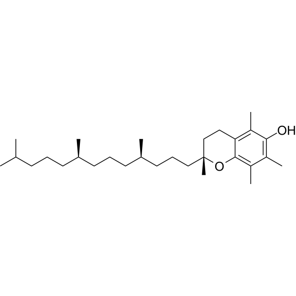
[Tocopherols]
-
Structure Classification
-
Initial Source
-
Shipping
Room temperature in continental US; may vary elsewhere.
-
Storage
Pure form -20°C 3 years In solvent -80°C 6 months -20°C 1 month


Solvent & Solubility
In Vitro:
DMSO : 100 mg/mL (Need ultrasonic; Hygroscopic DMSO has a significant impact on the solubility of product, please use newly opened DMSO)
In Vivo:
Select the appropriate dissolution method based on your experimental animal and administration route.
- For the following dissolution methods, please ensure to first prepare a clear stock solution using an In Vitro approach and then sequentially add co-solvents:
- To ensure reliable experimental results, the clarified stock solution can be appropriately stored based on storage conditions. As for the working solution for In Vivo experiments, it is recommended to prepare freshly and use it on the same day.
- The percentages shown for the solvents indicate their volumetric ratio in the final prepared solution. If precipitation or phase separation occurs during preparation, heat and/or sonication can be used to aid dissolution.
Add each solvent one by one: 10% DMSO 40% PEG300 5% Tween-80 45% Saline
Solubility: ≥ 5 mg/mL; Clear solution
This protocol yields a clear solution of ≥ 5 mg/mL (saturation unknown).
Taking 1 mL working solution as an example, add 100 μL DMSO stock solution (50.0 mg/mL) to 400 μL PEG300, and mix evenly; then add 50 μL Tween-80 and mix evenly; then add 450 μL Saline to adjust the volume to 1 mL.
Preparation of Saline: Dissolve 0.9 g sodium chloride in ddH₂O and dilute to 100 mL to obtain a clear Saline solution.
In Vivo Dissolution Calculator
Please enter the basic information of animal experiments:
-
-
-
-
Recommended: Prepare an additional quantity of animals to account for potential losses during experiments.
Please enter your animal formula composition:
-
%DMSO +
Recommended: Keep the proportion of DMSO in working solution below 2% if your animal is weak.
-
%+
-
+%Tween-80 + +
-
%Saline +
The co-solvents required include: DMSO, . All of co-solvents are available by MedChemExpress (MCE). , Tween 80. All of co-solvents are available by MedChemExpress (MCE).
Working solution concentration: 0.22 mg/mL
Method for preparing stock solution: mg drug dissolved in μL DMSO. Stock solution concentration: mg/mL.
1. Take μL DMSO stock solution;
2. Add μL .
μL , mix evenly;
3. Then add μL Tween 80, mix evenly;
4. Then add μL
Please ensure that the stock solution in the first step is dissolved to a clear state, and add co-solvents in sequence. You can use ultrasonic heating (ultrasonic cleaner, recommended frequency 20-40 kHz), vortexing, etc. to assist dissolution.

Protocols
-
Gene Editing
Gene editing modify specific sites within the genome through gene deletions, insertions or conversions to study functionally unknown genes or conduct gene therapy. It is also used to change the biological traits of organisms to establish new varieties. Gene editing techniques include zinc finger nuclease (ZFN), transcription activator-like effector nuclease (TALEN), and clustered regularly interspaced short palindromic repeats (CRISPR)/CRISPR-associated protein 9 (Cas 9) (CRISPR/Cas9).
-
ROS/oxidative-stress fluorescent staining
ROS/oxidative-stress fluorescent staining uses cell-permeant fluorogenic probes that become fluorescent after oxidation inside cells or tissues; commonly used examples include DCFH-DA/DCFDA for broad cellular oxidant detection, DHE for superoxide-related signal detection, MitoSOX for mitochondrial superoxide-related signal detection, and CellROX probes for oxidative-stress-associated fluorescence readouts. The assay detects probe oxidation rather than a single ROS species unless the probe and analysis method have been chemically validated for that species. DCFH-DA enters cells, is deacetylated by intracellular esterases to DCFH, and produces fluorescent DCF after oxidation, so the readout is used as an operational measure of total cellular oxidative stress rather than a species-specific ROS measurement. DHE and MitoSOX can report superoxide-related oxidation, but red fluorescence alone can include non-specific ethidium-like oxidation products; HPLC or optimized spectral approaches are
-
Research Protocol for Endocrine Diseases
Endocrine diseases often arise from disrupted hormone production, hormone signaling, or target-tissue responsiveness; for diabetes-focused endocrine disease models, insulin signaling regulates glucose uptake, hepatic glucose output, lipid metabolism, and β-cell compensation. Type 2 diabetes develops through interacting defects in insulin resistance, β-cell dysfunction, adipose inflammation, hepatic glucose overproduction, altered incretin signaling, and ectopic lipid metabolism. A major unresolved question is whether endocrine dysfunction is driven primarily by target-tissue insulin resistance, intrinsic β-cell failure, immune/inflammatory stress, or combined multi-organ failure that differs by disease stage.
-
Patient-Derived Xenograft (PDX)
Patient-derived xenograft (PDX) models are generated by engrafting primary human tumor tissue directly into immunodeficient mice, allowing in vivo propagation of patient tumor biology without initial in vitro adaptation. These models are used to preserve key histopathological and molecular characteristics of the original tumor and enable assessment of tumor growth dynamics and therapeutic response in a living organism. The biological readout is tumor engraftment and subsequent growth in the murine host, which reflects the ability of human tumor cells to survive, vascularize, and expand in an immunocompromised microenvironment.
-
Patient-Derived Orthotopic Xenograft (PDOX)
Patient-derived orthotopic xenograft (PDOX) modeling implants fresh patient tumor tissue or patient-derived tumor cells into the anatomically corresponding organ or tissue site of immunodeficient mice, usually by surgical orthotopic implantation, to preserve patient tumor histology, local microenvironmental context, invasion, metastatic behavior, and treatment-response features better than subcutaneous implantation. PDOX readouts include tumor engraftment, orthotopic tumor growth, local invasion, metastasis, recurrence after resection, histologic similarity to the donor tumor, biomarker retention, molecular concordance, survival, and response or resistance to therapy. PDOX models are used for preclinical drug testing and individualized therapy evaluation, but engraftment success varies by tumor type and specimen quality.
-
Ferroptosis Solutions
Ferroptosis is an iron-dependent, non-apoptotic form of regulated cell death characterized by lethal lipid peroxidation and sensitivity to suppression by iron chelators or lipophilic radical-trapping antioxidants. The core pathway links cystine uptake through system Xc−, glutathione availability, GPX4-dependent detoxification of phospholipid hydroperoxides, iron-dependent oxidative reactions, and polyunsaturated-phospholipid metabolism into a cell-death program that is biochemically and morphologically distinct from apoptosis, necrosis, and autophagy. The ferroptosis pathway is experimentally linked to phenotype through chemical and genetic perturbation. Erastin induces ferroptosis by inhibiting cystine uptake through system Xc− and weakening antioxidant defenses, while GPX4 inhibition or depletion causes lipid peroxide accumulation and ferroptotic cancer-cell death. ACSL4 and oxidizable arachidonoyl- or adrenoyl-containing phosphatidylethanolamines shape ferroptosis sensitivity by con
-
Subcutaneous Cell-Line-Derived Xenograft
Subcutaneous cell-line-derived xenograft (CDX) models are established by implanting cultured human cancer cell lines into immunodeficient mice, where the injected cells form localized tumors that can be monitored in vivo as a measure of tumorigenic potential, growth kinetics, and treatment response. These models are widely used in oncology research because they allow reproducible tumor formation and enable comparative assessment of tumor growth between different cell lines or genetic manipulations in a controlled in vivo microenvironment. Subcutaneous implantation of cancer cells in immunodeficient mice is a standard approach for evaluating tumor growth behavior and therapeutic response across multiple cancer types, including prostate, esophageal, pancreatic, and colon cancer models.
-
Breast Cancer Modeling
Breast cancer is a heterogeneous cancer, and it has been distinguished into four subtypes: luminal A, luminal B, HER2-positive and basal-like. Molecular mutations, epigenetic alterations, hormone exposure and immune microenvironment are related to the progression of breast cancer.
-
Genotoxicity/Mutagenicity Study
The bacterial reverse mutation assay detects point mutations that restore amino-acid prototrophy in auxotrophic Salmonella typhimurium or Escherichia coli tester strains; after exposure to a test article, mutagenic activity is read out as an increased number of revertant colonies on minimal agar compared with the vehicle control. The assay uses tester strains with different mutation targets so that base-substitution and frameshift mutagens can be detected, and testing is performed with and without exogenous mammalian metabolic activation because some chemicals require biotransformation to become mutagenic.
-
Orthotopic Cell-Line Xenograft
Orthotopic cell-line xenograft models involve implantation of human cancer cell lines into the anatomically corresponding organ of immunodeficient mice to reproduce tumor growth within a native microenvironment, enabling more clinically relevant tumor behavior compared with subcutaneous models. These models are widely used because orthotopic placement better recapitulates tumor progression, including invasion and metastatic spread, which are often underrepresented in heterotopic implantation systems. Compared with conventional xenografts, orthotopic implantation is described as more technically complex but provides improved simulation of tumor-microenvironment interactions and metastatic behavior, making it particularly valuable for translational oncology research. Surgical orthotopic implantation approaches have been emphasized as enabling faithful reproduction of clinical cancer features, including metastasis and disease progression patterns that align with the tumor’s organ of origi


Purity & Documentation
-
Data Sheet (283 KB)
-
SDS (392 KB)
- English - EN (392 KB)
- Français - FR (392 KB)
- Deutsch - DE (392 KB)
- Norwegian - NO (392 KB)
- Español - ES (392 KB)
- Swedish - SV (392 KB)
- Italian - IT (392 KB)
- Korean - KR (392 KB)
- Portuguese - PT (392 KB)
-
Handling Instructions (2659 KB)
References
[1]. Bak MJ, et al. Inhibitory Effects of γ- and δ-Tocopherols on Estrogen-Stimulated Breast Cancer In Vitro and In Vivo. Cancer Prev Res (Phila). 2017 Mar;10(3):188-197. [Content Brief]
[2]. Yang H, et al. Tocotrienols exhibit superior ferroptosis inhibition over tocopherols. Scientific reports. 2026 Jan 07;16(1):4497. [Content Brief]

Calculators
Concentration (start) × Volume (start) = Concentration (final) × Volume (final)


Tocopherols Related Classifications
HY-126400 Related Classifications
 Powered by Bioz