



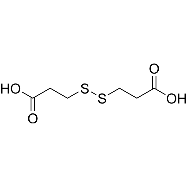
Dithiodipropionic acid
Based on 1 publication(s) in Google Scholar
Dithiodipropionic acid can interact with CPUL1 (HY-151802) (a TrxR inhibitor) to form nanoaggregates (CPUL1-DA NAs). CPUL1-DA NAs generates more abundant ROS to induce cell apoptosis than that of free CPUL1, and improves antitumor efficacy against HUH7 cancer cells.
For research use only. We do not sell to patients.
- Purity : 99.0%
- CAS No.: 1119-62-6
- Formula: C6H10O4S2
- Molecular Weight:210.26
-
Storage:Powder -20°C, 3 years , 4°C, 2 years ; In solvent -80°C, 6 months , -20°C, 1 month

To place orders, for customer services and technical support, please contact: MedChemExpress USA
Tel: 609-228-6898 E-mail: [email protected] [email protected]


-
 Biological Activity
-
Chemical Information
-
Solvent & Solubility
- Protocols
- Purity & Documentation
- References
-
 Help & FAQs


Publications Citing Use of MedChemExpress (MCE) Dithiodipropionic acid
More

Biological Activity
Description
In Vitro
CPUL1-DA NAs (molar ratio was 1:2) inhibits HUH7 hepatoma cell viability with an IC50 value of 4.3 μM, and has weak cytotoxicity against normal L02 cells[1].
CPUL1-DA NAs (2.5-10 μM, 6 h) can be more effectively enriched in HUH7 cells mitochondria and displays faster cellular uptake ability to deliver CPUL1 into cells than that of free CPUL1[1].
CPUL1-DA NAs (2.5-10 μM, 12 h) results in the accumulation of superoxides and mitochondrial membrane damage in HUH7 cells[1].
MedChemExpress (MCE) has not independently confirmed the accuracy of these methods. They are for reference only.


Chemical Information
-
CAS No. 1119-62-6
-
Appearance Solid
-
Molecular Weight 210.26
-
Formula C6H10O4S2
-
Color White to off-white
-
SMILES
O=C(O)CCSSCCC(O)=O
-
Shipping
Room temperature in continental US; may vary elsewhere.
-
Storage
Powder -20°C 3 years 4°C 2 years In solvent -80°C 6 months -20°C 1 month


Publications (1)
-
Journal Impact Factor
-
Most Recent


Solvent & Solubility
In Vitro:
DMSO : ≥ 100 mg/mL (475.60 mM; Hygroscopic DMSO has a significant impact on the solubility of product, please use newly opened DMSO)
* "≥" means soluble, but saturation unknown.
Please refer to the solubility information to select the appropriate solvent. Once prepared, please aliquot and store the solution to prevent product inactivation from repeated freeze-thaw cycles.
Storage method and period of stock solution: -80°C, 6 months; -20°C, 1 month. When stored at -80°C, please use it within 6 months. When stored at -20°C, please use it within 1 month.
Please refer to the solubility information to select the appropriate solvent. Once prepared, please aliquot and store the solution to prevent product inactivation from repeated freeze-thaw cycles.
Storage method and period of stock solution: -80°C, 6 months; -20°C, 1 month. When stored at -80°C, please use it within 6 months. When stored at -20°C, please use it within 1 month.
Concentration (start) × Volume (start) = Concentration (final) × Volume (final)


Purity & Documentation
-
Data Sheet (267 KB)
-
SDS (394 KB)
- English - EN (394 KB)
- Français - FR (394 KB)
- Deutsch - DE (394 KB)
- Norwegian - NO (394 KB)
- Español - ES (394 KB)
- Swedish - SV (394 KB)
- Italian - IT (394 KB)
- Korean - KR (394 KB)
- Portuguese - PT (394 KB)
-
Handling Instructions (2659 KB)
References


Complete Stock Solution Preparation Table
Please refer to the solubility information to select the appropriate solvent. Once prepared, please aliquot and store the solution to prevent product inactivation from repeated freeze-thaw cycles.
Storage method and period of stock solution: -80°C, 6 months; -20°C, 1 month. When stored at -80°C, please use it within 6 months. When stored at -20°C, please use it within 1 month.
| Optional Solvent | Concentration Solvent Mass | 1 mg | 5 mg | 10 mg | 25 mg |
|---|---|---|---|---|---|
| DMSO | 1 mM | 4.7560 mL | 23.7801 mL | 47.5602 mL | 118.9004 mL |
| 5 mM | 0.9512 mL | 4.7560 mL | 9.5120 mL | 23.7801 mL | |
| 10 mM | 0.4756 mL | 2.3780 mL | 4.7560 mL | 11.8900 mL | |
| 15 mM | 0.3171 mL | 1.5853 mL | 3.1707 mL | 7.9267 mL | |
| 20 mM | 0.2378 mL | 1.1890 mL | 2.3780 mL | 5.9450 mL | |
| 25 mM | 0.1902 mL | 0.9512 mL | 1.9024 mL | 4.7560 mL | |
| 30 mM | 0.1585 mL | 0.7927 mL | 1.5853 mL | 3.9633 mL | |
| 40 mM | 0.1189 mL | 0.5945 mL | 1.1890 mL | 2.9725 mL | |
| 50 mM | 0.0951 mL | 0.4756 mL | 0.9512 mL | 2.3780 mL | |
| 60 mM | 0.0793 mL | 0.3963 mL | 0.7927 mL | 1.9817 mL | |
| 80 mM | 0.0595 mL | 0.2973 mL | 0.5945 mL | 1.4863 mL | |
| 100 mM | 0.0476 mL | 0.2378 mL | 0.4756 mL | 1.1890 mL |


Dithiodipropionic acid Related Classifications
HY-W014395 Related Classifications
 Powered by Bioz