



Ramantamig

Based on 1 Customer Validation
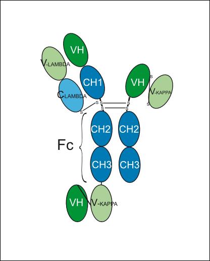
Ramantamig (JNJ-79635322) is a humanized monoclonal antibody targeting human CD3ε, GPRC5D, and TNFRSF17 (BCMA). Ramantamig binds to BCMA and GPRC5D on multiple myeloma cells, binds to CD3ε on T cells, forms immunological synapses, and enables T-cell-mediated cytotoxicity. Ramantamig activates T cells concomitantly with inducing myeloma cell cytotoxicity, with no nonspecific T-cell activation in the absence of target myeloma cells. Ramantamig carries mutations to reduce interaction with Fc receptors and disrupt protein A binding of monomeric and homodimerized chains. Ramantamig can be used for the research of multiple myeloma.
For research use only. We do not sell to patients.
- Purity : 95%
- CAS No.: 2988886-91-3
- Molecular Weight:153.279 kDa
-
Storage:
Please store the product under the recommended conditions in the Certificate of Analysis.


To place orders, for customer services and technical support, please contact: MedChemExpress USA
Tel: 609-228-6898 E-mail: [email protected] [email protected]


-
 Biological Activity
-
Chemical Information
- Protocols
- Purity & Documentation
- References
-
 Help & FAQs




Biological Activity
Description
Isotype
immunoglobulin (H-gamma1-scFvkh_L-lambda2)_scFvkh-G1(h-CH2-CH3)
Species Reactivity
Human
IC50 & Target
CD3E & GPRC5D & TNFRSF17
In Vitro
Ramantamig (72 h) potently induces T-cell-mediated cytotoxicity and activation in BCMA-GPRC5D dual-positive human multiple myeloma cell lines (MM.1S, H929, JIM-3, OPM-2) with sub-nanomolar to low nanomolar EC50 values, but has no effect on dual-negative myeloid cell lines[1].
Ramantamig (72 h) induces potent T-cell-mediated cytotoxicity and activation in H929-WT, H929-BCMA-KO, and H929-GPRC5D-KO human multiple myeloma cell lines, with significantly higher potency against dual-target-expressing H929-WT cells[1].
Ramantamig (48 h) potently induces T-cell-mediated cytotoxicity, T-cell activation, and proinflammatory cytokine production in H929 human multiple myeloma cells spiked into healthy human whole blood, with low nanomolar EC50 values[1].
Ramantamig (48 h) does not induce nonspecific T-cell activation or immune cell cytotoxicity in healthy human purified T cells or whole blood, except at very high concentrations (>10 nM)[1].
MedChemExpress (MCE) has not independently confirmed the accuracy of these methods. They are for reference only.
-
Cell Line:BCMA-GPRC5D dual-positive human multiple myeloma cell lines: MM.1S, H929, JIM-3, OPM-2; BCMA-GPRC5D dual-negative cell lines: MV-4-11, OCI-AML-3; pan-T cells from 6 healthy human donors
-
Concentration:dose-dependent (EC50 values reported)
-
Incubation Time:72 h
-
Result:Induced potent cytotoxicity in dual-positive MM cell lines with EC50 values of 0.002 nM (MM.1S), 0.005 nM (H929), 0.312 nM (JIM-3), and 0.019 nM (OPM-2).
Reached maximum cytotoxicity of 92.58% (MM.1S), 94.47% (H929), 61.22% (JIM-3), and 81.32% (OPM-2).
Induced T-cell activation with EC50 values of 0.010 nM (MM.1S), 0.008 nM (H929), 1.070 nM (JIM-3), and 0.062 nM (OPM-2).
Reached maximum T-cell activation of 75.03% (MM.1S), 85.90% (H929), 30.85% (JIM-3), and 45.95% (OPM-2).
Did not induce cytotoxicity or T-cell activation in dual-negative MV-4-11 or OCI-AML-3 cell lines.
In Vivo
Ramantamig (0.025-1 mg/kg; i.p.; twice weekly; 7 doses) induces dose-dependent tumor growth inhibition and complete regression in MM.1S multiple myeloma xenografts, with the 1 mg/kg dose achieving 109% ΔTGI and 100% tumor regression by day 34, and sustaining 100% complete regression by day 51[1].
MedChemExpress (MCE) has not independently confirmed the accuracy of these methods. They are for reference only.
-
Animal Model:NOD.Cg-Prkdcscid Il2rgtm1Wjl/SzJ (NSG) (female, aged ~5 to 8 weeks, weighing ~20 g, T-cell-humanized, subcutaneous xenograft of RPMI 8226 cells)[1]
-
Dosage:0.1 mg/kg; 0.4 mg/kg; 1 mg/kg; 2.5 mg/kg
-
Administration:i.p.; twice weekly; 8 doses
-
Result:Induced tumor growth inhibition (ΔTGI) values of 38.4% at 0.1 mg/kg, 90.5% at 0.4 mg/kg, 106.2% (with 51.7% tumor regression) at 1 mg/kg, and 111.2% (with 95.2% tumor regression) at 2.5 mg/kg on day 38.
Achieved complete tumor regression in 2 of 9 mice in the 0.4 mg/kg group, 8 of 10 mice in the 1 mg/kg group, and 9 of 9 mice in the 2.5 mg/kg group by day 60.
-
Animal Model:NOD.Cg-Prkdcscid Il2rgtm1Wjl/SzJ (NSG) (female, aged ~5 to 8 weeks, weighing ~20 g, T-cell-humanized, subcutaneous xenograft of MM.1S cells)[1]
-
Dosage:0.025 mg/kg; 0.1 mg/kg; 0.5 mg/kg; 1 mg/kg
-
Administration:i.p.; twice weekly; 7 doses
-
Result:Induced ΔTGI values of 72.9% at 0.025 mg/kg, 99.4% at 0.1 mg/kg, 108.6% (with 95.9% tumor regression) at 0.5 mg/kg, and 109% (with 100% tumor regression) at 1 mg/kg on day 34.
Gene ID
Accession
Conjugated
Unconjugated
Reconsititution
The product can be reconstituted/diluted with sterile PBS or saline.
Format
-
half-G1-lambda-scFvkh_scFvkh-h-CH2-CH3
Application
ELISA, FACS, Functional assay


Chemical Information
-
CAS No. 2988886-91-3
-
Appearance Liquid
-
Molecular Weight 153.279 kDa
-
Color Colorless to light yellow
-
SMILES
N/A
-
Synonyms
JNJ-79635322; JNJ-5322
-
Shipping
Shipping with dry ice.
-
Formulation
Please refer to the lot-specific COA for specific buffer information.
-
Storage
Please store the product under the recommended conditions in the Certificate of Analysis.


Purity & Documentation
-
Data Sheet (263 KB)
-
SDS (251 KB)
- English - EN (251 KB)
- Français - FR (251 KB)
- Deutsch - DE (251 KB)
- Norwegian - NO (251 KB)
- Español - ES (251 KB)
- Swedish - SV (251 KB)
- Italian - IT (251 KB)
- Korean - KR (251 KB)
- Portuguese - PT (251 KB)
-
Inhibitory Antibodies User Guide (603 KB)
References

Calculators
Concentration (start) × Volume (start) = Concentration (final) × Volume (final)


Ramantamig Related Classifications
HY-P991155 Related Classifications
 Powered by Bioz
Keywords