



Benzothiazole

Based on 1 Customer Validation
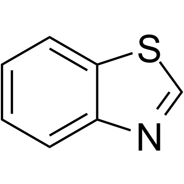
Benzothiazole is a natural occurring heterocyclic nuclei. Benzothiazole nucleus possesses a number of biological activities such as anticancer, antimicrobial, antidiabetic, anti-inflammatory, antileishmanial, and antiviral. Furthermore, Benzothiazole nucleus can function as an efficacious β-amyloid imaging agent[1][2][3][4].
For research use only. We do not sell to patients.
- Purity : 99.04%
- CAS No.: 95-16-9
- Formula: C7H5NS
- Molecular Weight:135.19
-
Storage:Pure form -20°C, 3 years , 4°C, 2 years ; In solvent -80°C, 6 months , -20°C, 1 month

To place orders, for customer services and technical support, please contact: MedChemExpress USA
Tel: 609-228-6898 E-mail: [email protected] [email protected]


-
 Biological Activity
-
Chemical Information
-
Solvent & Solubility
- Protocols
- Purity & Documentation
- References
-
 Help & FAQs


-

HY-W012634R
Benzothiazole (Standard)Heterocyclic Nuclei -
HY-W017424
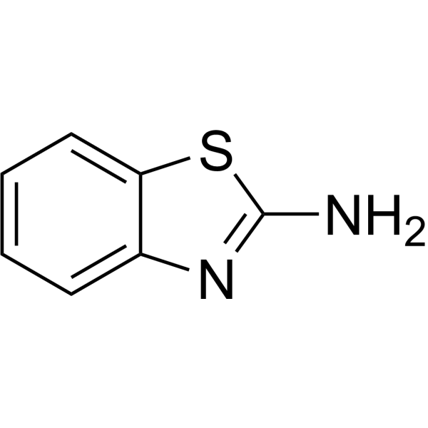
2-AminobenzothiazoleCaspase 3/7 Activator -

HY-W100945
2,4,5-TrimethylthiazoleBiochemical Reagent -
HY-W040269
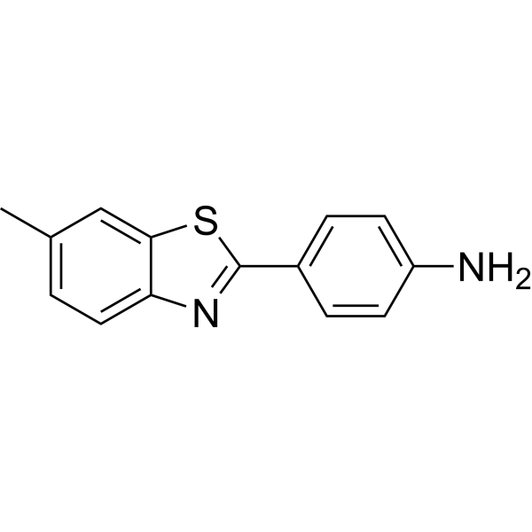
Dehydrothio-p-toluidineChemical Intermediate -
HY-59318
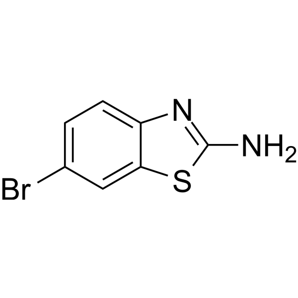
2-Amino-6-bromobenzothiazoleBiochemical Reagent -
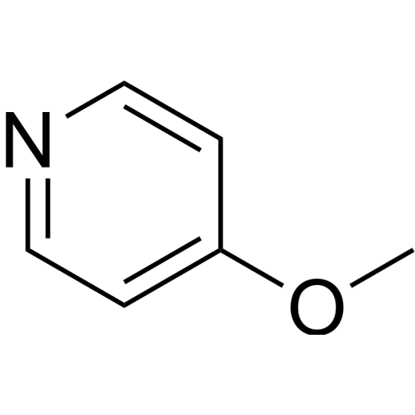
HY-W007634
4-MethoxypyridineBiochemical Reagent -

HY-W017424R
2-Aminobenzothiazole (Standard)Dinuclear Complexes Ligand -
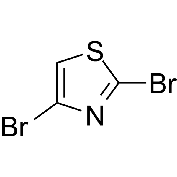
HY-59304
2,4-DibromothiazoleBiochemical Reagent -
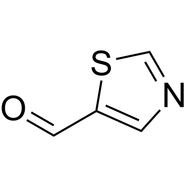
HY-69015
Thiazole-5-carboxyaldehydeBiochemical Reagent -
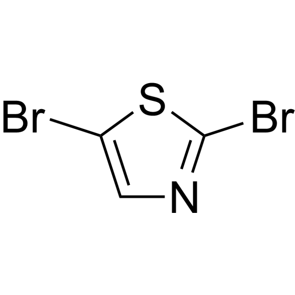
HY-69360
2,5-DibromothiazoleBiochemical Reagent -
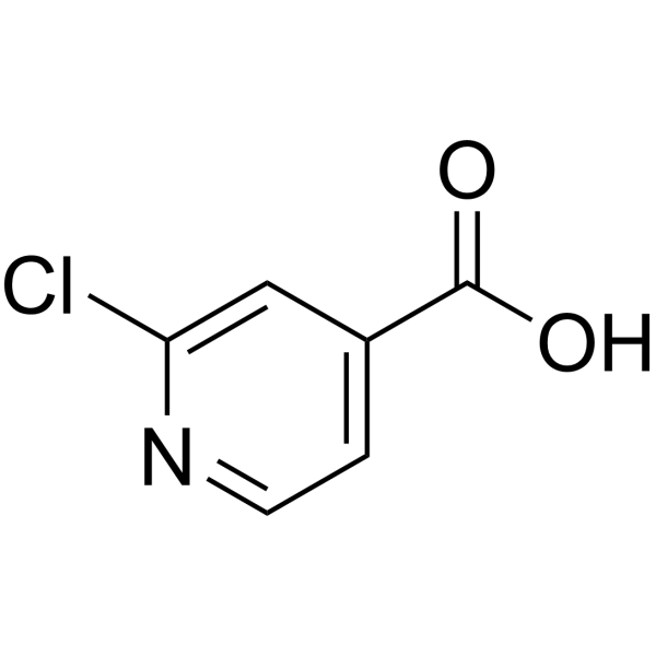
HY-65003
2-Chloroisonicotinic acidDrug Intermediate -

HY-Y1049
2-Amino-6-bromopyridinePqsR Ligand


Biological Activity
Description
Cellular Effect
|
Cell Line

|
Type | Value | Description | References |
|---|---|---|---|---|
| HCT-116 | IC50 |
>40 μM
Compound: 28
|
Cytotoxicity against human HCT-116 cells assessed as reduction in cell viability incubated for 3 days by methylene blue staining based analysis
Cytotoxicity against human HCT-116 cells assessed as reduction in cell viability incubated for 3 days by methylene blue staining based analysis
|
[PMID: 35984756] |
| NCI-H522 | IC50 |
>40 μM
Compound: 28
|
Cytotoxicity against human NCI-H522 cells assessed as reduction in cell viability incubated for 3 days by methylene blue staining based analysis
Cytotoxicity against human NCI-H522 cells assessed as reduction in cell viability incubated for 3 days by methylene blue staining based analysis
|
[PMID: 35984756] |
In Vitro
Benzothiazole (0-10-3 M, 21 h) don’t affect the phagocytic activity and efficiency of Hemocytes from blue mussels ( M. edulis )[2]. The benzothiazole derivatives (A01-A10) (10-100 μM, 48 h) exhibit potent cytotoxic against SiHa and C33-A cancer cell lines. All the compounds, except A09 result in apoptosis by inducing DNA breakage at multiple positions across chromosomal DNA. Almost all the compounds show accumulation of cells in the sub-G1 and S-phase of the cell cycle in SiHa and C33-A, respectively[3]. The benzothiazole derivatives PP-BTA-1 and PP-BTA-2 (61.1 nM 5.48 μM, 10 min) can inhibit the binding of thioflavin T / Aβ (1-42) aggregates with IC50 values of 0.12 and 0.11 μM, respectively. PP-BTA-1 and PP-BTA-2 can derivative as probes for detecting beta-amyloid plaques in Alzheimer's brains[4].
MedChemExpress (MCE) has not independently confirmed the accuracy of these methods. They are for reference only.
-
Cell Line:Hemocytes from blue mussels ( M. edulis )
-
Concentration:0, 10-3, 10-5, 10-7, 10-9 M
-
Incubation Time:21 h
-
Result:Observed no significant differences in phagocytic activity and efficiency as well as in viability of the hemocytes exposed from 10-9 up to 10-5 M.
-
Cell Line:SiHa and C33-A cervical cancer cells
-
Concentration:10–100 μM
-
Incubation Time:48 h
-
Result:The benzothiazole derivatives (A01 – A10) showed variable degrees of growth-inhibitory activity towards both cell lines, with IC50 values ranging from 5 to 50 μM.
-
Cell Line:SiHa and C33-A cervical cancer cells
-
Concentration:At their respective IC50 values
-
Incubation Time:48 h
-
Result:Observed a significant accumulation of the SiHa population in the sub-G1 phase of the cell cycle with all of the benzothiazole derivatives A01 – A10. Additionally, all benzothiazole derivatives A01 – A10, except A09, caused S-phase arrest in C33-A cells.
-
Cell Line:SiHa and C33-A cervical cancer cells
-
Concentration:At their respective IC50 values
-
Incubation Time:48 h
-
Result:Resulted in apoptosis by inducing DNA breakage at multiple positions across chromosomal DNA by all benzothiazole derivatives A01 – A10, except A09, in both SiHa and C33-A cells.


Chemical Information
-
CAS No. 95-16-9
-
Appearance Liquid (Density: 1.238 g/cm3)
-
Molecular Weight 135.19
-
Formula C7H5NS
-
Color Colorless to light yellow
-
SMILES
C12=CC=CC=C1SC=N2
-
Structure Classification
-
Initial Source
-
Shipping
Room temperature in continental US; may vary elsewhere.
-
Storage
Pure form -20°C 3 years 4°C 2 years In solvent -80°C 6 months -20°C 1 month


Solvent & Solubility
In Vitro:
DMSO : 100 mg/mL (739.70 mM; Need ultrasonic; Hygroscopic DMSO has a significant impact on the solubility of product, please use newly opened DMSO)
Please refer to the solubility information to select the appropriate solvent. Once prepared, please aliquot and store the solution to prevent product inactivation from repeated freeze-thaw cycles.
Storage method and period of stock solution: -80°C, 6 months; -20°C, 1 month. When stored at -80°C, please use it within 6 months. When stored at -20°C, please use it within 1 month.
Please refer to the solubility information to select the appropriate solvent. Once prepared, please aliquot and store the solution to prevent product inactivation from repeated freeze-thaw cycles.
Storage method and period of stock solution: -80°C, 6 months; -20°C, 1 month. When stored at -80°C, please use it within 6 months. When stored at -20°C, please use it within 1 month.
Concentration (start) × Volume (start) = Concentration (final) × Volume (final)
In Vivo:
Select the appropriate dissolution method based on your experimental animal and administration route.
- For the following dissolution methods, please ensure to first prepare a clear stock solution using an In Vitro approach and then sequentially add co-solvents:
- To ensure reliable experimental results, the clarified stock solution can be appropriately stored based on storage conditions. As for the working solution for In Vivo experiments, it is recommended to prepare freshly and use it on the same day.
- The percentages shown for the solvents indicate their volumetric ratio in the final prepared solution. If precipitation or phase separation occurs during preparation, heat and/or sonication can be used to aid dissolution.
Add each solvent one by one: 10% DMSO 40% PEG300 5% Tween-80 45% Saline
Solubility: ≥ 2.75 mg/mL (20.34 mM); Clear solution
This protocol yields a clear solution of ≥ 2.75 mg/mL (saturation unknown).
Taking 1 mL working solution as an example, add 100 μL DMSO stock solution (27.5 mg/mL) to 400 μL PEG300, and mix evenly; then add 50 μL Tween-80 and mix evenly; then add 450 μL Saline to adjust the volume to 1 mL.
Preparation of Saline: Dissolve 0.9 g sodium chloride in ddH₂O and dilute to 100 mL to obtain a clear Saline solution.
Add each solvent one by one: 10% DMSO 90% (20% SBE-β-CD in Saline)
Solubility: ≥ 2.75 mg/mL (20.34 mM); Clear solution
This protocol yields a clear solution of ≥ 2.75 mg/mL (saturation unknown).
Taking 1 mL working solution as an example, add 100 μL DMSO stock solution (27.5 mg/mL) to 900 μL 20% SBE-β-CD in Saline, and mix evenly.
Preparation of 20% SBE-β-CD in Saline (4°C, storage for one week): 2 g SBE-β-CD powder is dissolved in 10 mL Saline, completely dissolve until clear.
In Vivo Dissolution Calculator
Please enter the basic information of animal experiments:
-
-
-
-
Recommended: Prepare an additional quantity of animals to account for potential losses during experiments.
Please enter your animal formula composition:
-
%DMSO +
Recommended: Keep the proportion of DMSO in working solution below 2% if your animal is weak.
-
%+
-
+%Tween-80 + +
-
%Saline +
The co-solvents required include: DMSO, . All of co-solvents are available by MedChemExpress (MCE). , Tween 80. All of co-solvents are available by MedChemExpress (MCE).
Working solution concentration: 0.22 mg/mL
Method for preparing stock solution: mg drug dissolved in μL DMSO. Stock solution concentration: mg/mL.
1. Take μL DMSO stock solution;
2. Add μL .
μL , mix evenly;
3. Then add μL Tween 80, mix evenly;
4. Then add μL
Please ensure that the stock solution in the first step is dissolved to a clear state, and add co-solvents in sequence. You can use ultrasonic heating (ultrasonic cleaner, recommended frequency 20-40 kHz), vortexing, etc. to assist dissolution.


Purity & Documentation
-
Data Sheet (283 KB)
-
SDS (481 KB)
- English - EN (481 KB)
- Français - FR (481 KB)
- Deutsch - DE (481 KB)
- Norwegian - NO (481 KB)
- Español - ES (481 KB)
- Swedish - SV (481 KB)
- Italian - IT (481 KB)
- Korean - KR (481 KB)
- Portuguese - PT (481 KB)
-
Handling Instructions (2659 KB)
References
[2]. Côté C, et al. Immunotoxicity of Benzothiazole on Mytilus Edulis Following In Vitro Exposure of Hemocytes. J Xenobiot. 2016 Feb 10;5(2):5771. [Content Brief]
[3]. Singh M, et al. Benzothiazole derivatives bearing amide moiety: potential cytotoxic and apoptosis-inducing agents against cervical cancer. Anticancer Drugs. 2016;27(6):519-532. [Content Brief]
[4]. Ono M, et al. Push-pull benzothiazole derivatives as probes for detecting beta-amyloid plaques in Alzheimer's brains. Bioorg Med Chem. 2009;17(19):7002-7007. [Content Brief]


Complete Stock Solution Preparation Table
Please refer to the solubility information to select the appropriate solvent. Once prepared, please aliquot and store the solution to prevent product inactivation from repeated freeze-thaw cycles.
Storage method and period of stock solution: -80°C, 6 months; -20°C, 1 month. When stored at -80°C, please use it within 6 months. When stored at -20°C, please use it within 1 month.
| Optional Solvent | Concentration Solvent Mass | 1 mg | 5 mg | 10 mg | 25 mg |
|---|---|---|---|---|---|
| DMSO | 1 mM | 7.3970 mL | 36.9850 mL | 73.9700 mL | 184.9249 mL |
| 5 mM | 1.4794 mL | 7.3970 mL | 14.7940 mL | 36.9850 mL | |
| 10 mM | 0.7397 mL | 3.6985 mL | 7.3970 mL | 18.4925 mL | |
| 15 mM | 0.4931 mL | 2.4657 mL | 4.9313 mL | 12.3283 mL | |
| 20 mM | 0.3698 mL | 1.8492 mL | 3.6985 mL | 9.2462 mL | |
| 25 mM | 0.2959 mL | 1.4794 mL | 2.9588 mL | 7.3970 mL | |
| 30 mM | 0.2466 mL | 1.2328 mL | 2.4657 mL | 6.1642 mL | |
| 40 mM | 0.1849 mL | 0.9246 mL | 1.8492 mL | 4.6231 mL | |
| 50 mM | 0.1479 mL | 0.7397 mL | 1.4794 mL | 3.6985 mL | |
| 60 mM | 0.1233 mL | 0.6164 mL | 1.2328 mL | 3.0821 mL | |
| 80 mM | 0.0925 mL | 0.4623 mL | 0.9246 mL | 2.3116 mL | |
| 100 mM | 0.0740 mL | 0.3698 mL | 0.7397 mL | 1.8492 mL |


Benzothiazole Related Classifications
HY-W012634 Related Classifications
 Powered by Bioz