



Eurycomalactone

Based on 1 Customer Validation
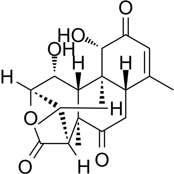
Eurycomalactone is an active quassinoid could be isolated from Eurycoma longifolia Jack. Eurycomalactone is a potent NF-κB inhibitor with an IC50 value of 0.5 μM. Eurycomalactone inhibits protein synthesis and depletes cyclin D1. Eurycomalactone enhances radiosensitivity through arrest cell cycle at G2/M phase and delayed DNA double-strand break repair. Eurycomalactone inhibits the activation of AKT/NF-κB signaling, induces apoptosis and enhances chemosensitivity to Cisplatin (HY-17394).
For research use only. We do not sell to patients.
- Purity : 99.74%
- CAS No.: 23062-24-0
- Formula: C19H24O6
- Molecular Weight:348.39
-
Storage:
4°C, protect from light
* In solvent : -80°C, 6 months; -20°C, 1 month (protect from light)

To place orders, for customer services and technical support, please contact: MedChemExpress USA
Tel: 609-228-6898 E-mail: [email protected] [email protected]


-
 Biological Activity
-
Chemical Information
-
Solvent & Solubility
- Protocols
- Purity & Documentation
- References
-
 Help & FAQs




Biological Activity
Description
IC50 & Target
IC50: 0.5 μM (NF-κB)[1]
Cellular Effect
|
Cell Line

|
Type | Value | Description | References |
|---|---|---|---|---|
| HT-1080 | IC50 |
0.98 μM
Compound: 11
|
Cytotoxicity against human HT1080 cells after 72 hrs by MTT assay
Cytotoxicity against human HT1080 cells after 72 hrs by MTT assay
|
[PMID: 19919052] |
In Vitro
Eurycomalactone (24, 48 and 72 h) selectively inhibits the viability of A549 and COR-L23 cells Eurycomalactone inhibits the viability of A549 cells with IC50 values of 20.17, 3.77, and 1.90 μM for 24, 48 and 72 hours, respectively. Eurycomalactone inhibits the viability of COR-L23 cells with IC50 values of 25.02, 2.74, and 1.80 μM for 24, 48 and 72 hours, respectively[1].
Eurycomalactone (2.29-156.3 μM; 24 h; A549 and Calu-1 cells) promotes Non-small cell lung cancer (NSCLC) cells apoptosis[2].
Eurycomalactone (0-25.05 μM; 24 h; A549 and COR-L23 cells) induces cell cycle arrest at the radiosensitive G2/M phase and induces apoptosis in irradiated Non-small cell lung cancer (NSCLC) cells. Eurycomalactone downregulated the key G2/M regulatory proteins in irradiated Non-small cell lung cancer (NSCLC) cells[1].
Eurycomalactone (2.5-25 μM; 24 h; A549 cells) suppressed the repair of radiation-Induced DNA double-strand breaks[1].
Eurycomalactone (2.29-156.3 μM; 24 h; A549 and Calu-1 cells) suppresses AKT/NF-κB activation in Non-small cell lung cancer (NSCLC) cells[2].
MedChemExpress (MCE) has not independently confirmed the accuracy of these methods. They are for reference only.
-
Cell Line:A549 and Calu-1 cells
-
Concentration:2.29, 10.14, 12.02, 20.81, 80.77, and 156.3 μM
-
Incubation Time:24 hours
-
Result:Increased the apoptotic rates in a dose-dependent manner.
-
Cell Line:A549 and COR-L23 cells
-
Concentration:1.57, 2.57, 20.17 and 25.05 μM
-
Incubation Time:24 hours
-
Result:Induced cell cycle arrest at G2/M phase in irradiated cells and increased the sub-G1 population. A549 and COR-L23 cells
-
Cell Line:A549 and Calu-1 cells
-
Concentration:2.29, 10.14, 12.02, 20.81, 80.77, and 156.3 μM
-
Incubation Time:24 hours
-
Result:Induced the expression levels of active caspase-3 and active PARP (cleaved form), while decreased Bcl-xL and surviving.
-
Cell Line:A549 and COR-L23 cells
-
Concentration:1.57, 2.57, 20.17 and 25.05 μM
-
Incubation Time:24 hours
-
Result:Downregulated the expression of both G2/M regulatory proteins in a dose-dependent manner.
-
Cell Line:A549 and COR-L23 cells
-
Concentration:2.29, 10.14, 12.02, 20.81, 80.77, and 156.3 μM
-
Incubation Time:24 hours
-
Result:inhibited the expression levels of p(S473)-AKT, total AKT, p(S536)-NF-κB p65 and total NF-κB p65.


Chemical Information
-
CAS No. 23062-24-0
-
Appearance Solid
-
Molecular Weight 348.39
-
Formula C19H24O6
-
Color White to off-white
-
SMILES
C[C@]12[C@@]([C@H]([C@@](O3)([H])[C@@](C)([H])[C@]2([H])C3=O)O)([H])[C@]([C@@](C(C)=CC4=O)([H])CC1=O)([C@@H]4O)C
-
Structure Classification
-
Initial Source
-
Shipping
Room temperature in continental US; may vary elsewhere.
-
Storage
4°C, protect from light
* In solvent : -80°C, 6 months; -20°C, 1 month (protect from light)


Solvent & Solubility
In Vitro:
DMSO : 100 mg/mL (287.03 mM; Need ultrasonic; Hygroscopic DMSO has a significant impact on the solubility of product, please use newly opened DMSO)
Please refer to the solubility information to select the appropriate solvent. Once prepared, please aliquot and store the solution to prevent product inactivation from repeated freeze-thaw cycles.
Storage method and period of stock solution: -80°C, 6 months; -20°C, 1 month (protect from light). When stored at -80°C, please use it within 6 months. When stored at -20°C, please use it within 1 month.
Please refer to the solubility information to select the appropriate solvent. Once prepared, please aliquot and store the solution to prevent product inactivation from repeated freeze-thaw cycles.
Storage method and period of stock solution: -80°C, 6 months; -20°C, 1 month (protect from light). When stored at -80°C, please use it within 6 months. When stored at -20°C, please use it within 1 month.
Concentration (start) × Volume (start) = Concentration (final) × Volume (final)
In Vivo:
Select the appropriate dissolution method based on your experimental animal and administration route.
- For the following dissolution methods, please ensure to first prepare a clear stock solution using an In Vitro approach and then sequentially add co-solvents:
- To ensure reliable experimental results, the clarified stock solution can be appropriately stored based on storage conditions. As for the working solution for In Vivo experiments, it is recommended to prepare freshly and use it on the same day.
- The percentages shown for the solvents indicate their volumetric ratio in the final prepared solution. If precipitation or phase separation occurs during preparation, heat and/or sonication can be used to aid dissolution.
Add each solvent one by one: 10% DMSO 40% PEG300 5% Tween-80 45% Saline
Solubility: ≥ 2.5 mg/mL (7.18 mM); Clear solution
This protocol yields a clear solution of ≥ 2.5 mg/mL (saturation unknown).
Taking 1 mL working solution as an example, add 100 μL DMSO stock solution (25.0 mg/mL) to 400 μL PEG300, and mix evenly; then add 50 μL Tween-80 and mix evenly; then add 450 μL Saline to adjust the volume to 1 mL.
Preparation of Saline: Dissolve 0.9 g sodium chloride in ddH₂O and dilute to 100 mL to obtain a clear Saline solution.
Add each solvent one by one: 10% DMSO 90% (20% SBE-β-CD in Saline)
Solubility: ≥ 2.5 mg/mL (7.18 mM); Clear solution
This protocol yields a clear solution of ≥ 2.5 mg/mL (saturation unknown).
Taking 1 mL working solution as an example, add 100 μL DMSO stock solution (25.0 mg/mL) to 900 μL 20% SBE-β-CD in Saline, and mix evenly.
Preparation of 20% SBE-β-CD in Saline (4°C, storage for one week): 2 g SBE-β-CD powder is dissolved in 10 mL Saline, completely dissolve until clear.
In Vivo Dissolution Calculator
Please enter the basic information of animal experiments:
-
-
-
-
Recommended: Prepare an additional quantity of animals to account for potential losses during experiments.
Please enter your animal formula composition:
-
%DMSO +
Recommended: Keep the proportion of DMSO in working solution below 2% if your animal is weak.
-
%+
-
+%Tween-80 + +
-
%Saline +
The co-solvents required include: DMSO, . All of co-solvents are available by MedChemExpress (MCE). , Tween 80. All of co-solvents are available by MedChemExpress (MCE).
Working solution concentration: 0.22 mg/mL
Method for preparing stock solution: mg drug dissolved in μL DMSO. Stock solution concentration: mg/mL. * In solvent : -80°C, 6 months; -20°C, 1 month (protect from light)
1. Take μL DMSO stock solution;
2. Add μL .
μL , mix evenly;
3. Then add μL Tween 80, mix evenly;
4. Then add μL
Please ensure that the stock solution in the first step is dissolved to a clear state, and add co-solvents in sequence. You can use ultrasonic heating (ultrasonic cleaner, recommended frequency 20-40 kHz), vortexing, etc. to assist dissolution.


Purity & Documentation
-
Data Sheet (290 KB)
-
SDS (252 KB)
- English - EN (252 KB)
- Français - FR (252 KB)
- Deutsch - DE (252 KB)
- Norwegian - NO (252 KB)
- Español - ES (252 KB)
- Swedish - SV (252 KB)
- Italian - IT (252 KB)
- Korean - KR (252 KB)
- Portuguese - PT (252 KB)
-
Handling Instructions (2659 KB)
References
[1]. Dukaew N, et, al. Enhancement of Radiosensitivity by Eurycomalactone in Human NSCLC Cells Through G₂/M Cell Cycle Arrest and Delayed DNA Double-Strand Break Repair. Oncol Res. 2020 Mar 27;28(2):161-175. [Content Brief]
[2]. Dukaew N, et, al. Inactivation of AKT/NF κB signaling by eurycomalactone decreases human NSCLC cell viability and improves the chemosensitivity to cisplatin. Oncol Rep. 2020 Oct;44(4):1441-1454. [Content Brief]
[3]. Malainer C, et, al. Eurycomalactone Inhibits Expression of Endothelial Adhesion Molecules at a Post-Transcriptional Level. J Nat Prod. 2017 Dec 22;80(12):3186-3193. [Content Brief]


Complete Stock Solution Preparation Table
Please refer to the solubility information to select the appropriate solvent. Once prepared, please aliquot and store the solution to prevent product inactivation from repeated freeze-thaw cycles.
Storage method and period of stock solution: -80°C, 6 months; -20°C, 1 month (protect from light). When stored at -80°C, please use it within 6 months. When stored at -20°C, please use it within 1 month.
| Optional Solvent | Concentration Solvent Mass | 1 mg | 5 mg | 10 mg | 25 mg |
|---|---|---|---|---|---|
| DMSO | 1 mM | 2.8703 mL | 14.3517 mL | 28.7035 mL | 71.7587 mL |
| 5 mM | 0.5741 mL | 2.8703 mL | 5.7407 mL | 14.3517 mL | |
| 10 mM | 0.2870 mL | 1.4352 mL | 2.8703 mL | 7.1759 mL | |
| 15 mM | 0.1914 mL | 0.9568 mL | 1.9136 mL | 4.7839 mL | |
| 20 mM | 0.1435 mL | 0.7176 mL | 1.4352 mL | 3.5879 mL | |
| 25 mM | 0.1148 mL | 0.5741 mL | 1.1481 mL | 2.8703 mL | |
| 30 mM | 0.0957 mL | 0.4784 mL | 0.9568 mL | 2.3920 mL | |
| 40 mM | 0.0718 mL | 0.3588 mL | 0.7176 mL | 1.7940 mL | |
| 50 mM | 0.0574 mL | 0.2870 mL | 0.5741 mL | 1.4352 mL | |
| 60 mM | 0.0478 mL | 0.2392 mL | 0.4784 mL | 1.1960 mL | |
| 80 mM | 0.0359 mL | 0.1794 mL | 0.3588 mL | 0.8970 mL | |
| 100 mM | 0.0287 mL | 0.1435 mL | 0.2870 mL | 0.7176 mL |


Eurycomalactone Related Classifications
HY-N4327 Related Classifications
 Powered by Bioz