



(Z)-Semaxanib
Based on 4 publication(s) in Google Scholar
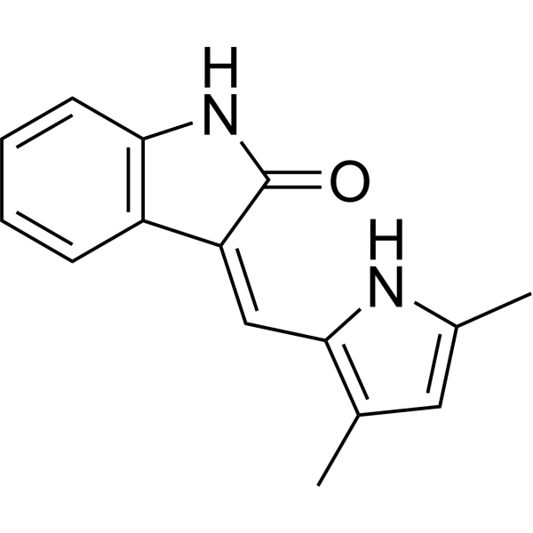
(Z)-Semaxanib ((Z)-SU5416) is a VEGF receptor tyrosine kinase inhibitor that blocks VEGF signaling through Flk-1/KDR. (Z)-Semaxanib inhibits angiogenesis by preventing new blood vessel formation. (Z)-Semaxanib induces cytotoxicity in liver cancer cells. (Z)-Semaxanib can be used in research on metastatic renal cell carcinoma, gastrointestinal stromal tumors, well-differentiated pancreatic neuroendocrine tumors, and solid tumors.
For research use only. We do not sell to patients.
- Purity : 99.72%
- CAS No.: 194413-58-6
- Formula: C15H14N2O
- Molecular Weight:238.29
-
Storage:Powder -20°C, 3 years , 4°C, 2 years ; In solvent -80°C, 6 months , -20°C, 1 month

To place orders, for customer services and technical support, please contact: MedChemExpress USA
Tel: 609-228-6898 E-mail: [email protected] [email protected]


-
 Biological Activity
-
Chemical Information
-
Solvent & Solubility
- Protocols
- Purity & Documentation
- References
-
 Help & FAQs



Publications Citing Use of MedChemExpress (MCE) (Z)-Semaxanib
MoreAll VEGFR Isoforms
More

Biological Activity
Description
Cellular Effect
|
Cell Line

|
Type | Value | Description | References |
|---|---|---|---|---|
| TAMH | IC50 |
6.28 μM
|
Cytotoxicity against TAMH cells assessed as reduction in cell viability incubated for 24 hrs by MTT assay.
Cytotoxicity against TAMH cells assessed as reduction in cell viability incubated for 24 hrs by MTT assay.
|
26593399 |
| HepG2 | IC50 |
8.17 μM
|
Cytotoxicity of the pure Z isomer against HepG2 cells assessed as reduction in cell viability incubated for 24 hrs by MTT assay.
Cytotoxicity of the pure Z isomer against HepG2 cells assessed as reduction in cell viability incubated for 24 hrs by MTT assay.
|
26593399 |
In Vitro
(Z)-Semaxanib ((Z)-SU5416) (10 mM; 0.25-24 h) undergoes photoisomerization in [D6]DMSO to form the E isomer and reverts to the Z isomer in the dark[1].
(Z)-Semaxanib (24 h) is cytotoxic to TAMH cells with an IC50 of 6.28 μM[1].
MedChemExpress (MCE) has not independently confirmed the accuracy of these methods. They are for reference only.
- Human pluripotent stem cell endothelial-cell differentiation
- Vascular/Branching Fractal Analysis
- Cell Cytotoxicity Assay
- Mammalian live/dead viability and cytotoxicity staining
- Endothelial Tube Formation Assay
- Liver Cancer Modeling
- Endothelial Cell Migration/Angiogenic Sprouting Assay
- Cell Viability Determination by MTT Colorimetric Assay


Chemical Information
-
CAS No. 194413-58-6
-
Appearance Solid
-
Molecular Weight 238.29
-
Formula C15H14N2O
-
Color Yellow to orange
-
SMILES
O=C(NC1=C/2C=CC=C1)C2=C/C3=C(C)C=C(C)N3
-
Synonyms
(Z)-SU5416
-
Shipping
Room temperature in continental US; may vary elsewhere.
-
Storage
Powder -20°C 3 years 4°C 2 years In solvent -80°C 6 months -20°C 1 month


Publications (4)
-
Journal Impact Factor
-
Most Recent
-
Sci Transl Med
PP2A inhibition is a druggable MEK inhibitor resistance mechanism in KRAS-mutant lung cancer cells. [Abstract]2018 Jul 18;10(450):eaaq1093. PMID: 30021885 -
Br J Pharmacol
VEGFR-2 blocker induces pulmonary vascular disease in rats with CRISPR-edited human non-deficient G6PD polymorphism: role of 3D genomic modifications and DNA methylation. [Abstract]2026 May 5. PMID: 42083756 -
Int J Mol Sci
Curcuminoids Inhibit Angiogenic Behaviors of Human Umbilical Vein Endothelial Cells via Endoglin/Smad1 Signaling. [Abstract]2022 Mar 31;23(7):3889. PMID: 35409247 -


Solvent & Solubility
In Vitro:
DMSO : 50 mg/mL (209.83 mM; Need ultrasonic; Hygroscopic DMSO has a significant impact on the solubility of product, please use newly opened DMSO)
Please refer to the solubility information to select the appropriate solvent. Once prepared, please aliquot and store the solution to prevent product inactivation from repeated freeze-thaw cycles.
Storage method and period of stock solution: -80°C, 6 months; -20°C, 1 month. When stored at -80°C, please use it within 6 months. When stored at -20°C, please use it within 1 month.
Please refer to the solubility information to select the appropriate solvent. Once prepared, please aliquot and store the solution to prevent product inactivation from repeated freeze-thaw cycles.
Storage method and period of stock solution: -80°C, 6 months; -20°C, 1 month. When stored at -80°C, please use it within 6 months. When stored at -20°C, please use it within 1 month.
Concentration (start) × Volume (start) = Concentration (final) × Volume (final)


Purity & Documentation
-
Data Sheet (278 KB)
-
SDS (393 KB)
- English - EN (393 KB)
- Français - FR (393 KB)
- Deutsch - DE (393 KB)
- Norwegian - NO (393 KB)
- Español - ES (393 KB)
- Swedish - SV (393 KB)
- Italian - IT (393 KB)
- Korean - KR (393 KB)
- Portuguese - PT (393 KB)
-
Handling Instructions (2659 KB)
References
[1]. Ngai MH, et al. Photoinduced Isomerization and Hepatoxicities of Semaxanib, Sunitinib and Related 3-Substituted Indolin-2-ones. ChemMedChem. 2016 Jan 05;11(1):72-80. [Content Brief]
[3]. Zhao Y, et al. Simultaneous determination of Z-SU5416 and its interconvertible geometric E-isomer in rat plasma by LC/MS/MS. Journal of pharmaceutical and biomedical analysis. 2004 May 28;35(3):513-22. [Content Brief]


Complete Stock Solution Preparation Table
Please refer to the solubility information to select the appropriate solvent. Once prepared, please aliquot and store the solution to prevent product inactivation from repeated freeze-thaw cycles.
Storage method and period of stock solution: -80°C, 6 months; -20°C, 1 month. When stored at -80°C, please use it within 6 months. When stored at -20°C, please use it within 1 month.
| Optional Solvent | Concentration Solvent Mass | 1 mg | 5 mg | 10 mg | 25 mg |
|---|---|---|---|---|---|
| DMSO | 1 mM | 4.1966 mL | 20.9828 mL | 41.9657 mL | 104.9142 mL |
| 5 mM | 0.8393 mL | 4.1966 mL | 8.3931 mL | 20.9828 mL | |
| 10 mM | 0.4197 mL | 2.0983 mL | 4.1966 mL | 10.4914 mL | |
| 15 mM | 0.2798 mL | 1.3989 mL | 2.7977 mL | 6.9943 mL | |
| 20 mM | 0.2098 mL | 1.0491 mL | 2.0983 mL | 5.2457 mL | |
| 25 mM | 0.1679 mL | 0.8393 mL | 1.6786 mL | 4.1966 mL | |
| 30 mM | 0.1399 mL | 0.6994 mL | 1.3989 mL | 3.4971 mL | |
| 40 mM | 0.1049 mL | 0.5246 mL | 1.0491 mL | 2.6229 mL | |
| 50 mM | 0.0839 mL | 0.4197 mL | 0.8393 mL | 2.0983 mL | |
| 60 mM | 0.0699 mL | 0.3497 mL | 0.6994 mL | 1.7486 mL | |
| 80 mM | 0.0525 mL | 0.2623 mL | 0.5246 mL | 1.3114 mL | |
| 100 mM | 0.0420 mL | 0.2098 mL | 0.4197 mL | 1.0491 mL |


(Z)-Semaxanib Related Classifications
HY-10332 Related Classifications
 Powered by Bioz