



9-SAHSA
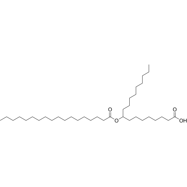
9-SAHSA is a fatty acid ester of hydroxy fatty acids (FAHFAs). 9-SAHSA is a protective molecule to prevent colon carcinoma cells from apoptotic cell death.
商品は「研究用試薬」です。人や動物の医療用・臨床診断用・食品用の製品ではありません。
研究用途以外に使用した場合、当社は一切の責任を負いかねます。
- CAS 番号: 1895916-79-6
- 分子式: C36H70O4
- 分子量:566.94
-
保管条件:
Please store the product under the recommended conditions in the Certificate of Analysis.

To place orders, for customer services and technical support, please contact: MedChemExpress USA
Tel: 609-228-6898 E-mail: [email protected] [email protected]


-
 生物活性
生物活性
-
化学情報
- プロトコル
- 純度とドキュメンテーション
- 参考文献
-
 Help & FAQs


-
HY-116193
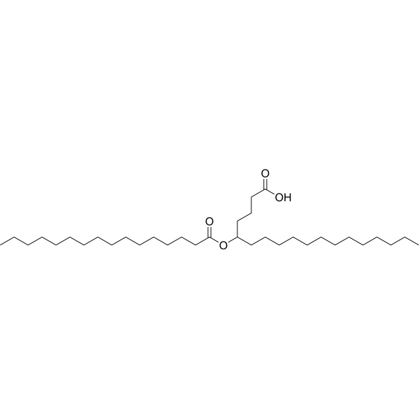
5-PAHSAAnti-Inflammatory Agent -
HY-117586
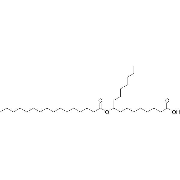
9-PAHPAAntidiabetic/Anti-inflammatory Agent -

HY-158584
13-OAHSAFAHFA -

HY-W709961
5-OAHSAEndogenous Metabolite -
HY-131668
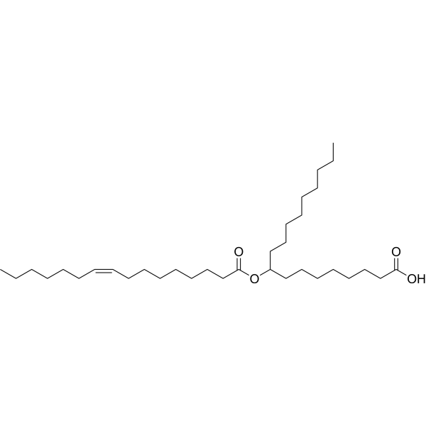
9-POHSAAnti-Inflammatory Agent -
HY-138177
Butyryl-Coenzyme A sodiumButyric Acid Derivative -
HY-113878
12-OAHSAAnti-Obese Agent -

HY-158583
13-POHSAFAHFA -
HY-131934
9-OAHSAAnti-Inflammatory reagent -
HY-130434
12-SAHSAFAHFA -

HY-116193R
5-PAHSA (Standard)Anti-Inflammatory Agent -

HY-133851
10-PAHSAFatty Ester
Endogenous Metabolite アイソフォーム固有の製品をすべて表示
More

生物活性
製品説明


化学情報
-
CAS 番号 1895916-79-6
-
分子量 566.94
-
分子式 C36H70O4
-
SMILES
CCCCCCCCCC(OC(CCCCCCCCCCCCCCCCC)=O)CCCCCCCC(O)=O
-
輸送条件
Room temperature in continental US; may vary elsewhere.
-
保管条件
Please store the product under the recommended conditions in the Certificate of Analysis.

プロトコル
-
Apoptosis
Apoptosis, also called programmed cell death, is generally characterized by distinct morphological characteristics.
-
Cell Cytotoxicity Assay
Cytotoxicity assays are usually based on the assessment of cell membrane damage, which can also be indirectly detected by measuring cell viability. Detection methods include MTT assay, CKK-8 assay, LDH assay and ATP assay, etc.
-
Mammalian live/dead viability and cytotoxicity staining
Live/dead viability and cytotoxicity staining assays are based on the simultaneous detection of intracellular esterase activity in metabolically active (viable) cells and membrane integrity loss in non-viable cells. In commonly used dual-staining approaches, membrane-permeant fluorogenic substrates are converted by intracellular esterases into fluorescent products in live cells, while impermeant DNA-binding dyes selectively enter cells with compromised plasma membranes and label nucleic acids in dead or dying cells, enabling discrimination between viable and non-viable populations by fluorescence microscopy or flow cytometry.
-
Apoptosis Solutions
Apoptosis is a regulated, generally non-lytic cell-death pathway that removes unwanted, damaged, infected, or abnormal cells through coordinated morphological changes, caspase activation, DNA fragmentation, and membrane remodeling. The intrinsic apoptosis pathway is controlled mainly by mitochondrial outer membrane permeabilization, BCL-2 family proteins, cytochrome c release, apoptosome formation, caspase-9 activation, and downstream executioner caspase-3/7 activation. The extrinsic apoptosis pathway is initiated by death receptors such as Fas, TNFR, and TRAIL receptors, which recruit adaptor proteins and activate caspase-8 before engaging executioner caspases or mitochondrial amplification through BID cleavage. Apoptosis is linked to many phenotypes, including cancer cell killing, tissue homeostasis, immune regulation, neurodegeneration, infection response, and treatment-induced cytotoxicity; unresolved questions include how apoptosis interacts with necroptosis, pyroptosis, ferroptos


純度とドキュメンテーション
参考文献
[1]. Zhu QF, et al. Highly sensitive determination of fatty acid esters of hydroxyl fatty acids by liquid chromatography-mass spectrometry. J Chromatogr B Analyt Technol Biomed Life Sci. 2017 Sep 1;1061-1062:34-40.2. [Content Brief]
[2]. Ding J, et al. In-Silico-Generated Library for Sensitive Detection of 2-Dimethylaminoethylamine Derivatized FAHFA Lipids Using High-Resolution Tandem Mass Spectrometry. Anal Chem. 2020 Apr 21;92(8):5960-5968. [Content Brief]

Calculators
濃度 (開始) × 体積 (開始) = 濃度 (終了) × 体積 (終了)


9-SAHSA 関連分類
HY-130352 関連分類
 Powered by Bioz