



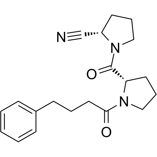
KYP-2047
Based on 1 publication(s) in Google Scholar
KYP-2047 is a potent and BBB-penetrating prolyl-oligopeptidase (POP) inhibitor, with an Ki value of 0.023 nM. KYP-2047 reduces glioblastoma proliferation through angiogenesis and apoptosis modulation.
商品は「研究用試薬」です。人や動物の医療用・臨床診断用・食品用の製品ではありません。
研究用途以外に使用した場合、当社は一切の責任を負いかねます。
- 純度 : 99.94%
- CAS 番号: 796874-99-2
- 分子式: C20H25N3O2
- 分子量:339.43
-
保管条件:Pure form -20°C, 3 years , 4°C, 2 years ; In solvent -80°C, 6 months , -20°C, 1 month

To place orders, for customer services and technical support, please contact: MedChemExpress USA
Tel: 609-228-6898 E-mail: [email protected] [email protected]


-
 生物活性
生物活性
-
化学情報
-
溶剤 & 溶解度
- プロトコル
- 純度とドキュメンテーション
- 参考文献
-
 Help & FAQs


-
HY-100684
UAMC-1110FAP Inhibitor -
HY-15114
S 17092PEP Inhibitor -
HY-145983
Suc-Gly-Pro-AMCFAP Substrate -
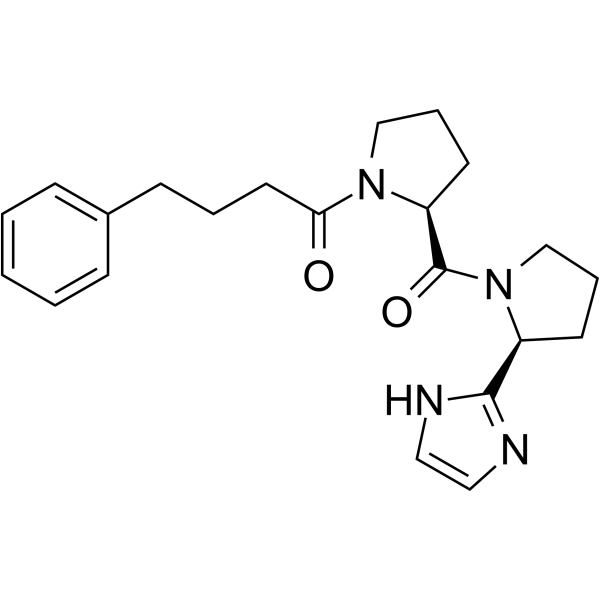
HY-139648
PREP inhibitor-1PREP Inhibitor -

HY-100475R
KYP-2047 (Standard)POP Inhibitor -
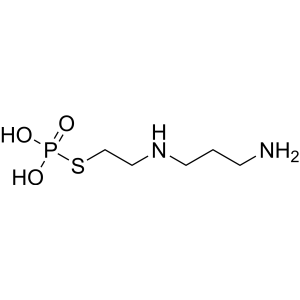
HY-B0639
AmifostineCytoprotective Agent -
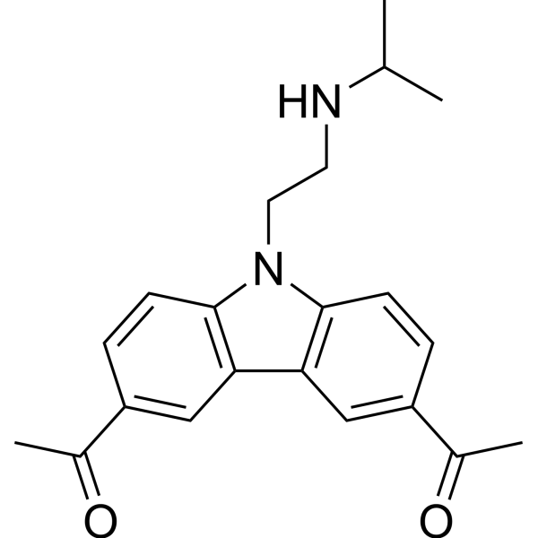
HY-18935
CBL0137FACT Inhibitor -
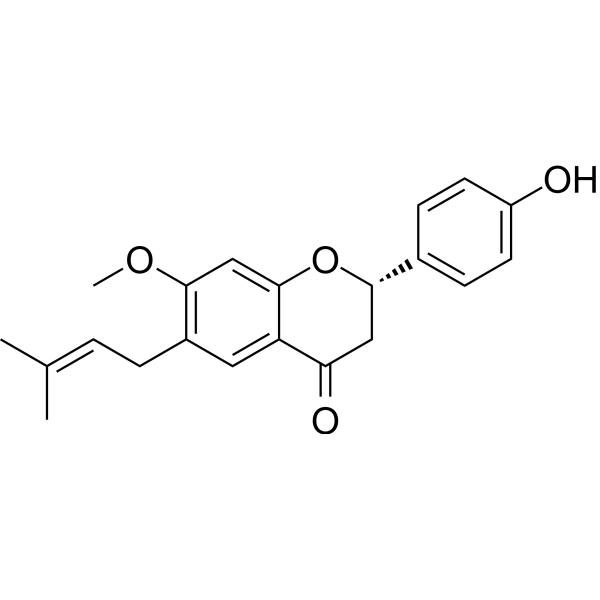
HY-N0234
BavachininPan-PPAR Agonist, HIF-1α Inhibitor -
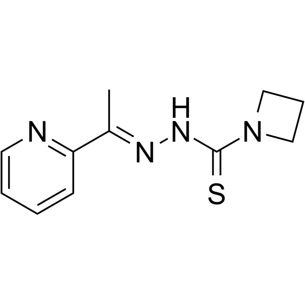
HY-18634
NSC319726MDM-2/p53 p53 Activator -
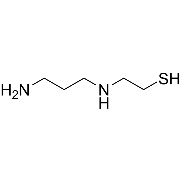
HY-137864
Amifostine thiolRadioprotector/p53 Activator -
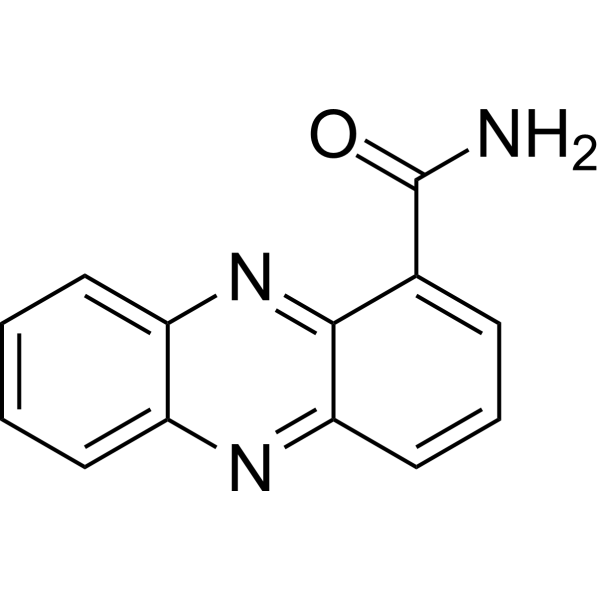
HY-W042191
OxychlororaphineAntifungal Agent -
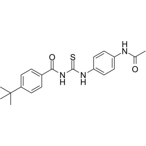
HY-13423
Tenovin-1p53 Activator
MedChemExpress(MCE)の使用を引用している文献 KYP-2047
More

生物活性
製品説明
Cellular Effect
|
Cell Line

|
Type | Value | Description | References |
|---|---|---|---|---|
| HEK293 | IC50 |
>10 μM
Compound: 17, KYP-2047
|
Inhibition of mouse recombinant FAP expressed in HEK293 cells assessed as pNA release from Ala-Pro-p-nitroanilide pre-incubated with enzyme for 15 mins prior to substrate addition by fluorescence technique
Inhibition of mouse recombinant FAP expressed in HEK293 cells assessed as pNA release from Ala-Pro-p-nitroanilide pre-incubated with enzyme for 15 mins prior to substrate addition by fluorescence technique
|
[PMID: 23121075] |
体外実験
KYP-2047 (0-100 μM) decreases U-87, U-138 and A-172 cell viability in a concentration-dependent manner[2].
KYP-2047 (0-100 μM) increases the pro-apoptotic protein Bax, p53 and caspase-3 expression whereas reduces Bcl-2 expression, and reduced significantly TGF-β expression[2].
MedChemExpress (MCE) has not independently confirmed the accuracy of these methods. They are for reference only.
-
Cell Line:U-87, U-138 and A-172 cell[2]
-
Concentration:0.01 μM, 0.1 μM, 0.5 μM, 1 μM, 10 μM, 30 μM, 50 μM and 100 μM
-
Incubation Time:24 h
-
Result:Decreased U-87, U-138 and A-172 cell viability in a concentration-dependent manner.
-
Cell Line:U-87 cell[2]
-
Concentration:0, 50, 100 μM
-
Incubation Time:24 h
-
Result:Increased the pro-apoptotic protein Bax, p53 and cleaved-caspase-3 expression, reduced significantly Bcl2 expression, reduced Ang1 and Ang2 expression, and decreased Ki-67 expression.
体内実験
KYP-2047 (9 or 27 μmol/kg; IP; once, 1 or 3 h before decapitation; two daily doses for 10 days) increases neurotensin concentration in the hypothalamus[1].
KYP-2047 (0-5 mg/kg) significantly reduces tumor mass and neutrophil infiltration[2].
KYP-2047 (0-5 mg/kg) significantly reduces vascular endothelial-growth-factor (VEGF), CD34, angiopoietins (Ang) and endothelial-nitric-oxide synthase (eNOS) expression[2].
MedChemExpress (MCE) has not independently confirmed the accuracy of these methods. They are for reference only.
- Human pluripotent stem cell endothelial-cell differentiation
- Detection of Apoptosis
- TUNEL staining for apoptotic DNA fragmentation
- Vascular/Branching Fractal Analysis
- Annexin V plus membrane-impermeant dye apoptosis staining
- Endothelial Tube Formation Assay
- Apoptosis Solutions
- Endothelial Cell Migration/Angiogenic Sprouting Assay
- Cell Viability Determination by MTT Colorimetric Assay


化学情報
-
CAS 番号 796874-99-2
-
性状 Solid-Liquid Mixture
-
分子量 339.43
-
分子式 C20H25N3O2
-
Color Colorless to light yellow
-
SMILES
N#C[C@H]1N(C([C@H]2N(C(CCCC3=CC=CC=C3)=O)CCC2)=O)CCC1
-
輸送条件
Room temperature in continental US; may vary elsewhere.
-
保管条件
Pure form -20°C 3 years 4°C 2 years In solvent -80°C 6 months -20°C 1 month


Publications (1)
-
Journal Impact Factor
-
Most Recent
-
J Med Chem
Structure-Guided Optimization of 4-Chloro-Pyrazolopyridine Analogs for Covalent PREP Inhibition. [Abstract]2026 Jan 22;69(2):1303-1321. PMID: 41512187


溶剤 & 溶解度
体外:
DMSO : 100 mg/mL (294.61 mM; Need ultrasonic; Hygroscopic DMSO has a significant impact on the solubility of product, please use newly opened DMSO)
Please refer to the solubility information to select the appropriate solvent. Once prepared, please aliquot and store the solution to prevent product inactivation from repeated freeze-thaw cycles.
Storage method and period of stock solution: -80°C, 6 months; -20°C, 1 month. When stored at -80°C, please use it within 6 months. When stored at -20°C, please use it within 1 month.
Please refer to the solubility information to select the appropriate solvent. Once prepared, please aliquot and store the solution to prevent product inactivation from repeated freeze-thaw cycles.
Storage method and period of stock solution: -80°C, 6 months; -20°C, 1 month. When stored at -80°C, please use it within 6 months. When stored at -20°C, please use it within 1 month.
濃度 (開始) × 体積 (開始) = 濃度 (終了) × 体積 (終了)
体内:
Select the appropriate dissolution method based on your experimental animal and administration route.
- For the following dissolution methods, please ensure to first prepare a clear stock solution using an In Vitro approach and then sequentially add co-solvents:
- To ensure reliable experimental results, the clarified stock solution can be appropriately stored based on storage conditions. As for the working solution for In Vivo experiments, it is recommended to prepare freshly and use it on the same day.
- The percentages shown for the solvents indicate their volumetric ratio in the final prepared solution. If precipitation or phase separation occurs during preparation, heat and/or sonication can be used to aid dissolution.
Add each solvent one by one: 10% DMSO 40% PEG300 5% Tween-80 45% Saline
Solubility: 2.5 mg/mL (7.37 mM); Clear solution; Need ultrasonic
This protocol yields a clear solution of 2.5 mg/mL.
Taking 1 mL working solution as an example, add 100 μL DMSO stock solution (25.0 mg/mL) to 400 μL PEG300, and mix evenly; then add 50 μL Tween-80 and mix evenly; then add 450 μL Saline to adjust the volume to 1 mL.
Preparation of Saline: Dissolve 0.9 g sodium chloride in ddH₂O and dilute to 100 mL to obtain a clear Saline solution.
Add each solvent one by one: 10% DMSO 90% (20% SBE-β-CD in Saline)
Solubility: 2.5 mg/mL (7.37 mM); Clear solution; Need ultrasonic
This protocol yields a clear solution of 2.5 mg/mL.
Taking 1 mL working solution as an example, add 100 μL DMSO stock solution (25.0 mg/mL) to 900 μL 20% SBE-β-CD in Saline, and mix evenly.
Preparation of 20% SBE-β-CD in Saline (4°C, storage for one week): 2 g SBE-β-CD powder is dissolved in 10 mL Saline, completely dissolve until clear.
In Vivo Dissolution Calculator
Please enter the basic information of animal experiments:
-
-
-
-
Recommended: Prepare an additional quantity of animals to account for potential losses during experiments.
Please enter your animal formula composition:
-
%DMSO +
Recommended: Keep the proportion of DMSO in working solution below 2% if your animal is weak.
-
%+
-
+%Tween-80 + +
-
%Saline +
The co-solvents required include: DMSO, . All of co-solvents are available by MedChemExpress (MCE). , Tween 80. All of co-solvents are available by MedChemExpress (MCE).
Working solution concentration: 0.22 mg/mL
Method for preparing stock solution: mg drug dissolved in μL DMSO. Stock solution concentration: mg/mL.
1. Take μL DMSO stock solution;
2. Add μL .
μL , mix evenly;
3. Then add μL Tween 80, mix evenly;
4. Then add μL
Please ensure that the stock solution in the first step is dissolved to a clear state, and add co-solvents in sequence. You can use ultrasonic heating (ultrasonic cleaner, recommended frequency 20-40 kHz), vortexing, etc. to assist dissolution.


純度とドキュメンテーション
-
データシート (276 KB)
-
SDS (393 KB)
- English - EN (393 KB)
- Français - FR (393 KB)
- Deutsch - DE (393 KB)
- Norwegian - NO (393 KB)
- Español - ES (393 KB)
- Swedish - SV (393 KB)
- Italian - IT (393 KB)
- Korean - KR (393 KB)
- Portuguese - PT (393 KB)
-
取扱説明書 (2659 KB)
参考文献
[1]. Jalkanen AJ, et al. Beneficial effect of prolyl oligopeptidase inhibition on spatial memory in young but not in old scopolamine-treated rats. Basic Clin Pharmacol Toxicol. 2007 Feb;100(2):132-8. [Content Brief]
[2]. Scuderi SA, et al. KYP-2047, an Inhibitor of Prolyl-Oligopeptidase, Reduces GlioBlastoma Proliferation through Angiogenesis and Apoptosis Modulation. Cancers (Basel). 2021 Jul 9;13(14):3444. [Content Brief]


Complete Stock Solution Preparation Table
Please refer to the solubility information to select the appropriate solvent. Once prepared, please aliquot and store the solution to prevent product inactivation from repeated freeze-thaw cycles.
Storage method and period of stock solution: -80°C, 6 months; -20°C, 1 month. When stored at -80°C, please use it within 6 months. When stored at -20°C, please use it within 1 month.
| Optional Solvent | Concentration Solvent Mass | 1 mg | 5 mg | 10 mg | 25 mg |
|---|---|---|---|---|---|
| DMSO | 1 mM | 2.9461 mL | 14.7306 mL | 29.4612 mL | 73.6529 mL |
| 5 mM | 0.5892 mL | 2.9461 mL | 5.8922 mL | 14.7306 mL | |
| 10 mM | 0.2946 mL | 1.4731 mL | 2.9461 mL | 7.3653 mL | |
| 15 mM | 0.1964 mL | 0.9820 mL | 1.9641 mL | 4.9102 mL | |
| 20 mM | 0.1473 mL | 0.7365 mL | 1.4731 mL | 3.6826 mL | |
| 25 mM | 0.1178 mL | 0.5892 mL | 1.1784 mL | 2.9461 mL | |
| 30 mM | 0.0982 mL | 0.4910 mL | 0.9820 mL | 2.4551 mL | |
| 40 mM | 0.0737 mL | 0.3683 mL | 0.7365 mL | 1.8413 mL | |
| 50 mM | 0.0589 mL | 0.2946 mL | 0.5892 mL | 1.4731 mL | |
| 60 mM | 0.0491 mL | 0.2455 mL | 0.4910 mL | 1.2275 mL | |
| 80 mM | 0.0368 mL | 0.1841 mL | 0.3683 mL | 0.9207 mL | |
| 100 mM | 0.0295 mL | 0.1473 mL | 0.2946 mL | 0.7365 mL |


KYP-2047 関連分類
HY-100475 関連分類
 Powered by Bioz