Resolvin E1
Based on 2 publication(s) in Google Scholar
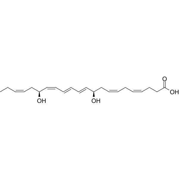
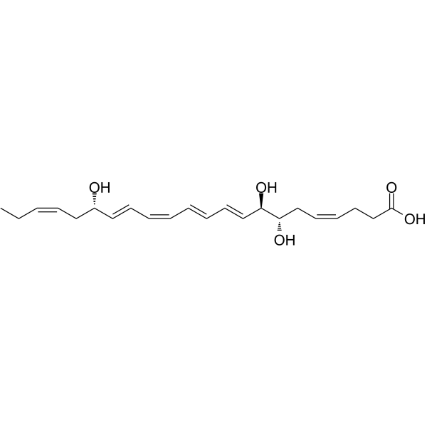
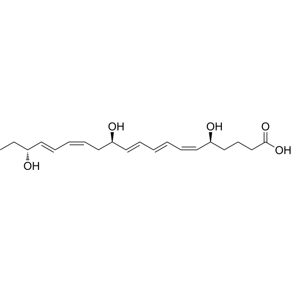
Resolvin E1 (RvE1), a potent endogenous pro-resolving mediator of inflammation, is derived from omega-3 fatty acid eicosapentaenoic acid (EPA). Resolvin E1 is endogenously biosynthesized from EPA in the presence of Aspirin during the spontaneous resolution phase of acute inflammation, where specific cell-cell interactions occur. Resolvin E1 possesses unique counterregulatory actions that inhibit polymorphonuclear leukocyte (PMN) transendothelial migration. Resolvin E1 also acts as a potent inhibitor of leukocyte infiltration, dendritic cell migration, and IL-12 production.
商品は「研究用試薬」です。人や動物の医療用・臨床診断用・食品用の製品ではありません。
研究用途以外に使用した場合、当社は一切の責任を負いかねます。
- 純度 : 98.1%
- CAS 番号: 552830-51-0
- 分子式: C20H30O5
- 分子量:350.45
-
保管条件:
Solution, -20°C, 2 years
MedChemExpress(MCE)の使用を引用している文献 Resolvin E1
More-
Histological Imaging/Staining
-
WB
-
IF
-
ELISA
Endogenous Metabolite アイソフォーム固有の製品をすべて表示
More
生物活性
製品説明
IC50 & Target
[1]|
Human Endogenous Metabolite |
体外実験
Resolvin E1 (0.1-100 nM) gives concentration-dependent inhibition of TNF-α–induced NF-κB activation with an EC50 of ~1.0 nM in ChemR23-transfected cells. Resolvin E1 (100 nM) increases phosphorylation of extracellular signal-regulated kinase (ERK) mitogen-activated protein (MAP) kinase both in peripheral blood monocytes and HEK-ChemR23 cells[2]. Resolvin E1 specifically interacts with the LTB4 receptor BLT1 on neutrophils and ChemR23 on monocytes to regulate leukocytes during inflammation. Resolvin E1 also stimulates the uptake and clearance of local cytokine[3].
MedChemExpress (MCE) has not independently confirmed the accuracy of these methods. They are for reference only. Further protocols information, click here.
体内実験
MedChemExpress (MCE) has not independently confirmed the accuracy of these methods. They are for reference only.
-
Animal Model:6- to 8-week-old female BALB/c mice (TNBS colitis model)[1]
-
Dosage:1.0 μg per mouse; 50 μg/kg
-
Administration:i.p.; on days -8, -1, and 0 before the induction of colitis
-
Result:Reduced overall mortality, which was 25% and 62.5% at 1.0 μg per mouse and 50 μg/kg, respectively.
化学情報
-
CAS 番号 552830-51-0
-
性状 Liquid
-
分子量 350.45
-
分子式 C20H30O5
-
Color Colorless to light yellow
-
SMILES
CC[C@@H](O)/C=C/C=C\C[C@@H](O)/C=C/C=C/C=C\[C@@H](O)CCCC(O)=O
-
別名
RvE1
-
Structure Classification
-
Initial Source
-
輸送条件
Room temperature in continental US; may vary elsewhere.
-
保管条件
Solution, -20°C, 2 years
Publications (2)
-
Journal Impact Factor
-
Most Recent
-
Exp Neurol
RvE1/ChemR23 facilitates hematoma clearance and promotes M2 polarization of macrophages/microglia in intracerebral hemorrhage. [Abstract]2025 Apr:386:115173. PMID: 39892453 -
Immunol Invest
Resolvin E1 and Inhibition of BLT2 Signaling Attenuate the Inflammatory Response and Improve One-Lung Ventilation-Induced Lung Injury. [Abstract]2024 Nov;53(8):1293-1307. PMID: 39230105
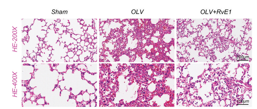
Resolvin E1 purchased from MedChemExpress. Usage Cited in: Immunol Invest. 2024 Nov;53(8):1293-1307. [Abstract]
Pathological alterations in lung tissues analyzed via H&E staining with Resolvin E1 (RvE1) treatment.
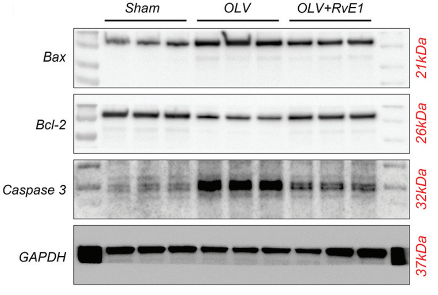
Resolvin E1 purchased from MedChemExpress. Usage Cited in: Immunol Invest. 2024 Nov;53(8):1293-1307. [Abstract]
Expression levels of apoptotic markers Bax, bcl-2, and caspase-3 were evaluated through Western blot analysis with Resolvin E1 (RvE1) treatment.
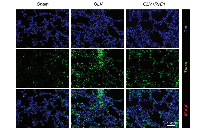
Resolvin E1 purchased from MedChemExpress. Usage Cited in: Immunol Invest. 2024 Nov;53(8):1293-1307. [Abstract]
Apoptosis in lung tissues quantified via TUNEL staining with Resolvin E1 (RvE1) treatment.
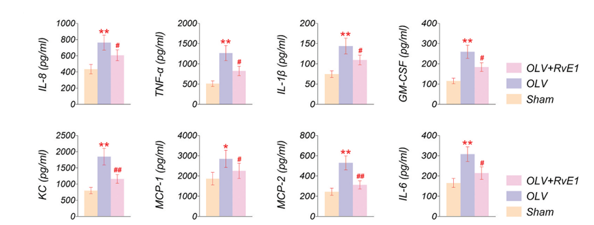
Resolvin E1 purchased from MedChemExpress. Usage Cited in: Immunol Invest. 2024 Nov;53(8):1293-1307. [Abstract]
Quantification of inflammatory cytokines IL-8, tnf-α, IL-1β, GM-CSF (CSF2, colony stimulating factor 2), KC (CXCL1, chemokine ligand 1), MCP-1 (CCL2, Monocyte Chemotactic and activating factor), MIP-2 (CXCL2, chemokine ligand 2), and IL-6, as determined by ELISA with Resolvin E1 (RvE1) treatment.
プロトコル
-
Scratch/Wound-Healing Migration Assay
The scratch/wound-healing migration assay measures collective migration of adherent cells into an experimentally created cell-free gap in a confluent monolayer. The readout is generated by imaging the gap immediately after scratching and at later time points, then quantifying reduction in wound area, wound width, or percentage closure as cells move into the denuded region. Gap closure reflects cell migration but may also include cell proliferation, so interpretation should distinguish migration-focused conditions from proliferation-driven closure when possible, such as by using short assay windows, serum-controlled conditions, cell counting, or proliferation controls reported in published protocols.
-
Cell migration
Cell migration is a method that plays an important role in wound healing, cell differentiation, embryonic development, etc.
-
Chemotaxis Gradient Chamber Assay 1
The chemotaxis gradient chamber assay is based on the principle of observing directional cell migration in response to a stable, linear or quasi-linear concentration gradient of a chemoattractant formed between two fluid reservoirs separated by a narrow observation chamber. Cells placed within the chamber respond to the gradient by polarized movement toward higher chemoattractant concentrations, allowing quantification of chemotactic behavior in real time under microscopy. The classic Zigmond chamber design enables simultaneous visualization of gradient formation and individual cell trajectories, making it suitable for studying leukocyte chemotaxis and other motile cell types in vitro.
-
Carrageenan-Induced Paw Edema
Carrageenan-induced paw edema is an acute inflammation model in which intraplantar injection of carrageenan induces localized inflammatory swelling characterized by vascular permeability, leukocyte infiltration, and production of inflammatory mediators such as prostaglandins and cytokines, making it widely used to evaluate anti-inflammatory agents in vivo. The resulting paw volume or thickness increase is quantified over time as a direct readout of inflammatory intensity and drug efficacy, typically reflecting cyclooxygenase-mediated prostaglandin-driven edema formation and immune cell recruitment in peripheral tissue[20].
-
Under-Agarose Cell Migration Assay
The under-agarose cell migration assay is a classical in vitro chemotaxis method designed to evaluate directed cell movement through a semi-solid agarose matrix toward soluble chemoattractant gradients, enabling visualization and quantification of leukocyte or motile cell migration in a confined 2D-like environment. In this system, cells and chemoattractants are placed in separate wells cut into an agarose gel, allowing diffusion-driven gradient formation that guides directional migration, which is typically assessed by measuring migration distance, cell morphology changes, and accumulation toward the chemoattractant source. This assay has been widely used to study neutrophil and leukocyte chemotaxis as a simple alternative to filter-based migration systems and allows direct microscopic observation of migrating cells under near-physiological confinement conditions.
-
Neural Crest/Neuronal Cell Migration Explant Assay
Neural crest (NC) and neuronal cell migration explant assays are in vitro systems in which neural tube-derived tissues are cultured to allow premigratory or newly emigrated neural crest cells to undergo epithelial-to-mesenchymal transition (EMT), migrate away from the explant, and form a measurable radial outgrowth that reflects migratory capacity and environmental responsiveness. These assays typically quantify migration by measuring the expansion of cell outgrowth from neural tube or neural plate border explants over time, often comparing early and later timepoints to derive a migration index such as a radius ratio, which reflects net cell dispersal from the explant core. Neural tube explant cultures preserve key aspects of neural crest behavior, including EMT, migration, and early differentiation, making them suitable for assessing intrinsic migratory ability and extrinsic cue dependence. However, studies emphasize that migratory outgrowth from neural tube explants may include non-n
-
Transwell/Boyden Chamber Migration Assay
The Transwell/Boyden chamber migration assay measures cell movement through a porous membrane separating an upper and lower chamber, usually after a chemoattractant gradient is established by placing cells in the upper chamber and chemoattractant-containing medium in the lower chamber. The readout is generated by quantifying cells that traverse the membrane and appear on the lower membrane surface or in the lower chamber, depending on whether the cell type is adherent or non-adherent. This assay reflects chemotactic or haptotactic migration rather than matrix invasion unless an extracellular-matrix barrier is added to the membrane.
-
Zymosan-Induced Peritonitis
Zymosan-induced peritonitis is a sterile acute-inflammation model produced by intraperitoneal injection of zymosan, a yeast cell-wall particle preparation, followed by quantification of leukocyte recruitment and soluble inflammatory mediators in peritoneal lavage fluid. Low-dose zymosan peritonitis is commonly used as a self-resolving acute inflammation model in which neutrophil recruitment occurs early and monocyte/macrophage accumulation follows later. The assay readouts include total peritoneal leukocyte number, differential neutrophil and monocyte/macrophage counts, peritoneal cytokines and chemokines, plasma or peritoneal exudation, and optional lipidomic or metabolomic changes during inflammation and resolution. Early neutrophil recruitment after zymosan depends strongly on complement and mast-cell C5a receptor signaling, whereas later monocyte recruitment is linked to MCP-1/CCL2 production.
-
3D Collagen/Hydrogel Matrix Migration Assay
The 3D collagen/hydrogel matrix migration assay is based on embedding cells within or on top of a fibrillar collagen type I-rich three-dimensional matrix to model in vivo-like extracellular matrix (ECM) architecture, enabling analysis of cell migration through a physically and biochemically relevant scaffold. In contrast to 2D migration systems, cells in 3D matrices interact with fibrillar collagen networks, requiring coordinated adhesion remodeling and proteolytic or non-proteolytic deformation mechanisms to move through confined spaces, thereby providing a more physiologically relevant readout of invasive and migratory behavior in tissue-like environments. Cell movement in 3D collagen matrices is typically quantified by tracking single-cell trajectories, invasion depth, or matrix penetration over time, reflecting combined effects of cytoskeletal dynamics, cell-ECM adhesion turnover, and ECM remodeling. These systems are widely used to study tumor cell invasion and stromal cell motili
-
Research Protocol for Inflammation-related Diseases
The NLRP3 inflammasome is a cytosolic innate immune signaling platform that integrates priming signals and danger-signal activation to promote caspase-1 activation, maturation of IL-1β and IL-18, and gasdermin D-mediated pyroptotic cell death. The core experimental logic is to determine whether inflammatory disease phenotypes are driven by increased NLRP3 expression, ASC-containing inflammasome assembly, caspase-1 cleavage, GSDMD cleavage, and extracellular release of IL-1β/IL-18 rather than by nonspecific cell injury alone. The pathway is strongly linked to inflammation-related disease phenotypes because monosodium urate crystals activate NALP3/NLRP3 inflammasome signaling in gout-like crystal inflammation, cholesterol crystals activate NLRP3 inflammasomes in atherogenesis models, and DSS-induced intestinal inflammation has been reported to involve NLRP3 inflammasome activity. However, experimental colitis studies also show context-dependent protective effects of NLRP3 inflammasome co
-
Cell-Exclusion Zone Migration Assay
The Cell-Exclusion Zone (CEZ) migration assay is an in vitro 2D cell migration method in which a defined cell-free area is created using removable physical barriers such as silicone stoppers, allowing cells to be seeded around the barrier and subsequently migrate into the cleared zone after barrier removal. This approach enables quantification of collective cell migration by monitoring repopulation of the initially cell-free region over time using microscopy-based imaging. Compared with scratch-based wound healing assays, barrier-based exclusion methods are designed to avoid mechanical damage to the extracellular matrix and reduce injury-induced effects on boundary cells, thereby improving interpretability of migration behavior in vitro. The assay readout is typically the progressive reduction in the cell-free area or the number of cells invading the exclusion zone, reflecting coordinated cell motility relevant to physiological processes such as wound healing, epithelial repair, and ca
-
Chemotaxis Gradient Chamber Assay 2
Chemotaxis gradient chamber assays measure directional cell migration in response to a soluble chemical gradient by imaging cells as they move across a defined observation region; the readout is generated from time-lapse cell trajectories, displacement toward the gradient, forward migration index, trajectory plots, rose/polar plots, and statistical tests of non-random directionality. The Dunn chamber is a direct-viewing glass chamber in which cells migrate across a bridge between control and chemoattractant wells, allowing observation of cells in a linear concentration gradient; related direct-viewing formats include the Insall chamber, which supports defined unidirectional gradients and high numerical-aperture microscopy, and the μ-Slide Chemotaxis chamber, which supports long-term live-cell imaging and gradient characterization with fluorescent dye.
純度とドキュメンテーション
-
データシート (268 KB)
-
SDS (394 KB)
- English - EN (394 KB)
- Français - FR (394 KB)
- Deutsch - DE (394 KB)
- Norwegian - NO (394 KB)
- Español - ES (394 KB)
- Swedish - SV (394 KB)
- Italian - IT (394 KB)
- Korean - KR (394 KB)
- Portuguese - PT (394 KB)
-
取扱説明書 (2659 KB)
参考文献
[1]. Arita M, et al. Resolvin E1, an endogenous lipid mediator derived from omega-3 eicosapentaenoic acid, protects against 2,4,6-trinitrobenzene sulfonic acid-induced colitis. Proc Natl Acad Sci U S A. 2005 May 24;102(21):7671-6. [Content Brief]
[2]. Schwab JM, et al. Resolvin E1 and protectin D1 activate inflammation-resolution programmes. Nature. 2007 Jun 14;447(7146):869-74. [Content Brief]
[3]. Hasturk H, et al. Resolvin E1 regulates inflammation at the cellular and tissue level and restores tissuehomeostasis in vivo. J Immunol. 2007 Nov 15;179(10):7021-9. [Content Brief]
Calculators
濃度 (開始) × 体積 (開始) = 濃度 (終了) × 体積 (終了)