



K6PC-5
Based on 4 publication(s) in Google Scholar
K6PC-5, a ceramide derivative, is a sphingosine kinase 1(SPHK1) activator and elicites a rapid transient increase in intracellular calcium levels. K6PC-5 has the potential for skin diseases involving abnormal keratinocyte, and neurodegeneration and virus infection research.
For research use only. We do not sell to patients.
- Purity : 99.27%
- CAS No.: 756875-51-1
- Formula: C19H37NO4
- Molecular Weight:343.50
-
Storage:Powder -20°C, 3 years , 4°C, 2 years ; In solvent -80°C, 6 months , -20°C, 1 month


To place orders, for customer services and technical support, please contact: MedChemExpress USA
Tel: 609-228-6898 E-mail: [email protected] [email protected]


-
 Biological Activity
-
Chemical Information
-
Solvent & Solubility
- Protocols
- Purity & Documentation
- References
-
 Help & FAQs


-
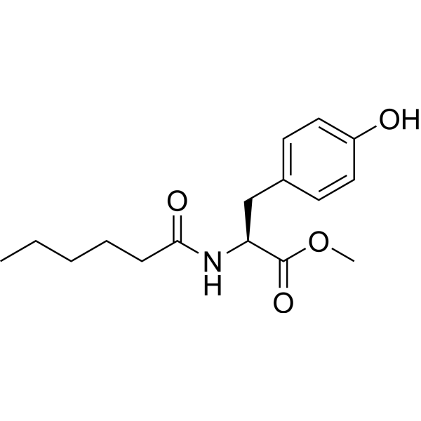
HY-101572
MHPSPHK1 Activator -
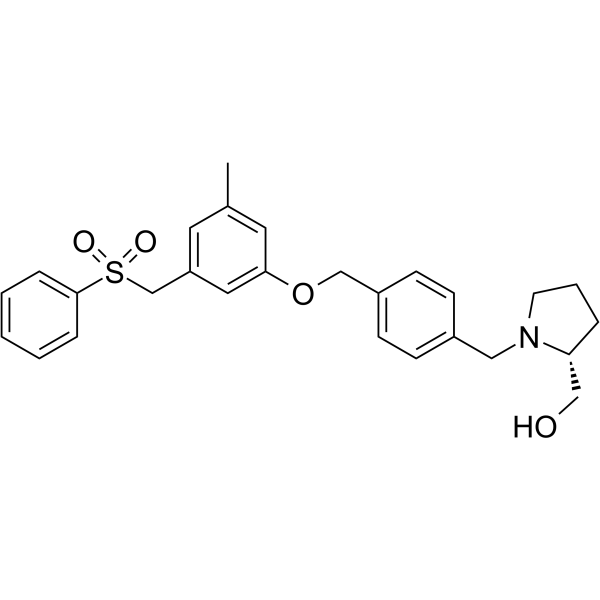
HY-15425
PF-543SPHK1 Inhibitor -
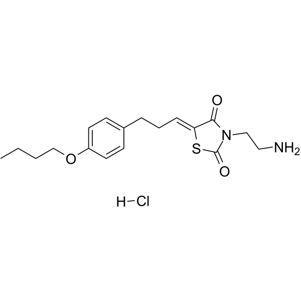
HY-15779A
K145 hydrochlorideSphK2 Inhibitor -
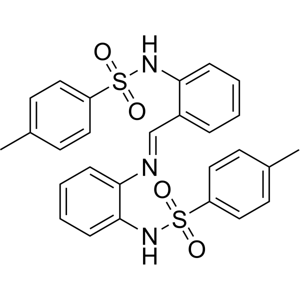
HY-19794
MP-A08SphK Inhibitor -
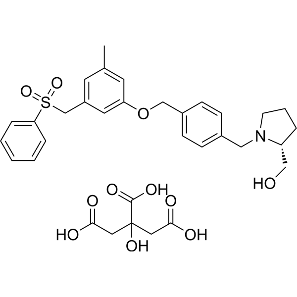
HY-15425A
PF-543 CitrateSPHK1 Inhibitor -
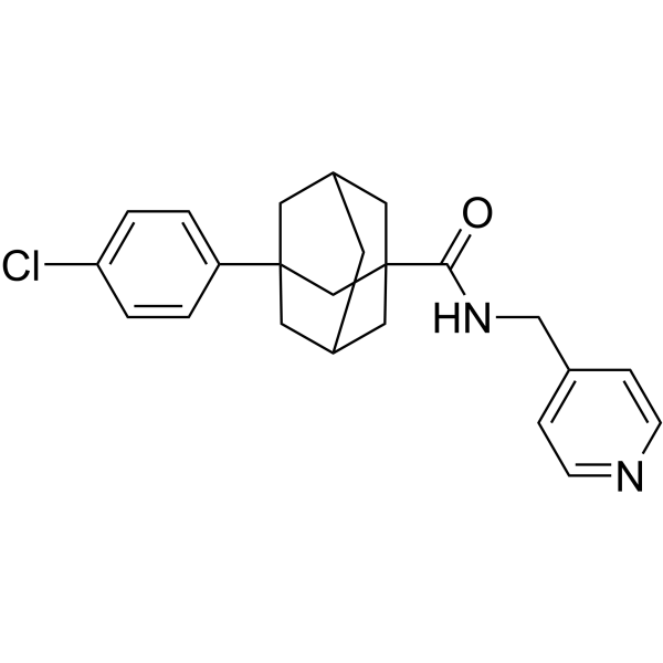
HY-16015
Opaganib -
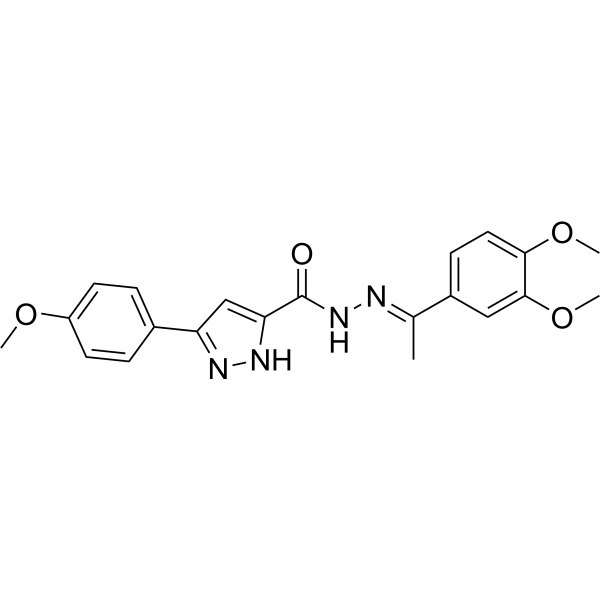
HY-12892
SKI-178SphK1/SphK2 Inhibitor -
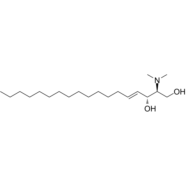
HY-108491
N,N-DimethylsphingosineSphingosine Kinase Inhibitor -

HY-101572R
MHP (Standard)SPHK1 Activator -
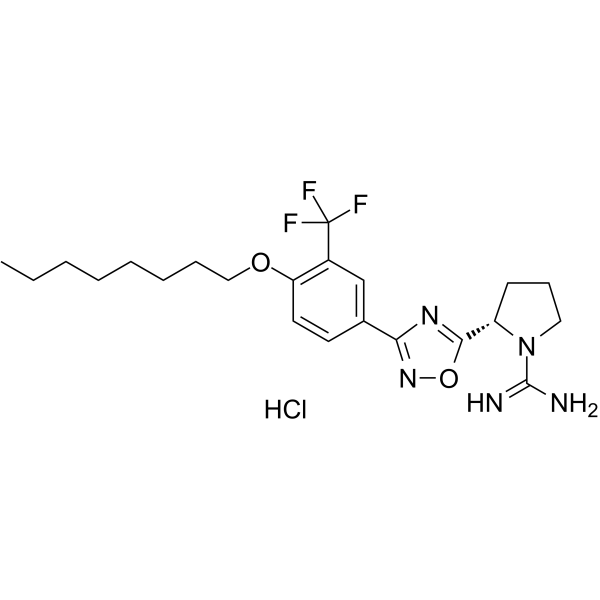
HY-120268A
SLM6031434 hydrochlorideSphK2 Inhibitor -
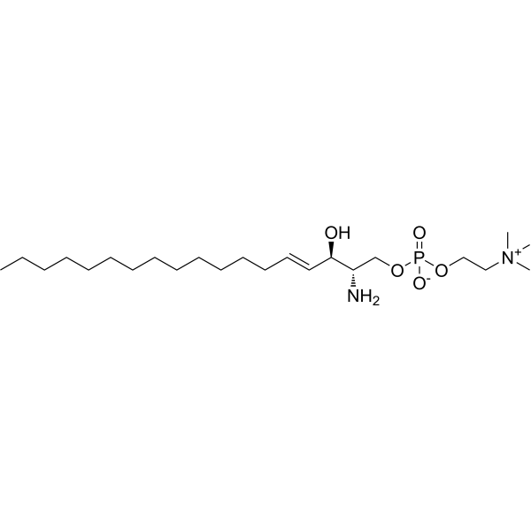
HY-132187
SphingosylphosphorylcholineBioactive Lipid -
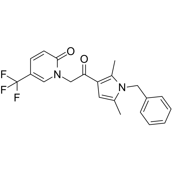
HY-111253
CYM-5478S1P2 Agonist
Publications Citing Use of MedChemExpress (MCE) K6PC-5
More

Biological Activity
Description
IC50 & Target
|
SphK1 |
EBOV |
In Vitro
K6PC-5 (1-10 μM; 24 h) increases the involucrin and loricrin levels in a dose-dependent manner in normal human epidermal keratinocytes (NHEKs). K6PC-5 promotes differentiation and proliferation of keratinocytes via intracellular Ca2+ signaling. In addition, K6PC-5 stimulates the phosphorylation of p42/44 extracellular signal-regulated kinase and c-Jun N-terminal kinase[1].
K6PC-5 (1-10 μM; 24 h) promotes fibroblasts proliferation and collagen synthesis in human fibroblasts. K6PC-5 induces intracellular Ca2+ concentration ([Ca2+]i) oscillations in human fibroblasts[2].
K6PC-5 (10, 25, and 50 μM; 48 h) significantly attenuates EBOV-induced infection in EBOV-infected EA.hy926 cells. K6PC-5 significantly reduces the virus titers in supernatants of infected cells and strikingly decreased the amount of VP40 in a concentration-dependent manner[3].
MedChemExpress (MCE) has not independently confirmed the accuracy of these methods. They are for reference only. Further protocols information, click here.
-
Cell Line:Normal human epidermal keratinocytes (NHEKs)
-
Concentration:1 μM, 5 μM, 10 μM
-
Incubation Time:24 h
-
Result:Increased the involucrin and loricrin levels in a dose-dependent manner.
-
Cell Line:Human fibroblasts
-
Concentration:1 μM, 5 μM, 10 μM
-
Incubation Time:24 h
-
Result:Promoted fibroblast proliferation and procollagen production in human fibroblasts significantly.
In Vivo
MedChemExpress (MCE) has not independently confirmed the accuracy of these methods. They are for reference only.
-
Animal Model:Intrinsically aged hairless mice (56 weeks old)[2]
-
Dosage:1% (vehicle (PEG:EtOH = 7:3))
-
Administration:Topical application; twice daily for 2 weeks
-
Result:Enhanced fibroblast proliferation, collagen production, and eventually increased dermal thickness.


Chemical Information
-
CAS No. 756875-51-1
-
Appearance Solid
-
Molecular Weight 343.50
-
Formula C19H37NO4
-
Color White to off-white
-
SMILES
CCCCCCCC(C(C(NC(CO)CO)=O)CCCCCC)=O
-
Shipping
Room temperature in continental US; may vary elsewhere.
-
Storage
Powder -20°C 3 years 4°C 2 years In solvent -80°C 6 months -20°C 1 month


Publications (4)
-
Journal Impact Factor
-
Most Recent
-
Phytomedicine
Novel mechanism by which geniposide alleviates rheumatoid arthritis: Targeted inhibition of SphK1 negatively regulates S1P signaling, promotes HIF-1α acetylation-mediated degradation and blocks the VEGF-induced synovial angiogenesis. [Abstract]2026 Jan:150:157739. PMID: 41456528 -
Phytother Res
Geniposide Improves Glycolysis Driven Angiogenesis in Experimentary Arthritis by Inhibiting SphK1-PI3K-Akt-PFKFB3 Signal. [Abstract]2025 Aug;39(8):3419-3431. PMID: 40583637 -
Int Immunopharmacol
Liquiritigenin attenuates alcohol-induced liver inflammation by regulating the SphK1/S1P/SPNS2 signaling pathway. [Abstract]2026 Jan 1;168(Pt 2):115881. PMID: 41297343 -
Discov Oncol
Ritanserin suppresses acute myeloid leukemia by inhibiting DGKα to downregulate phospholipase D and the Jak-Stat/MAPK pathway. [Abstract]2023 Jul 1;14(1):118. PMID: 37392305


Solvent & Solubility
In Vitro:
DMSO : 100 mg/mL (291.12 mM; Need ultrasonic; Hygroscopic DMSO has a significant impact on the solubility of product, please use newly opened DMSO)
Please refer to the solubility information to select the appropriate solvent. Once prepared, please aliquot and store the solution to prevent product inactivation from repeated freeze-thaw cycles.
Storage method and period of stock solution: -80°C, 6 months; -20°C, 1 month. When stored at -80°C, please use it within 6 months. When stored at -20°C, please use it within 1 month.
Please refer to the solubility information to select the appropriate solvent. Once prepared, please aliquot and store the solution to prevent product inactivation from repeated freeze-thaw cycles.
Storage method and period of stock solution: -80°C, 6 months; -20°C, 1 month. When stored at -80°C, please use it within 6 months. When stored at -20°C, please use it within 1 month.
Concentration (start) × Volume (start) = Concentration (final) × Volume (final)
In Vivo:
Select the appropriate dissolution method based on your experimental animal and administration route.
- For the following dissolution methods, please ensure to first prepare a clear stock solution using an In Vitro approach and then sequentially add co-solvents:
- To ensure reliable experimental results, the clarified stock solution can be appropriately stored based on storage conditions. As for the working solution for In Vivo experiments, it is recommended to prepare freshly and use it on the same day.
- The percentages shown for the solvents indicate their volumetric ratio in the final prepared solution. If precipitation or phase separation occurs during preparation, heat and/or sonication can be used to aid dissolution.
Add each solvent one by one: 10% DMSO 40% PEG300 5% Tween-80 45% Saline
Solubility: ≥ 2.5 mg/mL (7.28 mM); Clear solution
This protocol yields a clear solution of ≥ 2.5 mg/mL (saturation unknown).
Taking 1 mL working solution as an example, add 100 μL DMSO stock solution (25.0 mg/mL) to 400 μL PEG300, and mix evenly; then add 50 μL Tween-80 and mix evenly; then add 450 μL Saline to adjust the volume to 1 mL.
Preparation of Saline: Dissolve 0.9 g sodium chloride in ddH₂O and dilute to 100 mL to obtain a clear Saline solution.
Add each solvent one by one: 10% DMSO 90% (20% SBE-β-CD in Saline)
Solubility: ≥ 2.5 mg/mL (7.28 mM); Clear solution
This protocol yields a clear solution of ≥ 2.5 mg/mL (saturation unknown).
Taking 1 mL working solution as an example, add 100 μL DMSO stock solution (25.0 mg/mL) to 900 μL 20% SBE-β-CD in Saline, and mix evenly.
Preparation of 20% SBE-β-CD in Saline (4°C, storage for one week): 2 g SBE-β-CD powder is dissolved in 10 mL Saline, completely dissolve until clear.
In Vivo Dissolution Calculator
Please enter the basic information of animal experiments:
-
-
-
-
Recommended: Prepare an additional quantity of animals to account for potential losses during experiments.
Please enter your animal formula composition:
-
%DMSO +
Recommended: Keep the proportion of DMSO in working solution below 2% if your animal is weak.
-
%+
-
+%Tween-80 + +
-
%Saline +
The co-solvents required include: DMSO, . All of co-solvents are available by MedChemExpress (MCE). , Tween 80. All of co-solvents are available by MedChemExpress (MCE).
Working solution concentration: 0.22 mg/mL
Method for preparing stock solution: mg drug dissolved in μL DMSO. Stock solution concentration: mg/mL.
1. Take μL DMSO stock solution;
2. Add μL .
μL , mix evenly;
3. Then add μL Tween 80, mix evenly;
4. Then add μL
Please ensure that the stock solution in the first step is dissolved to a clear state, and add co-solvents in sequence. You can use ultrasonic heating (ultrasonic cleaner, recommended frequency 20-40 kHz), vortexing, etc. to assist dissolution.

Protocols
-
Neurotoxicity Study
This protocol assesses in vitro neurotoxicity by combining neuronal viability, mitochondrial/metabolic activity, neurite outgrowth, and optional neuronal network function readouts. Calcein-AM or resazurin/PrestoBlue readouts estimate viable or metabolically active cells; βIII-tubulin immunofluorescence detects neuronal morphology and neurite networks; TMRE detects mitochondrial membrane potential; and MEA recordings detect functional changes in neuronal network activity.
-
Research Protocol for Infectious Diseases
Infectious-disease experiments test how pathogens interact with host barriers, innate immune receptors, inflammatory signaling, pathogen replication, and tissue injury; pattern-recognition receptors such as TLRs, RIG-I-like receptors, NOD-like receptors, and inflammasomes detect microbial molecules and activate NF-κB, interferon, and cytokine responses. The central hypothesis is that infection severity reflects the balance between pathogen burden and host response: protective inflammation restricts pathogen growth, whereas excessive or mislocalized inflammation contributes to tissue damage and disease phenotype. Unresolved questions include which host pathways are protective versus pathogenic, why some infection models fail to translate to human disease, and which combined readouts best predict clinically relevant infection outcomes.
-
Calcium Spark Assay
Calcium sparks are localized, transient increases in intracellular calcium concentration ([Ca2+]i) that occur in cardiac myocytes and represent elementary events underlying excitation-contraction coupling. These events are generated by the coordinated opening of clusters of ryanodine receptors (RyRs) on the sarcoplasmic reticulum membrane, leading to a brief release of Ca2+ into the cytosol. The detection and analysis of calcium sparks provide insights into the mechanisms of calcium handling and signaling in cardiac cells. Imaging techniques using fluorescent calcium indicators such as Fluo-3 are employed to visualize these subcellular calcium transients with high spatial and temporal resolution. The protocol is based on established methodologies described in primary literature for both experimental measurement and automated analysis of calcium sparks.
-
Ca2+ Staining Technique
Ca2+ staining is an experimental technique that utilizes specific fluorescent probes (such as Fluo-4 AM, Fura-2, etc.) to qualitatively or quantitatively detect dynamic changes in intracellular Ca2+ concentrations; this is achieved by monitoring the changes in fluorescent signals generated when these probes bind to free intracellular calcium ions. The underlying principle relies primarily on the presence of chelating groups within the probe's molecular structure that possess high affinity for calcium ions.


Purity & Documentation
-
Data Sheet (276 KB)
-
SDS (393 KB)
- English - EN (393 KB)
- Français - FR (393 KB)
- Deutsch - DE (393 KB)
- Norwegian - NO (393 KB)
- Español - ES (393 KB)
- Swedish - SV (393 KB)
- Italian - IT (393 KB)
- Korean - KR (393 KB)
- Portuguese - PT (393 KB)
-
Handling Instructions (2659 KB)
References
[1]. Kwon YB, et al. Novel synthetic ceramide derivatives increase intracellular calcium levels and promote epidermal keratinocyte differentiation. J Lipid Res. 2007 Sep;48(9):1936-43. [Content Brief]
[2]. Jong-Kyung Youm, et al. K6PC-5, a sphingosine kinase activator, induces anti-aging effects in intrinsically aged skin through intracellular Ca2+ signaling. J Dermatol Sci. 2008 Aug;51(2):89-102. [Content Brief]
[3]. Imre G, et al. The sphingosine kinase 1 activator, K6PC-5, attenuates Ebola virus infection. iScience. 2021 Mar 5;24(4):102266. [Content Brief]


Complete Stock Solution Preparation Table
Please refer to the solubility information to select the appropriate solvent. Once prepared, please aliquot and store the solution to prevent product inactivation from repeated freeze-thaw cycles.
Storage method and period of stock solution: -80°C, 6 months; -20°C, 1 month. When stored at -80°C, please use it within 6 months. When stored at -20°C, please use it within 1 month.
| Optional Solvent | Concentration Solvent Mass | 1 mg | 5 mg | 10 mg | 25 mg |
|---|---|---|---|---|---|
| DMSO | 1 mM | 2.9112 mL | 14.5560 mL | 29.1121 mL | 72.7802 mL |
| 5 mM | 0.5822 mL | 2.9112 mL | 5.8224 mL | 14.5560 mL | |
| 10 mM | 0.2911 mL | 1.4556 mL | 2.9112 mL | 7.2780 mL | |
| 15 mM | 0.1941 mL | 0.9704 mL | 1.9408 mL | 4.8520 mL | |
| 20 mM | 0.1456 mL | 0.7278 mL | 1.4556 mL | 3.6390 mL | |
| 25 mM | 0.1164 mL | 0.5822 mL | 1.1645 mL | 2.9112 mL | |
| 30 mM | 0.0970 mL | 0.4852 mL | 0.9704 mL | 2.4260 mL | |
| 40 mM | 0.0728 mL | 0.3639 mL | 0.7278 mL | 1.8195 mL | |
| 50 mM | 0.0582 mL | 0.2911 mL | 0.5822 mL | 1.4556 mL | |
| 60 mM | 0.0485 mL | 0.2426 mL | 0.4852 mL | 1.2130 mL | |
| 80 mM | 0.0364 mL | 0.1820 mL | 0.3639 mL | 0.9098 mL | |
| 100 mM | 0.0291 mL | 0.1456 mL | 0.2911 mL | 0.7278 mL |


K6PC-5 Related Classifications
HY-124042 Related Classifications
 Powered by Bioz