



AMPK activator 10
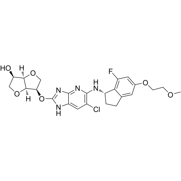
AMPK activator 10 is an orally active, potent AMPK activator with EC150 of 44.3 nM by cell-ELISA. AMPK activator 10 increases the phosphorylation levels of ACC. AMPK activator 10 exhibits a glucose lowering effect.
연구목적의 판매만을 진행합니다. 환자를 대상으로 한 판매는 하지 않습니다.
- CAS No.: 1914176-03-6
- 화학식: C24H26ClFN4O6
- 분자량:520.94
-
보관:
Please store the product under the recommended conditions in the Certificate of Analysis.

To place orders, for customer services and technical support, please contact: MedChemExpress USA
Tel: 609-228-6898 E-mail: [email protected] [email protected]


-
 Biological Activity
-
Chemical Information
- Protocol
- 순도&문서
- References
-
 Help & FAQs


All AMPK Isoforms
More

Biological Activity
제품 설명
IC50 & Target
[1]|
AMPK |
In Vivo
AMPK activator 10 (0.06, 0.19, 0.56, 1.67, 5 mg/kg; orally; single dose) leads to increase in the phosphorylation levels of ACC, which is a downstream target molecule of AMPK, in a dose-dependent manner[1].
AMPK activator 10 (0.5 mg/kg by iv or 1.0 mg/kg by po) has a CL of 9.02 mL/min•kg, a Cmax of 65.4 ng/mL and an AUC of 347 ng•h/mL for rats[1].
MedChemExpress (MCE) has not independently confirmed the accuracy of these methods. They are for reference only.
-
Animal Model:KKAy mice[1]
-
Dosage:0.3, 1, 3 mg/kg
-
Administration:Orally; bid; for 21 days
-
Result:Exhibited a glucose lowering effect.
The increase in hemoglobin A1c (ΔHbA1c) between day 0 and day 21 was dose-dependently suppressed in the 14d dosed group.
-
Animal Model:Rats[1]
-
Dosage:0.5 mg/kg by iv or 1.0 mg/kg by po
-
Administration:IV or po
-
Result:Had a CL of 9.02 mL/min•kg, a Cmax of 65.4 ng/mL and an AUC of 347 ng•h/mL.


Chemical Information
-
CAS No. 1914176-03-6
-
분자량 520.94
-
화학식 C24H26ClFN4O6
-
SMILES
COCCOC1=CC(F)=C(C(CC2)=C1)[C@H]2NC3=C(Cl)C=C(NC(O[C@H]4[C@]5([H])[C@]([C@@H](CO5)O)([H])OC4)=N6)C6=N3
-
선적
Room temperature in continental US; may vary elsewhere.
-
보관
Please store the product under the recommended conditions in the Certificate of Analysis.

Protocol
-
Kinase activity and phosphorylation assays
Kinase activity assays measure the ability of kinases to transfer phosphate groups from ATP to specific substrates, while phosphorylation assays detect the presence and levels of phosphorylated proteins. Common methods include radiolabeled ATP incorporation (e. g. ,), ADP release detection via bioluminescence (e. g. ,[3]), enzyme-linked immunosorbent assays (ELISA) for phospho-specific epitopes (e. g. ,[6]), and microtiter-based formats for high-throughput screening (e. g. ,[8]). The ADP-Glo assay quantifies kinase activity by measuring ADP produced during phosphorylation using a luciferase-based system. Radiometric assays involve autoradiography or scintillation counting after incorporation of 32P-labeled ATP into substrate proteins. ELISA-based approaches rely on phospho-specific antibodies to detect activated kinases in cell lysates or purified samples.
-
Western Blot
Western blotting (WB) is a commonly used experimental method in molecular biology, biochemistry, and immunogenetics for identifying and quantifying target proteins. It combines gel electrophoresis with immunoassay, enabling researchers to analyze protein expression, post-translational modifications, and molecular weight.
-
Enzyme-Linked Immunosorbent Assay (ELISA)
Enzyme-linked immunosorbent assay (ELISA) is a biochemical technology widely used to detect and quantify proteins, antibodies, hormones and other biomolecules. This method uses enzyme-labeled antibodies or antigens to specifically bind to the test substance, and generates a measurable signal through an enzyme-catalyzed reaction. ELISA has the characteristics of high sensitivity, specificity and high-throughput, and is suitable for medical diagnosis, biomedical research and drug development and other fields.
-
Protocol for Kinase activity and phosphorylation assays
Kinase activity assays measure transfer of phosphate from ATP to a protein or peptide substrate, generating phosphorylated substrate, ADP, or incorporated radiolabeled phosphate as the readout; phosphorylation assays measure site-specific phosphorylation in cells or tissues as a proxy for kinase-pathway activation, inhibition, or substrate regulation. Phosphorylation can be detected by phospho-specific Western blot, immunoprecipitation kinase assay, phospho-immunofluorescence, phospho-flow cytometry, luminescent ADP detection, radiolabeled ATP incorporation, or reporter-based pathway assays, and these readouts can be applied to cancer cells, primary neurons, mouse tumors, organoids, inflammatory macrophages, ferroptosis studies, and mitophagy studies when the kinase target is biologically relevant.


순도&문서
References

Calculators
Concentration (start) × Volume (start) = Concentration (final) × Volume (final)


AMPK activator 10 Related Classifications
HY-148684 Related Classifications
 Powered by Bioz