



BOLD-100
Based on 1 publication(s) in Google Scholar
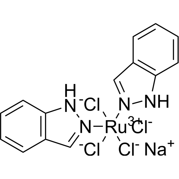
BOLD-100 (NKP-1339; IT-139) is the first-in-class ruthenium-based anticancer agent in development against solid cancer with limited side effects. BOLD-100 induces G2/M cell cycle arrest, blockage of DNA synthesis, and induction of apoptosis via the mitochondrial pathway. BOLD-100 has a high tumor targeting potential, strongly binds to serum proteins such as albumin and transferrin and activates in the reductive tumor milieu.
연구목적의 판매만을 진행합니다. 환자를 대상으로 한 판매는 하지 않습니다.
- Purity : 95.20%
- CAS No.: 197723-00-5
- 화학식: C14H12Cl4N4NaRu
- 분자량:502.14
-
보관:
4°C, sealed storage, away from moisture
* In solvent : -80°C, 6 months; -20°C, 1 month (sealed storage, away from moisture)

To place orders, for customer services and technical support, please contact: MedChemExpress USA
Tel: 609-228-6898 E-mail: [email protected] [email protected]


-
 Biological Activity
-
Chemical Information
-
용액&용해도
- Protocol
- 순도&문서
- References
-
 Help & FAQs


-
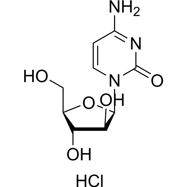
HY-13605A
Cytarabine hydrochlorideDNA Synthesis Inhibitor -
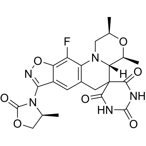
HY-17647
ZoliflodacinDNA gyrase/topoisomerase Inhibitor -
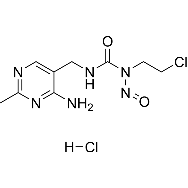
HY-13703A
Nimustine hydrochlorideApoptosis Inducer -
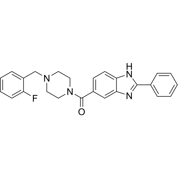
HY-126020
BractoppinRAD51 Inhibitor -

HY-17647R
Zoliflodacin (Standard)DNA gyrase/topoisomerase Inhibitor -

HY-13605AR
Cytarabine hydrochloride (Standard)DNA Synthesis Inhibitor
Publications Citing Use of MedChemExpress (MCE) BOLD-100
MoreAll DNA/RNA Synthesis Isoforms
More

Biological Activity
제품 설명
In Vitro
BOLD-100 (0-200 μM; 72 hours) has the anticancer activity against malignant cell lines of diverse origin, exhibits IC50 values of 45-200 μM for KP1339 mono-therapy. It against Hepatoma cell line, Hep3B, HepG2, PLC/PRF/5 and HCC2 cells with the Mean IC50 value of 186.3 μM, 165.4 μM, 124.4 μM, and 69.4 μM, respectively. It against Melanoma cell line, VM-1, VM-21,VM-48 with IC50 values of 178 μM, 111 μM, and 143 μM, respectively. It against Lung cancer and Colon cancer cell lines, inhibits A549, VL-8, SW480 and HCT116 cells, respectively[2].BOLD-100 (0-150 μM; 24 hours) induces cell apoptosis alone. When it combines with sorafenib, it increases the numbers of the apopotic cells. Additionally, the p-PARP and caspase 7 cleavage is promoted either[2].BOLD-100 (0-150 μM; 24 hours) can promote phosphorylation of STAT3 and CREB expression, however, the decreasation is inhibited by sorafenib cotreatment[2].
MedChemExpress (MCE) has not independently confirmed the accuracy of these methods. They are for reference only.
-
Cell Line:Hepatoma, Melanoma, Lung cancer and Colon cancer cell lines
-
Concentration:0 μM, 50 μM, 100 μM, 150 μM and 200 μM
-
Incubation Time:72 hours
-
Result:Has anti-cancer activity in diverse malignant tumour cell types.
-
Cell Line:Hep3B cells
-
Concentration:0 μM, 75 μM, 150 μM
-
Incubation Time:72 hours
-
Result:Promoted cell apoptosis as a concentration manner.
-
Cell Line:Hep3B cells
-
Concentration:0 μM, 75 μM, 150 μM
-
Incubation Time:72 hours
-
Result:Increased p-STAT3 and p-CREB expression in cells without sorafenib cotreatment.
In Vivo
MedChemExpress (MCE) has not independently confirmed the accuracy of these methods. They are for reference only.
-
Animal Model:Hep3B xenograft in Balb/c mice[2]
-
Dosage:30 mg/kg
-
Administration:Intravenous injection
-
Result:Had synergistic activity of KP1339 with sorafenib in vivo.
- EdU Incorporation Assay (Click Chemistry-Based DNA Synthesis Measurement)
- Detection of Apoptosis
- TUNEL staining for apoptotic DNA fragmentation
- Flow cytometric DNA-content cell-cycle staining
- Annexin V plus membrane-impermeant dye apoptosis staining
- BrdU Incorporation Assay
- Apoptosis Solutions
- Protocol for Cell Cycle


Chemical Information
-
CAS No. 197723-00-5
-
Appearance Solid
-
분자량 502.14
-
화학식 C14H12Cl4N4NaRu
-
Color Brown to dark brown
-
SMILES
[Cl-][Ru+3]([Cl-])([Cl-])([N]1=CC2=C(N1)C=CC=C2)([N]3=CC4=C(N3)C=CC=C4)[Cl-].[Na+]
-
Synonyms
NKP-1339; IT-139; KP-1339
-
선적
Room temperature in continental US; may vary elsewhere.
-
보관
4°C, sealed storage, away from moisture
* In solvent : -80°C, 6 months; -20°C, 1 month (sealed storage, away from moisture)


Publications (1)
-
Journal Impact Factor
-
Most Recent


용액&용해도
In Vitro:
DMSO : ≥ 59 mg/mL (117.50 mM; Hygroscopic DMSO has a significant impact on the solubility of product, please use newly opened DMSO)
* "≥" means soluble, but saturation unknown.
Please refer to the solubility information to select the appropriate solvent. Once prepared, please aliquot and store the solution to prevent product inactivation from repeated freeze-thaw cycles.
Storage method and period of stock solution: -80°C, 6 months; -20°C, 1 month (sealed storage, away from moisture). When stored at -80°C, please use it within 6 months. When stored at -20°C, please use it within 1 month.
Please refer to the solubility information to select the appropriate solvent. Once prepared, please aliquot and store the solution to prevent product inactivation from repeated freeze-thaw cycles.
Storage method and period of stock solution: -80°C, 6 months; -20°C, 1 month (sealed storage, away from moisture). When stored at -80°C, please use it within 6 months. When stored at -20°C, please use it within 1 month.
Concentration (start) × Volume (start) = Concentration (final) × Volume (final)


순도&문서
-
Data Sheet (285 KB)
-
SDS (393 KB)
- English - EN (393 KB)
- Français - FR (393 KB)
- Deutsch - DE (393 KB)
- Norwegian - NO (393 KB)
- Español - ES (393 KB)
- Swedish - SV (393 KB)
- Italian - IT (393 KB)
- Korean - KR (393 KB)
- Portuguese - PT (393 KB)
-
Handling Instructions (2659 KB)
References


Complete Stock Solution Preparation Table
Please refer to the solubility information to select the appropriate solvent. Once prepared, please aliquot and store the solution to prevent product inactivation from repeated freeze-thaw cycles.
Storage method and period of stock solution: -80°C, 6 months; -20°C, 1 month (sealed storage, away from moisture). When stored at -80°C, please use it within 6 months. When stored at -20°C, please use it within 1 month.
| Optional Solvent | Concentration Solvent Mass | 1 mg | 5 mg | 10 mg | 25 mg |
|---|---|---|---|---|---|
| DMSO | 1 mM | 1.9915 mL | 9.9574 mL | 19.9148 mL | 49.7869 mL |
| 5 mM | 0.3983 mL | 1.9915 mL | 3.9830 mL | 9.9574 mL | |
| 10 mM | 0.1991 mL | 0.9957 mL | 1.9915 mL | 4.9787 mL | |
| 15 mM | 0.1328 mL | 0.6638 mL | 1.3277 mL | 3.3191 mL | |
| 20 mM | 0.0996 mL | 0.4979 mL | 0.9957 mL | 2.4893 mL | |
| 25 mM | 0.0797 mL | 0.3983 mL | 0.7966 mL | 1.9915 mL | |
| 30 mM | 0.0664 mL | 0.3319 mL | 0.6638 mL | 1.6596 mL | |
| 40 mM | 0.0498 mL | 0.2489 mL | 0.4979 mL | 1.2447 mL | |
| 50 mM | 0.0398 mL | 0.1991 mL | 0.3983 mL | 0.9957 mL | |
| 60 mM | 0.0332 mL | 0.1660 mL | 0.3319 mL | 0.8298 mL | |
| 80 mM | 0.0249 mL | 0.1245 mL | 0.2489 mL | 0.6223 mL | |
| 100 mM | 0.0199 mL | 0.0996 mL | 0.1991 mL | 0.4979 mL |


BOLD-100 Related Classifications
HY-16350 Related Classifications
 Powered by Bioz