



Rifamycin S
Based on 2 publication(s) in Google Scholar
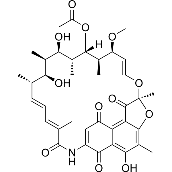
Rifamycin S, a quinone, is an antibiotic against Gram-positive bacteria (including MRSA). Rifamycin S is the oxidized forms of a reversible oxidation-reduction system involving two electrons. Rifamycin S generates reactive oxygen species (ROS) and inhibits microsomal lipid peroxidation. Rifamycin S can be used for tuberculosis and leprosy.
연구목적의 판매만을 진행합니다. 환자를 대상으로 한 판매는 하지 않습니다.
- Purity : 98.80%
- CAS No.: 13553-79-2
- 화학식: C37H45NO12
- 분자량:695.75
-
보관:Powder -20°C, 3 years , 4°C, 2 years ; In solvent -80°C, 2 years , -20°C, 1 year

To place orders, for customer services and technical support, please contact: MedChemExpress USA
Tel: 609-228-6898 E-mail: [email protected] [email protected]


-
 Biological Activity
-
Chemical Information
-
용액&용해도
- Protocol
- 순도&문서
- References
-
 Help & FAQs



Publications Citing Use of MedChemExpress (MCE) Rifamycin S
MoreAll Antibiotic Isoforms
More

Biological Activity
제품 설명
IC50 & Target
In Vitro
The inhibition of bacterial growth by Rifamycin SV is due to the production of active species of oxygen resulting from the oxidation-reduction cycle of Rifamycin SV in the cells. The aerobic oxidation of Rifamycin SV to Rifamycin S is induced by metal ions, such as Mn2+, Cu2+, and Co2+. The most effective metal ion is Mn2+[2].
MedChemExpress (MCE) has not independently confirmed the accuracy of these methods. They are for reference only. Further protocols information, click here.
In Vivo
MedChemExpress (MCE) has not independently confirmed the accuracy of these methods. They are for reference only.
Clinical Trial
| NCT Number | Sponsor | Condition | Start Date |
Phase

|
|---|---|---|---|---|
| NCT01329991 | Plexxikon| | 2011-05 | PHASE1 |


Chemical Information
-
CAS No. 13553-79-2
-
Appearance Solid
-
분자량 695.75
-
화학식 C37H45NO12
-
Color Light yellow to orange
-
SMILES
C[C@H]([C@H](/C=C/O[C@@]1(C)OC(C(C)=C(O)C(C(C(N2)=C3)=O)=C4C3=O)=C4C1=O)OC)[C@H]([C@H](C)[C@H](O)[C@H](C)[C@@H](O)[C@@H](C)/C=C/C=C(C)\C2=O)OC(C)=O
-
Structure Classification
-
Initial Source
Curvularia lunata
-
선적
Room temperature in continental US; may vary elsewhere.
-
보관
Powder -20°C 3 years 4°C 2 years In solvent -80°C 2 years -20°C 1 year


Publications (2)
-
Journal Impact Factor
-
Most Recent
-
Adv Sci (Weinh)
2026 Mar;13(16):e13310. PMID: 41589654 -
Proc Natl Acad Sci U S A
Structure-guided approach to modulate small molecule binding to a promiscuous ligand-activated protein. [Abstract]2023 Mar 7;120(10):e2217804120. PMID: 36848571


용액&용해도
In Vitro:
DMSO : 100 mg/mL (143.73 mM; Need ultrasonic; Hygroscopic DMSO has a significant impact on the solubility of product, please use newly opened DMSO)
Please refer to the solubility information to select the appropriate solvent. Once prepared, please aliquot and store the solution to prevent product inactivation from repeated freeze-thaw cycles.
Storage method and period of stock solution: -80°C, 2 years; -20°C, 1 year. When stored at -80°C, please use it within 2 years. When stored at -20°C, please use it within 1 year.
Please refer to the solubility information to select the appropriate solvent. Once prepared, please aliquot and store the solution to prevent product inactivation from repeated freeze-thaw cycles.
Storage method and period of stock solution: -80°C, 2 years; -20°C, 1 year. When stored at -80°C, please use it within 2 years. When stored at -20°C, please use it within 1 year.
Concentration (start) × Volume (start) = Concentration (final) × Volume (final)
In Vivo:
Select the appropriate dissolution method based on your experimental animal and administration route.
- For the following dissolution methods, please ensure to first prepare a clear stock solution using an In Vitro approach and then sequentially add co-solvents:
- To ensure reliable experimental results, the clarified stock solution can be appropriately stored based on storage conditions. As for the working solution for In Vivo experiments, it is recommended to prepare freshly and use it on the same day.
- The percentages shown for the solvents indicate their volumetric ratio in the final prepared solution. If precipitation or phase separation occurs during preparation, heat and/or sonication can be used to aid dissolution.
Add each solvent one by one: 10% DMSO 40% PEG300 5% Tween-80 45% Saline
Solubility: 2.5 mg/mL (3.59 mM); Suspended solution; Need ultrasonic
This protocol yields a suspended solution of 2.5 mg/mL. Suspended solution can be used for oral and intraperitoneal injection.
Taking 1 mL working solution as an example, add 100 μL DMSO stock solution (25.0 mg/mL) to 400 μL PEG300, and mix evenly; then add 50 μL Tween-80 and mix evenly; then add 450 μL Saline to adjust the volume to 1 mL.
Preparation of Saline: Dissolve 0.9 g sodium chloride in ddH₂O and dilute to 100 mL to obtain a clear Saline solution.
In Vivo Dissolution Calculator
Please enter the basic information of animal experiments:
-
-
-
-
Recommended: Prepare an additional quantity of animals to account for potential losses during experiments.
Please enter your animal formula composition:
-
%DMSO +
Recommended: Keep the proportion of DMSO in working solution below 2% if your animal is weak.
-
%+
-
+%Tween-80 + +
-
%Saline +
The co-solvents required include: DMSO, . All of co-solvents are available by MedChemExpress (MCE). , Tween 80. All of co-solvents are available by MedChemExpress (MCE).
Working solution concentration: 0.22 mg/mL
Method for preparing stock solution: mg drug dissolved in μL DMSO. Stock solution concentration: mg/mL.
1. Take μL DMSO stock solution;
2. Add μL .
μL , mix evenly;
3. Then add μL Tween 80, mix evenly;
4. Then add μL
Please ensure that the stock solution in the first step is dissolved to a clear state, and add co-solvents in sequence. You can use ultrasonic heating (ultrasonic cleaner, recommended frequency 20-40 kHz), vortexing, etc. to assist dissolution.

Protocol
-
ROS/oxidative-stress fluorescent staining
ROS/oxidative-stress fluorescent staining uses cell-permeant fluorogenic probes that become fluorescent after oxidation inside cells or tissues; commonly used examples include DCFH-DA/DCFDA for broad cellular oxidant detection, DHE for superoxide-related signal detection, MitoSOX for mitochondrial superoxide-related signal detection, and CellROX probes for oxidative-stress-associated fluorescence readouts. The assay detects probe oxidation rather than a single ROS species unless the probe and analysis method have been chemically validated for that species. DCFH-DA enters cells, is deacetylated by intracellular esterases to DCFH, and produces fluorescent DCF after oxidation, so the readout is used as an operational measure of total cellular oxidative stress rather than a species-specific ROS measurement. DHE and MitoSOX can report superoxide-related oxidation, but red fluorescence alone can include non-specific ethidium-like oxidation products; HPLC or optimized spectral approaches are
-
Gram Staining of Tissue Sections
Gram staining of tissue sections is a histochemical technique used to differentiate Gram-positive and Gram-negative bacteria within histological specimens based on differences in bacterial cell wall structure and dye retention, adapted from classical bacteriological Gram staining into tissue-compatible “histological Gram stain” variants. In tissue applications, modifications of the Brown-Hopps and Brown-Brenn methods are commonly used to improve differentiation of microorganisms embedded within host connective tissue and to reduce overstaining or loss of Gram-negative signal, which are known limitations of earlier approaches. The principle relies on crystal violet-iodine complex retention in Gram-positive organisms and subsequent decolorization and counterstaining steps that allow contrast visualization of Gram-negative organisms against tissue background.
-
Ferroptosis Solutions
Ferroptosis is an iron-dependent, non-apoptotic form of regulated cell death characterized by lethal lipid peroxidation and sensitivity to suppression by iron chelators or lipophilic radical-trapping antioxidants. The core pathway links cystine uptake through system Xc−, glutathione availability, GPX4-dependent detoxification of phospholipid hydroperoxides, iron-dependent oxidative reactions, and polyunsaturated-phospholipid metabolism into a cell-death program that is biochemically and morphologically distinct from apoptosis, necrosis, and autophagy. The ferroptosis pathway is experimentally linked to phenotype through chemical and genetic perturbation. Erastin induces ferroptosis by inhibiting cystine uptake through system Xc− and weakening antioxidant defenses, while GPX4 inhibition or depletion causes lipid peroxide accumulation and ferroptotic cancer-cell death. ACSL4 and oxidizable arachidonoyl- or adrenoyl-containing phosphatidylethanolamines shape ferroptosis sensitivity by con


순도&문서
-
Data Sheet (282 KB)
-
SDS (623 KB)
- English - EN (623 KB)
- Français - FR (623 KB)
- Deutsch - DE (623 KB)
- Norwegian - NO (623 KB)
- Español - ES (623 KB)
- Swedish - SV (623 KB)
- Italian - IT (623 KB)
- Korean - KR (623 KB)
- Portuguese - PT (623 KB)
-
Handling Instructions (2659 KB)
References
[1]. Rao DN, et al. A comparative study of the redox-cycling of a quinone (rifamycin S) and a quinonimine (rifabutin) antibiotic by rat liver microsomes. Free Radic Biol Med. 1997;22(3):439-46. [Content Brief]
[2]. Kono Y. Oxygen Enhancement of bactericidal activity of rifamycin SV on Escherichia coli and aerobic oxidation of rifamycin SV to rifamycin S catalyzed by manganous ions: the role of superoxide. J Biochem. 1982 Jan;91(1):381-95. [Content Brief]
[3]. Huang H, et al. Rifamycin S and its geometric isomer produced by a newly found actinomycete, Micromonospora rifamycinica. Antonie Van Leeuwenhoek. 2009 Feb;95(2):143-8. [Content Brief]


Complete Stock Solution Preparation Table
Please refer to the solubility information to select the appropriate solvent. Once prepared, please aliquot and store the solution to prevent product inactivation from repeated freeze-thaw cycles.
Storage method and period of stock solution: -80°C, 2 years; -20°C, 1 year. When stored at -80°C, please use it within 2 years. When stored at -20°C, please use it within 1 year.
| Optional Solvent | Concentration Solvent Mass | 1 mg | 5 mg | 10 mg | 25 mg |
|---|---|---|---|---|---|
| DMSO | 1 mM | 1.4373 mL | 7.1865 mL | 14.3730 mL | 35.9324 mL |
| 5 mM | 0.2875 mL | 1.4373 mL | 2.8746 mL | 7.1865 mL | |
| 10 mM | 0.1437 mL | 0.7186 mL | 1.4373 mL | 3.5932 mL | |
| 15 mM | 0.0958 mL | 0.4791 mL | 0.9582 mL | 2.3955 mL | |
| 20 mM | 0.0719 mL | 0.3593 mL | 0.7186 mL | 1.7966 mL | |
| 25 mM | 0.0575 mL | 0.2875 mL | 0.5749 mL | 1.4373 mL | |
| 30 mM | 0.0479 mL | 0.2395 mL | 0.4791 mL | 1.1977 mL | |
| 40 mM | 0.0359 mL | 0.1797 mL | 0.3593 mL | 0.8983 mL | |
| 50 mM | 0.0287 mL | 0.1437 mL | 0.2875 mL | 0.7186 mL | |
| 60 mM | 0.0240 mL | 0.1198 mL | 0.2395 mL | 0.5989 mL | |
| 80 mM | 0.0180 mL | 0.0898 mL | 0.1797 mL | 0.4492 mL | |
| 100 mM | 0.0144 mL | 0.0719 mL | 0.1437 mL | 0.3593 mL |


Rifamycin S Related Classifications
HY-125365 Related Classifications
 Powered by Bioz