



ZIKV-IN-6
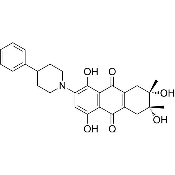
ZIKV-IN-6 (compound 22) is an inhibitor of Zika virus (ZIKV), with low cytotoxicity (CC50>50 μM). ZIKV-IN-6 binds to ZIKV RdRp directly and inhibits viral RNA synthesis by ZIKV NS5. ZIKV-IN-6 suppresses the excessive inflammatory response and pyroptosis.
연구목적의 판매만을 진행합니다. 환자를 대상으로 한 판매는 하지 않습니다.
- 화학식: C27H29NO6
- 분자량:463.52
-
보관:
Please store the product under the recommended conditions in the Certificate of Analysis.

To place orders, for customer services and technical support, please contact: MedChemExpress USA
Tel: 609-228-6898 E-mail: [email protected] [email protected]


-
 Biological Activity
-
Chemical Information
- Protocol
- 순도&문서
- References
-
 Help & FAQs


All DNA/RNA Synthesis Isoforms
More

Biological Activity
제품 설명
IC50 & Target
Zika virus (ZIKV)[1]
Cellular Effect
|
Cell Line

|
Type | Value | Description | References |
|---|---|---|---|---|
| A549 | CC50 |
>50 μM
Compound: 22
|
Cytotoxicity against human A549 cells assessed as reduction in cell viability incubated for 48 hrs by MTT assay
Cytotoxicity against human A549 cells assessed as reduction in cell viability incubated for 48 hrs by MTT assay
|
[PMID: 37421888] |
| JEG-3 | CC50 |
54.7 μM
Compound: 22
|
Cytotoxicity against human JEG-3 cells assessed as reduction in cell viability incubated for 48 hrs by MTT assay
Cytotoxicity against human JEG-3 cells assessed as reduction in cell viability incubated for 48 hrs by MTT assay
|
[PMID: 37421888] |
| SNB-19 | CC50 |
66.1 μM
Compound: 22
|
Cytotoxicity against human SNB-19 cells assessed as reduction in cell viability incubated for 48 hrs by MTT assay
Cytotoxicity against human SNB-19 cells assessed as reduction in cell viability incubated for 48 hrs by MTT assay
|
[PMID: 37421888] |
| Vero | CC50 |
65.4 μM
Compound: 22
|
Cytotoxicity against african green monkey Vero cells assessed as reduction in cell viability incubated for 48 hrs by MTT assay
Cytotoxicity against african green monkey Vero cells assessed as reduction in cell viability incubated for 48 hrs by MTT assay
|
[PMID: 37421888] |


Chemical Information
-
분자량 463.52
-
화학식 C27H29NO6
-
SMILES
O=C1C2=C(C(O)=C(N3CCC(C4=CC=CC=C4)CC3)C=C2O)C(C5=C1C[C@@](C)(O)[C@@](C)(O)C5)=O
-
선적
Room temperature in continental US; may vary elsewhere.
-
보관
Please store the product under the recommended conditions in the Certificate of Analysis.

Protocol
-
Cell Cytotoxicity Assay
Cytotoxicity assays are usually based on the assessment of cell membrane damage, which can also be indirectly detected by measuring cell viability. Detection methods include MTT assay, CKK-8 assay, LDH assay and ATP assay, etc.
-
Mammalian live/dead viability and cytotoxicity staining
Live/dead viability and cytotoxicity staining assays are based on the simultaneous detection of intracellular esterase activity in metabolically active (viable) cells and membrane integrity loss in non-viable cells. In commonly used dual-staining approaches, membrane-permeant fluorogenic substrates are converted by intracellular esterases into fluorescent products in live cells, while impermeant DNA-binding dyes selectively enter cells with compromised plasma membranes and label nucleic acids in dead or dying cells, enabling discrimination between viable and non-viable populations by fluorescence microscopy or flow cytometry.
-
Research Protocol for Inflammation-related Diseases
The NLRP3 inflammasome is a cytosolic innate immune signaling platform that integrates priming signals and danger-signal activation to promote caspase-1 activation, maturation of IL-1β and IL-18, and gasdermin D-mediated pyroptotic cell death. The core experimental logic is to determine whether inflammatory disease phenotypes are driven by increased NLRP3 expression, ASC-containing inflammasome assembly, caspase-1 cleavage, GSDMD cleavage, and extracellular release of IL-1β/IL-18 rather than by nonspecific cell injury alone. The pathway is strongly linked to inflammation-related disease phenotypes because monosodium urate crystals activate NALP3/NLRP3 inflammasome signaling in gout-like crystal inflammation, cholesterol crystals activate NLRP3 inflammasomes in atherogenesis models, and DSS-induced intestinal inflammation has been reported to involve NLRP3 inflammasome activity. However, experimental colitis studies also show context-dependent protective effects of NLRP3 inflammasome co
-
Cell Viability Determination by MTT Colorimetric Assay
The following protocol uses the MTT colorimetric assay as a classic literature-established method for assessing cell viability/metabolic activity in cultured mammalian cells. MTT[3-(4,5-dimethylthiazol-2-yl)-2,5-diphenyltetrazolium bromide] is reduced by metabolically active cells to a colored formazan product; the amount of formazan is quantified spectrophotometrically and provides an indirect measure of metabolically active viable cells. Importantly, MTT reduction reflects cellular oxidoreductase/metabolic activity rather than an absolute direct count of living cells, so changes in cellular metabolism can alter the signal independently of cell number.
-
Pyroptosis Solutions
Pyroptosis is a lytic inflammatory cell-death pathway executed by gasdermin pores, most classically through inflammasome-mediated activation of caspase-1, cleavage of gasdermin D, membrane pore formation, LDH release, and secretion of IL-1β and IL-18. The canonical pathway is commonly modeled by priming cells with an inflammatory signal such as LPS to induce pro-IL-1β and inflammasome components, followed by an activation signal such as ATP or nigericin to activate NLRP3, ASC speck formation, caspase-1 cleavage, GSDMD cleavage, cytokine release, and pyroptotic membrane rupture. The non-canonical pathway is triggered when cytosolic LPS activates mouse caspase-11 or human caspase-4/5, leading to GSDMD cleavage and pyroptosis, and this can secondarily activate NLRP3-dependent IL-1β release. Pyroptosis is linked to inflammatory injury, infection, cancer, liver disease, ocular disease, placental inflammation, and other disease phenotypes, but unresolved questions include which gasdermin fam


순도&문서
References

Calculators
Concentration (start) × Volume (start) = Concentration (final) × Volume (final)


ZIKV-IN-6 Related Classifications
HY-155053 Related Classifications
 Powered by Bioz