



LysoTracker Red (solution)

Based on 1 Customer Validation
LysoTracker Red (solution) is a Red fluorescently labeled lysosomal probe with a maximum excitation/emission wavelength of 577/590 nm. The structure is composed of a fluorescein group and linked weak bases, which can freely cross the cell membrane and gather on spherical organelles. It is suitable for observing the internal biosynthesis and related pathogenesis of lysosomes.
Solvent and concentration: DMSO: 1 mM
For research use only. We do not sell to patients.
- CAS No.: 231946-72-8
- Formula: C20H24BF2N5O
- Molecular Weight:399.25
-
Storage:
Please store the product under the recommended conditions in the Certificate of Analysis.


To place orders, for customer services and technical support, please contact: MedChemExpress USA
Tel: 609-228-6898 E-mail: [email protected] [email protected]


-
 Biological Activity
-
Chemical Information
- Protocols
- Purity & Documentation
- References
-
 Help & FAQs


-

HY-DY1032
MitoTracker Green FM (solution)Mitochondrial Dye -
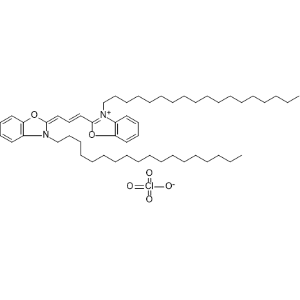
HY-D0969
DiOFluorescent Probe -
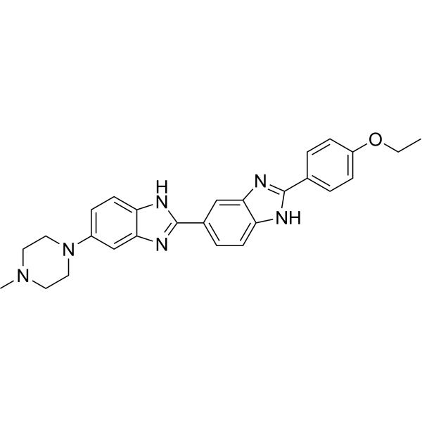
HY-15559
Hoechst 33342DNA Fluorescent Stain -
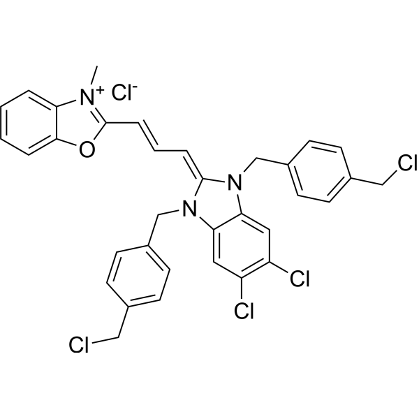
HY-135056
MitoTracker Green FMMitochondrial Dye -
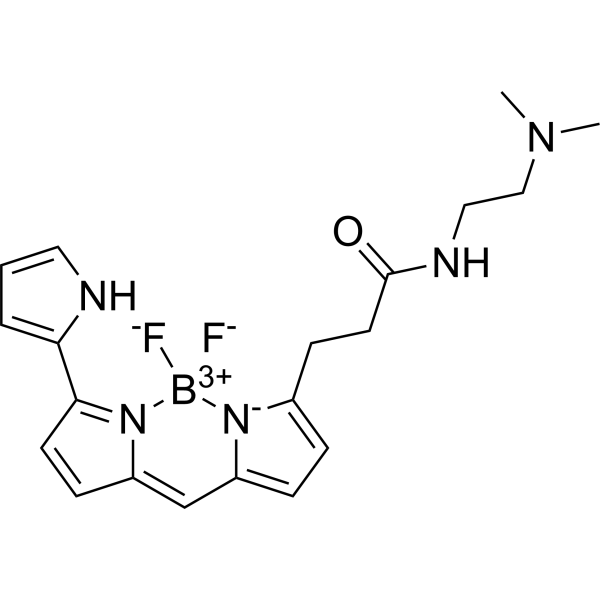
HY-D1300
LysoTracker RedCell Death Indicator -
HY-D1783
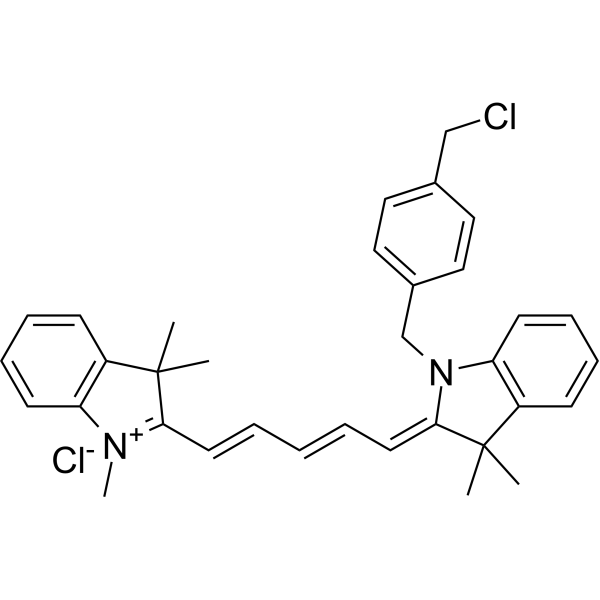
MitoTracker Deep Red FMRed Fluorescent Dye -
HY-15534
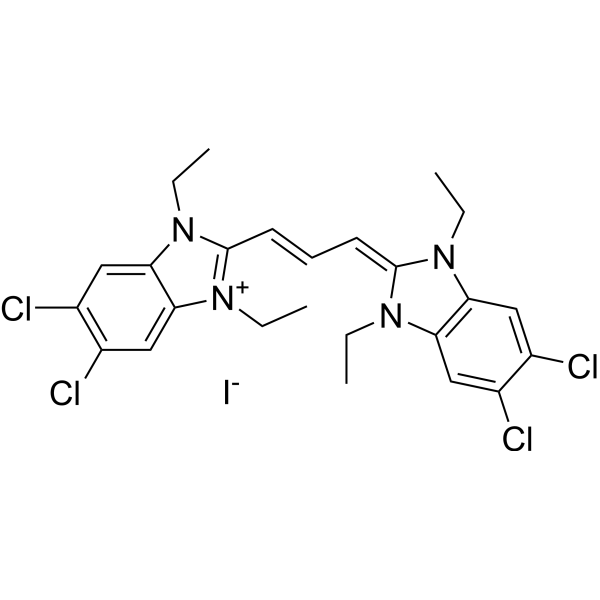
JC-1Mitochondrial Membrane Potential Probe -

HY-10219
RapamycinmTOR Inhibitor -

HY-Y0320
Dimethyl sulfoxide, meets analytical specification of Ch.P.Aprotic Solvent -

HY-100558
Bafilomycin A1V-ATPase Inhibitor -
HY-100941
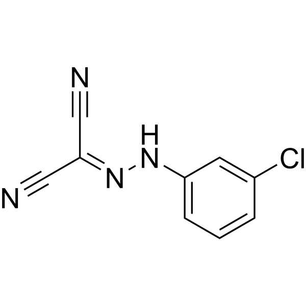
CCCPOXPHOS Uncoupler -
HY-100116A
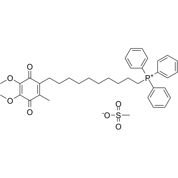
Mitoquinone mesylateAntioxidant


Biological Activity
Description
Solvent and concentration: DMSO: 1 mM
In Vitro
Guide (The following is our recommended protocol. This protocol is only a guide and should be modified according to your specific needs).
Preparation of LysoTracker Red working solution
1. Working solution preparation:
1.1 Restore the solution to room temperature and concentrate it at the bottom of the tube by instantaneous centrifugation.
1.2 Dilute 1 mM storage solution to the working solution using a medium or appropriate buffer (such as PBS) . The recommended working solution is 50-100 nM;
Note: Please adjust the concentration of the LysoTracker Red working solution according to the actual situation, and use it now.
2. Cell staining
2.1 For suspension cells: Centrifuge at 1000 g at 4°C for 3-5 minutes and then discard the supernatant. Wash twice with PBS, 5 minutes each time.
For adherent cells: Discard the cell culture medium, and add trypsin to dissociate cells to make a single-cell suspension. Centrifuge at 1000 g at 4°C for 3-5 minutes and then discard the supernatant. Wash twice with PBS, 5 minutes each time.
2.2 Add 1 mL of LysoTracker Red working solution, and then incubate at room temperature for 5-30 minutes.
2.3 Centrifuge at 400 g at 4°C for 3-4 minutes and then discard the supernatant.
2.4 Wash twice with PBS, 5 minutes each time.
2.5 Resuspend cells with serum-free cell culture medium or PBS, and then detect by fluorescence microscope or flow cytometer.
MedChemExpress (MCE) has not independently confirmed the accuracy of these methods. They are for reference only.


Chemical Information
-
CAS No. 231946-72-8
-
Appearance Liquid
-
Molecular Weight 399.25
-
Formula C20H24BF2N5O
-
Color Purple to purplish red
-
SMILES
O=C(CCC1=CC=C2C=C3C=CC(C4=CC=CN4)=[N]3[B+3]([F-])([N-]21)[F-])NCCN(C)C
-
Shipping
Room temperature in continental US; may vary elsewhere.
-
Storage
Please store the product under the recommended conditions in the Certificate of Analysis.


Purity & Documentation
-
Data Sheet (275 KB)
-
SDS (251 KB)
- English - EN (251 KB)
- Français - FR (251 KB)
- Deutsch - DE (251 KB)
- Norwegian - NO (251 KB)
- Español - ES (251 KB)
- Swedish - SV (251 KB)
- Italian - IT (251 KB)
- Korean - KR (251 KB)
- Portuguese - PT (251 KB)
-
Handling Instructions (2659 KB)
References
[1]. Price OT, et al. Quantitative fluorescence of 5-FU-treated fetal rat limbs using confocal laser scanning microscopy and Lysotracker Red. Cytometry A. 2003;53(1):9-21. [Content Brief]
[2]. Pereira LC, et al. An autophagic process is activated in HepG2 cells to mediate BDE-100-induced toxicity. Toxicology. 2017;376:59-65. [Content Brief]
[3]. Nagahama M, et al. Cellular vacuolation induced by Clostridium perfringens epsilon-toxin. FEBS J. 2011;278(18):3395-3407. [Content Brief]

Calculators
Concentration (start) × Volume (start) = Concentration (final) × Volume (final)


LysoTracker Red (solution) Related Classifications
HY-DY1040 Related Classifications
 Powered by Bioz