



MC0704

Based on 1 Customer Validation
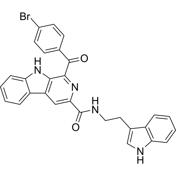
MC0704 is a STAT3 inhibitor with an IC50 value of 2.13 μM. MC0704 induces cell apoptosis and cell cycle arrest. MC0704 shows antitumor activity in mouse breast cancer models. MC0704 can be used for the research of metastatic triple-negative breast cancer (mTNBC).
Nur für Forschungszwecke. Wir verkaufen nicht an Patienten.
- Reinheit : 98.34%
- CAS. Nr.: 3034176-57-0
- Formel: C29H21BrN4O2
- Molecular Weight:537.41
-
Speicherung:Powder -20°C, 3 years ; In solvent -80°C, 6 months , -20°C, 1 month

To place orders, for customer services and technical support, please contact: MedChemExpress USA
Tel: 609-228-6898 E-mail: [email protected] [email protected]


-
 Biologische Aktivität
-
Chemical Information
-
Lösungsmittel & Löslichkeit
- Protokoll
- Reinheit & Dokumentation
- Verweise
-
 Help & FAQs




Biologische Aktivität
Beschreibung
Cellular Effect
|
Cell Line

|
Type | Value | Description | References |
|---|---|---|---|---|
| MDA-MB-231 | IC50 |
2.96 μM
Compound: MC0704
|
Antiproliferative activity against docetaxel-resistant human MDA-MB-231-DTR cells assessed as reduction in cell viability incubated for 72 hrs by sulforhodamine B assay
Antiproliferative activity against docetaxel-resistant human MDA-MB-231-DTR cells assessed as reduction in cell viability incubated for 72 hrs by sulforhodamine B assay
|
[PMID: 36786551] |
| MDA-MB-231 | IC50 |
2.98 μM
Compound: MC0704
|
Antiproliferative activity against human MDA-MB-231 cells assessed as reduction in cell viability incubated for 72 hrs by MTT assay
Antiproliferative activity against human MDA-MB-231 cells assessed as reduction in cell viability incubated for 72 hrs by MTT assay
|
[PMID: 36786551] |
| MDA-MB-231 | IC50 |
3.11 μM
Compound: MC0704
|
Antiproliferative activity against human MDA-MB-231 cells assessed as reduction in cell viability incubated for 72 hrs by sulforhodamine B assay
Antiproliferative activity against human MDA-MB-231 cells assessed as reduction in cell viability incubated for 72 hrs by sulforhodamine B assay
|
[PMID: 36786551] |
| MDA-MB-231 | IC50 |
3.24 μM
Compound: MC0704
|
Antiproliferative activity against docetaxel-resistant human MDA-MB-231-DTR cells assessed as reduction in cell viability incubated for 72 hrs by MTT assay
Antiproliferative activity against docetaxel-resistant human MDA-MB-231-DTR cells assessed as reduction in cell viability incubated for 72 hrs by MTT assay
|
[PMID: 36786551] |
| MRC5 | IC50 |
33.25 μM
Compound: MC0704
|
Cytotoxicity against human MRC5 cells assessed as reduction in cell viability incubated for 72 hrs by sulforhodamine B assay
Cytotoxicity against human MRC5 cells assessed as reduction in cell viability incubated for 72 hrs by sulforhodamine B assay
|
[PMID: 36786551] |
In Vitro
MC0704 (0-50 μM; 72 h) shows cytotoxicity to HEK-293, lung epithelial normal (MRC-5), MDA-MB 231, MDA-MB-231 paclitaxel-resistant (MDA-MB-231-PTR) and MDA-MB-231 docetaxel-resistant (MDA-MB-231-DTR) cells with IC50 values of 3.11, 33.25, 2.98, 3.24 and 2.87 μM, respectively[1]. MC0704 (0, 0.4, 2, 10 and 50 μM; 24 h) inhibits activity of STAT3 with an IC50 value of 2.13 μM[1]. MC0704 (5 μM; 24 h) suppresses the activation of STAT3[1]. MC0704 (0, 2.5, 5 and 10 μM; 36-48 h) increases the cell cycle arrest at the G2/M phase and induces apoptosis in MDA-MB-231-DTR cells[1].
MedChemExpress (MCE) has not independently confirmed the accuracy of these methods. They are for reference only.
-
Cell Line:HEK-293 and MRC-5 cell lines
-
Concentration:0-50 μM
-
Incubation Time:72 hours
-
Result:Showed cytotoxicity to HEK-293 and MRC-5 cells.
-
Cell Line:MDA-MB-231 TNBC cell line
-
Concentration:5 μM
-
Incubation Time:24 hour
-
Result:Inhibited the expression of p-STAT3.
In Vivo
MedChemExpress (MCE) has not independently confirmed the accuracy of these methods. They are for reference only.
-
Animal Model:Female BALB/c-nu mice with MDA-MB-231-DTR cells transplant[1]
-
Dosage:10 mg/kg
-
Administration:Intraperitoneal injection; 10 mg/kg, once daily for 12 days
-
Result:Significantly inhibited tumor growth without overt toxicity, and reversed the mesenchymal marker vimentin and epithelial marker E-cadherin expression changes.
- Detection of Apoptosis
- TUNEL staining for apoptotic DNA fragmentation
- Flow cytometric DNA-content cell-cycle staining
- Annexin V plus membrane-impermeant dye apoptosis staining
- BrdU Incorporation Assay
- Apoptosis Solutions
- Protocol for Cell Cycle
- Subcutaneous Cell-Line-Derived Xenograft
- Breast Cancer Modeling


Chemical Information
-
CAS. Nr. 3034176-57-0
-
Appearance Solid
-
Molecular Weight 537.41
-
Formel C29H21BrN4O2
-
Color Light yellow to yellow
-
SMILES
O=C(C1=NC(C(NCCC2=CNC3=C2C=CC=C3)=O)=CC4=C1NC5=CC=CC=C54)C6=CC=C(C=C6)Br
-
Versand
Room temperature in continental US; may vary elsewhere.
-
Speicherung
Powder -20°C 3 years In solvent -80°C 6 months -20°C 1 month


Lösungsmittel & Löslichkeit
In Vitro:
DMSO : 25 mg/mL (46.52 mM; Need ultrasonic; Hygroscopic DMSO has a significant impact on the solubility of product, please use newly opened DMSO)
Please refer to the solubility information to select the appropriate solvent. Once prepared, please aliquot and store the solution to prevent product inactivation from repeated freeze-thaw cycles.
Storage method and period of stock solution: -80°C, 6 months; -20°C, 1 month. When stored at -80°C, please use it within 6 months. When stored at -20°C, please use it within 1 month.
Please refer to the solubility information to select the appropriate solvent. Once prepared, please aliquot and store the solution to prevent product inactivation from repeated freeze-thaw cycles.
Storage method and period of stock solution: -80°C, 6 months; -20°C, 1 month. When stored at -80°C, please use it within 6 months. When stored at -20°C, please use it within 1 month.
Konzentration (Stammlösung) × Volumen (Stammlösung) = Konzentration (Ziellösung) × Volumen (Ziellösung)


Reinheit & Dokumentation
-
Data Sheet (278 KB)
-
SDS (251 KB)
- English - EN (251 KB)
- Français - FR (251 KB)
- Deutsch - DE (251 KB)
- Norwegian - NO (251 KB)
- Español - ES (251 KB)
- Swedish - SV (251 KB)
- Italian - IT (251 KB)
- Korean - KR (251 KB)
- Portuguese - PT (251 KB)
-
Handling Instructions (2659 KB)
Verweise


Complete Stock Solution Preparation Table
Please refer to the solubility information to select the appropriate solvent. Once prepared, please aliquot and store the solution to prevent product inactivation from repeated freeze-thaw cycles.
Storage method and period of stock solution: -80°C, 6 months; -20°C, 1 month. When stored at -80°C, please use it within 6 months. When stored at -20°C, please use it within 1 month.
| Optional Solvent | Concentration Solvent Mass | 1 mg | 5 mg | 10 mg | 25 mg |
|---|---|---|---|---|---|
| DMSO | 1 mM | 1.8608 mL | 9.3039 mL | 18.6078 mL | 46.5194 mL |
| 5 mM | 0.3722 mL | 1.8608 mL | 3.7216 mL | 9.3039 mL | |
| 10 mM | 0.1861 mL | 0.9304 mL | 1.8608 mL | 4.6519 mL | |
| 15 mM | 0.1241 mL | 0.6203 mL | 1.2405 mL | 3.1013 mL | |
| 20 mM | 0.0930 mL | 0.4652 mL | 0.9304 mL | 2.3260 mL | |
| 25 mM | 0.0744 mL | 0.3722 mL | 0.7443 mL | 1.8608 mL | |
| 30 mM | 0.0620 mL | 0.3101 mL | 0.6203 mL | 1.5506 mL | |
| 40 mM | 0.0465 mL | 0.2326 mL | 0.4652 mL | 1.1630 mL |


MC0704 Related Classifications
HY-148923 Related Classifications
 Powered by Bioz