



Myxochelin A
Myxochelin A is a microbial metabolite that has been found in A. disciformis and has diverse biological activities. It is active against Gram-positive bacteria, including B. cereus, S. aureus, and M. luteus, but not Gram-negative bacteria or fungi in an agar diffusion assay when used at a concentration of 80 μg/disc. Myxochelin A inhibits 5-lipoxygenase (5-LO) activity with an IC50 value of 1.9 μM for the recombinant human enzyme. It is cytotoxic to 26-L5 colon cancer cells when used at a concentration of 3 μg/mL.
商品は「研究用試薬」です。人や動物の医療用・臨床診断用・食品用の製品ではありません。
研究用途以外に使用した場合、当社は一切の責任を負いかねます。
- CAS 番号: 120243-02-9
- 分子式: C20H24N2O7
- 分子量:404.41
-
保管条件:
Please store the product under the recommended conditions in the Certificate of Analysis.


To place orders, for customer services and technical support, please contact: MedChemExpress USA
Tel: 609-228-6898 E-mail: [email protected] [email protected]


-
 生物活性
生物活性
-
化学情報
- プロトコル
- 純度とドキュメンテーション
- 参考文献
-
 Help & FAQs


-
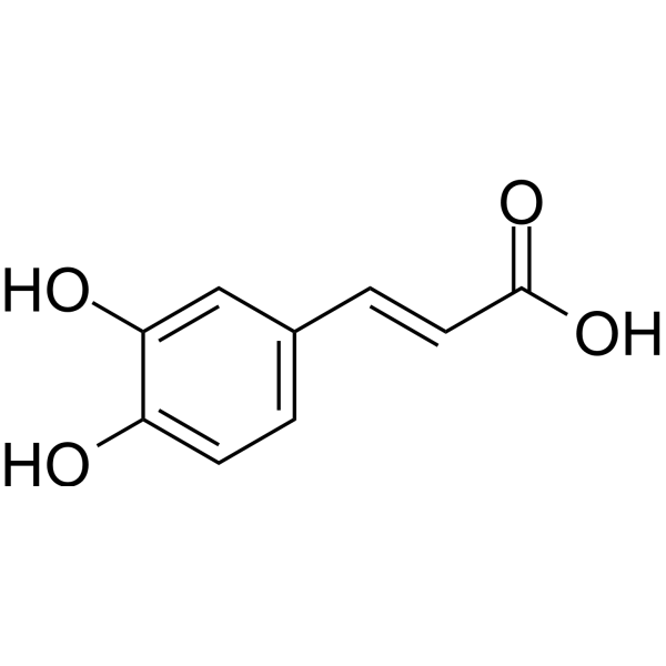
HY-N0172
Caffeic acidEndogenous Metabolite -

HY-N0172R
Caffeic acid (Standard)Endogenous Metabolite -
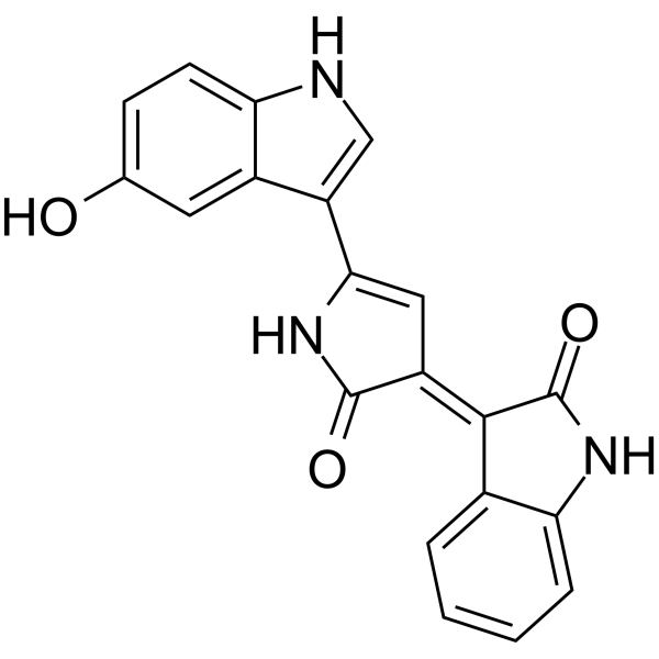
HY-119809
ViolaceinAnti-cancer Agent -
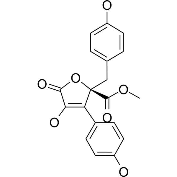
HY-111238
Butyrolactone IIFungal Metabolite -
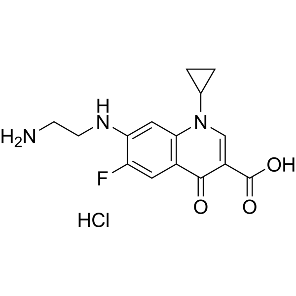
HY-122139
Desethylene Ciprofloxacin hydrochlorideDrug Metabolite -

HY-122139R
Desethylene Ciprofloxacin hydrochloride (Standard)Reference Standard -
HY-135468
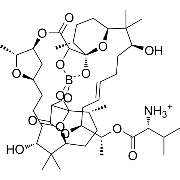
BoromycinAntibiotic


生物活性
製品説明


化学情報
-
CAS 番号 120243-02-9
-
分子量 404.41
-
分子式 C20H24N2O7
-
SMILES
OC[C@@H](NC(C1=CC=CC(O)=C1O)=O)CCCCNC(C2=CC=CC(O)=C2O)=O
-
別名
(S)-Myxochelin A
-
輸送条件
Room temperature in continental US; may vary elsewhere.
-
保管条件
Please store the product under the recommended conditions in the Certificate of Analysis.


純度とドキュメンテーション
参考文献

Calculators
濃度 (開始) × 体積 (開始) = 濃度 (終了) × 体積 (終了)


Myxochelin A 関連分類
HY-122980 関連分類
 Powered by Bioz