



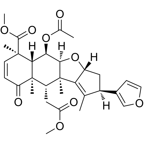
Nimbin

Based on 1 Customer Validation
Nimbin is an orally active intermediate limonoid found in Azadirachta. Nimbin prevents tau aggregation and increases cell viability. Nimbin is effective inhibits the envelope protein of dengue virus. Nimbin has anti-inflammatory, antipyretic, antifungal, antihistamine, antiseptic, antioxidant, anti-cancer and anti-viral properties. Nimbin is promising for research of neurodegenerative diseases and viral infections.
For research use only. We do not sell to patients.
- Purity : 99.59%
- CAS No.: 5945-86-8
- Formula: C30H36O9
- Molecular Weight:540.60
-
Storage:Powder -20°C, 3 years ; In solvent -80°C, 6 months , -20°C, 1 month

To place orders, for customer services and technical support, please contact: MedChemExpress USA
Tel: 609-228-6898 E-mail: [email protected] [email protected]


-
 Biological Activity
-
Chemical Information
- Protocols
- Purity & Documentation
- References
-
 Help & FAQs


-
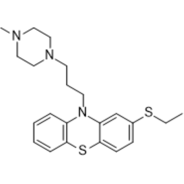
HY-B1794
ThiethylperazineD2/H1 Antagonist -
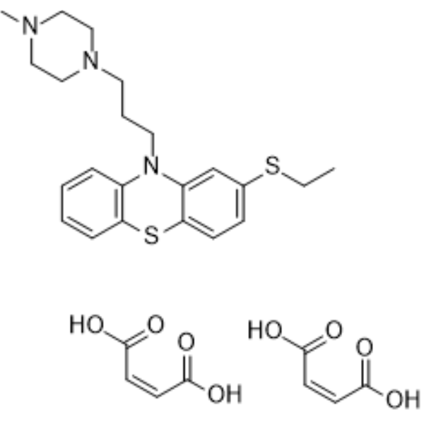
HY-B1794A
Thiethylperazine dimaleateAnti-emetic Agent -
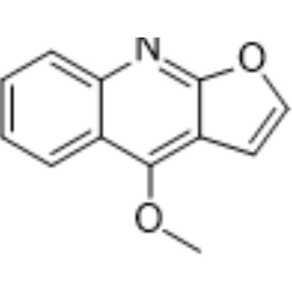
HY-N0849
DictamineBacterial Inhibitor -
HY-N1347
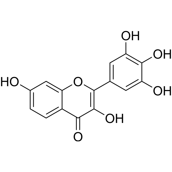
RobinetinBacterial Inhibitor -

HY-B1794R
Thiethylperazine (Standard)D2/H1 Antagonist -
HY-N10113
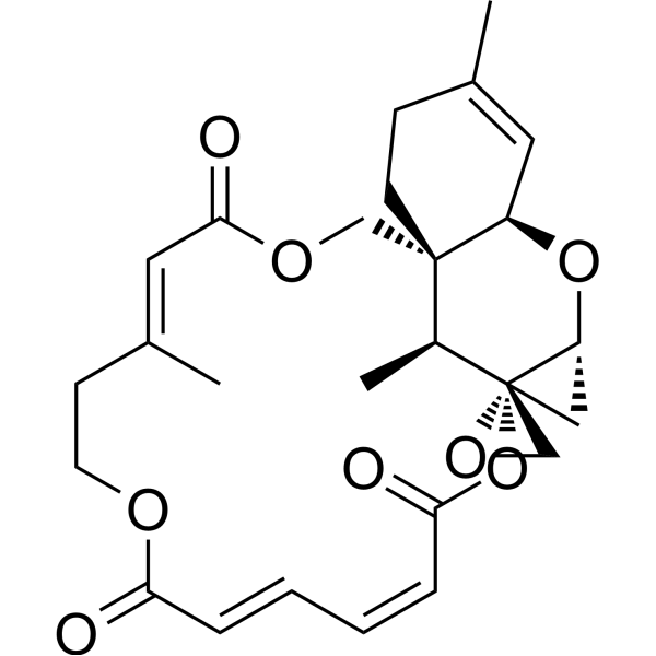
Verrucarin JMyrothecium Fungus Metabolite -

HY-B1794AR
Thiethylperazine dimaleate (Standard)Anti-emetic Agent -

HY-N0849R
Dictamine (Standard)Bacterial Inhibitor -

HY-N1347R
Robinetin (Standard)Bacterial Inhibitor
All Histamine Receptor Isoforms
More

Biological Activity
Description
Cellular Effect
|
Cell Line

|
Type | Value | Description | References |
|---|---|---|---|---|
| A549 | IC50 |
>20 μM
Compound: 25
|
Cytotoxicity against human A549 cells assessed as cell viability after 48 hrs by MTT assay
Cytotoxicity against human A549 cells assessed as cell viability after 48 hrs by MTT assay
|
[PMID: 21381696] |
| AZ-521 cell line | IC50 |
>20 μM
Compound: 25
|
Cytotoxicity against human AZ-521 cells assessed as cell viability after 48 hrs by MTT assay
Cytotoxicity against human AZ-521 cells assessed as cell viability after 48 hrs by MTT assay
|
[PMID: 21381696] |
| SK-BR-3 | IC50 |
>20 μM
Compound: 25
|
Cytotoxicity against human SK-BR-3 cells assessed as cell viability after 48 hrs by MTT assay
Cytotoxicity against human SK-BR-3 cells assessed as cell viability after 48 hrs by MTT assay
|
[PMID: 21381696] |
In Vitro
Nimbin (400 μM; 0-120 h) inhibits β-sheet formation and reduces hydrophobicity in hTau40wt protein in heparin-induced tau aggregation assay[1].
Nimbin (50 nM-50 μM; 24 h) shows no cytotoxicity and rescues tau aggregate-induced cell death in HEK293T cells[1].
MedChemExpress (MCE) has not independently confirmed the accuracy of these methods. They are for reference only. Further protocols information, click here.
-
Cell Line:HEK293T cells
-
Concentration:1 μM, 2 μM, 5 μM
-
Incubation Time:24 h
-
Result:Significantly reversed tau aggregate-induced cytotoxicity at 2 μM and 5 μM, restoring cell viability to 100% and 95%, respectively, compared to 32% viability in control groups.
In Vivo
MedChemExpress (MCE) has not independently confirmed the accuracy of these methods. They are for reference only.


Chemical Information
-
CAS No. 5945-86-8
-
Appearance Solid
-
Molecular Weight 540.60
-
Formula C30H36O9
-
Color White to off-white
-
SMILES
C[C@@]([C@@H]([C@]1(C(C=C2)=O)C)CC(OC)=O)(C3=C4C)[C@@](O[C@]3([H])C[C@@]4([H])C5=COC=C5)([H])[C@@H]([C@@]1([H])[C@]2(C)C(OC)=O)OC(C)=O
-
Structure Classification
-
Initial Source
-
Shipping
Room temperature in continental US; may vary elsewhere.
-
Storage
Powder -20°C 3 years In solvent -80°C 6 months -20°C 1 month

Protocols
-
Cell Cytotoxicity Assay
Cytotoxicity assays are usually based on the assessment of cell membrane damage, which can also be indirectly detected by measuring cell viability. Detection methods include MTT assay, CKK-8 assay, LDH assay and ATP assay, etc.
-
Mammalian live/dead viability and cytotoxicity staining
Live/dead viability and cytotoxicity staining assays are based on the simultaneous detection of intracellular esterase activity in metabolically active (viable) cells and membrane integrity loss in non-viable cells. In commonly used dual-staining approaches, membrane-permeant fluorogenic substrates are converted by intracellular esterases into fluorescent products in live cells, while impermeant DNA-binding dyes selectively enter cells with compromised plasma membranes and label nucleic acids in dead or dying cells, enabling discrimination between viable and non-viable populations by fluorescence microscopy or flow cytometry.
-
CCK-8/WST-8 Cell Proliferation Assay
The CCK-8/WST-8 assay is based on the reduction of the water-soluble tetrazolium salt WST-8 to a water-soluble formazan product by cellular dehydrogenases in metabolically active cells, where the generated formazan amount is proportional to the number of living cells and is quantified by measuring absorbance in the visible range, providing a colorimetric readout for cell viability and proliferation assessment. This class of tetrazolium-based assays improves upon earlier MTT-based systems by producing a water-soluble formazan, eliminating the need for organic solubilization steps and enabling direct spectrophotometric measurement in culture medium.
-
Research Protocol for Inflammation-related Diseases
The NLRP3 inflammasome is a cytosolic innate immune signaling platform that integrates priming signals and danger-signal activation to promote caspase-1 activation, maturation of IL-1β and IL-18, and gasdermin D-mediated pyroptotic cell death. The core experimental logic is to determine whether inflammatory disease phenotypes are driven by increased NLRP3 expression, ASC-containing inflammasome assembly, caspase-1 cleavage, GSDMD cleavage, and extracellular release of IL-1β/IL-18 rather than by nonspecific cell injury alone. The pathway is strongly linked to inflammation-related disease phenotypes because monosodium urate crystals activate NALP3/NLRP3 inflammasome signaling in gout-like crystal inflammation, cholesterol crystals activate NLRP3 inflammasomes in atherogenesis models, and DSS-induced intestinal inflammation has been reported to involve NLRP3 inflammasome activity. However, experimental colitis studies also show context-dependent protective effects of NLRP3 inflammasome co
-
MTT Cell Proliferation Assay
The MTT assay is a colorimetric endpoint assay for estimating viable cell number, cell growth, cytotoxicity, or cell activation in cultured mammalian cells. Living cells reduce the yellow tetrazolium salt MTT into purple/blue formazan, while dead cells do not generate the same signal; the resulting color can be quantified with a multiwell spectrophotometer. MTT reduction is commonly interpreted as a readout of metabolic activity that often correlates with viable cell number, but it should not be treated as a direct cell-counting method unless the assay is optimized for the cell type and experimental condition. Studies show that MTT reduction can involve mitochondrial and non-mitochondrial reducing systems, and formazan may accumulate in intracellular lipid droplets rather than simply marking mitochondria.
-
Cell Viability Determination by MTT Colorimetric Assay
The following protocol uses the MTT colorimetric assay as a classic literature-established method for assessing cell viability/metabolic activity in cultured mammalian cells. MTT[3-(4,5-dimethylthiazol-2-yl)-2,5-diphenyltetrazolium bromide] is reduced by metabolically active cells to a colored formazan product; the amount of formazan is quantified spectrophotometrically and provides an indirect measure of metabolically active viable cells. Importantly, MTT reduction reflects cellular oxidoreductase/metabolic activity rather than an absolute direct count of living cells, so changes in cellular metabolism can alter the signal independently of cell number.


Purity & Documentation
-
Data Sheet (281 KB)
-
SDS (251 KB)
- English - EN (251 KB)
- Français - FR (251 KB)
- Deutsch - DE (251 KB)
- Norwegian - NO (251 KB)
- Español - ES (251 KB)
- Swedish - SV (251 KB)
- Italian - IT (251 KB)
- Korean - KR (251 KB)
- Portuguese - PT (251 KB)
-
Handling Instructions (2659 KB)
References
[1]. Nalini V Gorantla, et al. Neem Derivatives Inhibits Tau Aggregation. J Alzheimers Dis Rep. 2019 Jun 14;3(1):169-178. [Content Brief]
[2]. P Lavanya, et al. Computational Analysis Reveal Inhibitory Action of Nimbin Against Dengue Viral Envelope Protein. Virusdisease. 2015 Dec;26(4):243-54. [Content Brief]
[3]. Li-Juan Yang, et al. Host-guest System of Nimbin and Beta-Cyclodextrin or Its Derivatives: Preparation, Characterization, Inclusion Mode, and Solubilization. J Agric Food Chem. 2010 Aug 11;58(15):8545-52. [Content Brief]

Calculators
Concentration (start) × Volume (start) = Concentration (final) × Volume (final)


Nimbin Related Classifications
HY-N3187 Related Classifications
 Powered by Bioz