



NLS-StAx-h
Based on 1 publication(s) in Google Scholar
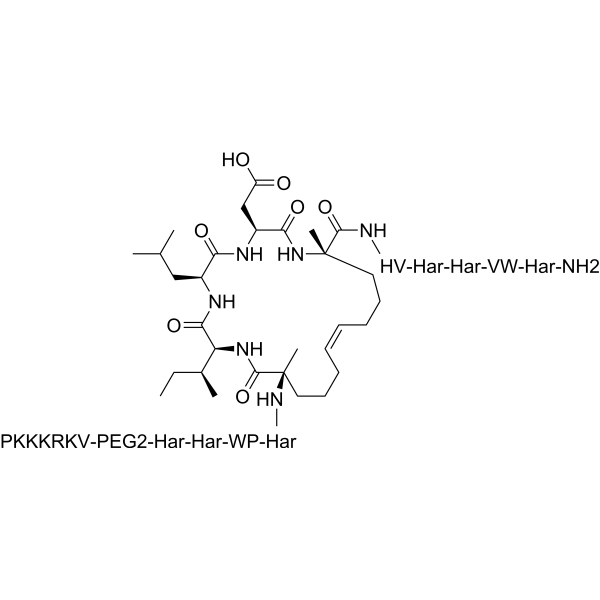
NLS-StAx-h is a selective, cell permeable, stapled peptide Wnt signaling inhibitor with an IC50 of 1.4 μM. NLS-StAx-h efficiently inhibits β-catenin-transcription factor interactions. NLS-StAx-h shows anti-proliferation of cancer cells.
For research use only. We do not sell to patients.
- CAS No.: 2872559-21-0
- Formula: C161H275N55O29
- Molecular Weight:3445.25
-
Storage:
Please store the product under the recommended conditions in the Certificate of Analysis.

To place orders, for customer services and technical support, please contact: MedChemExpress USA
Tel: 609-228-6898 E-mail: [email protected] [email protected]


-
 Biological Activity
-
Chemical Information
- Protocols
- Purity & Documentation
- References
-
 Help & FAQs


Publications Citing Use of MedChemExpress (MCE) NLS-StAx-h
More

Biological Activity
Description
IC50 & Target
IC50: 1.4 μM (Wnt)[1]
In Vitro
NLS-StAx-h (10 μM; 72 h) inhibits proliferation of colorectal cancer cells[1].
NLS-StAx-h (5 and 10 μM; 24 h) inhibits migration of colorectal cancer cells[1].
MedChemExpress (MCE) has not independently confirmed the accuracy of these methods. They are for reference only. Further protocols information, click here.
-
Cell Line:SW-480 and DLD-1 cells
-
Concentration:10 μM
-
Incubation Time:72 hr
-
Result:Reduced the viability of both SW-480 and DLD-1 by more than 80%.
-
Cell Line:DLD-1 cells
-
Concentration:5 and 10 μM
-
Incubation Time:24 hr
-
Result:Resulted in dose-dependent inhibition of wound closure (wound closure: 52% at 5 μM, and 24% at 10 μM).


Chemical Information
-
CAS No. 2872559-21-0
-
Molecular Weight 3445.25
-
Formula C161H275N55O29
-
Shipping
Room temperature in continental US; may vary elsewhere.
-
Storage
Please store the product under the recommended conditions in the Certificate of Analysis.


Publications (1)
-
Journal Impact Factor
-
Most Recent
-
Exp Cell Res
Ketohexokinase-A deficiency attenuates the proliferation via reducing β-catenin in gastric cancer cells. [Abstract]2024 Apr 16;438(1):114038. PMID: 38614422

Protocols
-
Nuclear Protein Extraction (High-Salt/Hypotonic Fractionation)
The high-salt/hypotonic fractionation method for nuclear protein extraction is based on the differential solubility of cellular components. Cytoplasmic proteins are extracted first using a hypotonic buffer that causes cell swelling and membrane rupture, followed by centrifugation to separate the cytoplasmic supernatant from the nuclear pellet. The nuclear pellet is then subjected to high-salt extraction (e. g. , 0. 4 M (NH4)2SO4 or 1 M NaCl) to solubilize tightly bound nuclear matrix proteins, including transcription factors, histones, and structural proteins associated with chromatin and the nuclear scaffold. This approach allows for the isolation of both soluble cytoplasmic proteins and salt-resistant nuclear proteins while minimizing cross-contamination.
-
Cell Cytotoxicity Assay
Cytotoxicity assays are usually based on the assessment of cell membrane damage, which can also be indirectly detected by measuring cell viability. Detection methods include MTT assay, CKK-8 assay, LDH assay and ATP assay, etc.
-
Cell Viability Determination by MTT Colorimetric Assay
The following protocol uses the MTT colorimetric assay as a classic literature-established method for assessing cell viability/metabolic activity in cultured mammalian cells. MTT[3-(4,5-dimethylthiazol-2-yl)-2,5-diphenyltetrazolium bromide] is reduced by metabolically active cells to a colored formazan product; the amount of formazan is quantified spectrophotometrically and provides an indirect measure of metabolically active viable cells. Importantly, MTT reduction reflects cellular oxidoreductase/metabolic activity rather than an absolute direct count of living cells, so changes in cellular metabolism can alter the signal independently of cell number.


Purity & Documentation
References
[1]. Laura Dietrich, et al. Cell Permeable Stapled Peptide Inhibitor of Wnt Signaling That Targets β-Catenin Protein-Protein Interactions [Content Brief]
[2]. ang Z, et al. Direct targeting of β-catenin in the Wnt signaling pathway: Current progress and perspectives. Med Res Rev. 2021 Jul;41(4):2109-2129. [Content Brief]

Calculators
Concentration (start) × Volume (start) = Concentration (final) × Volume (final)


NLS-StAx-h Related Classifications
HY-P2272 Related Classifications
 Powered by Bioz