



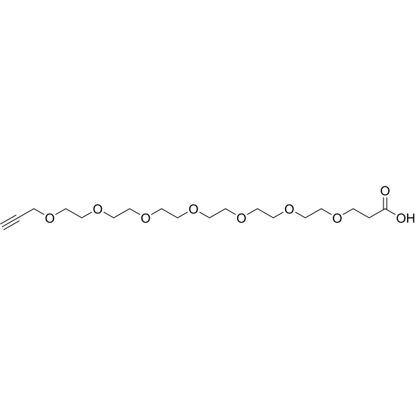
Propargyl-PEG8-acid

Based on 1 Customer Validation
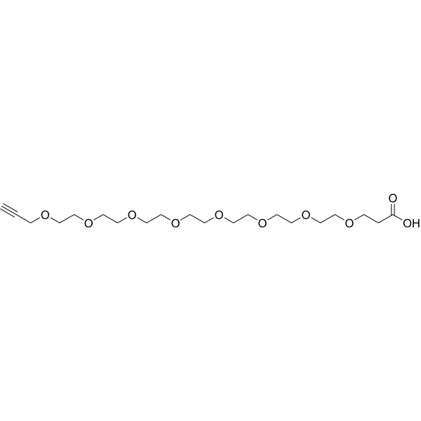
Propargyl-PEG8-acid is a PEG-based PROTAC linker can be used in the synthesis of PROTACs. Propargyl-PEG8-acid is a cleavable ADC linker used in the synthesis of antibody-drug conjugates (ADCs). The ADCs can be used in bacterial infections caused by Gram-negative bacteria. Propargyl-PEG8-acid is a click chemistry reagent, it contains an Alkyne group and can undergo copper-catalyzed azide-alkyne cycloaddition (CuAAc) with molecules containing Azide groups.
For research use only. We do not sell to patients.
- Purity : 98.0%
- CAS No.: 2055014-94-1
- Formula: C20H36O10
- Molecular Weight:436.49
-
Storage:
-20°C, stored under nitrogen
* In solvent : -80°C, 6 months; -20°C, 1 month (stored under nitrogen)

To place orders, for customer services and technical support, please contact: MedChemExpress USA
Tel: 609-228-6898 E-mail: [email protected] [email protected]


-
 Biological Activity
-
Chemical Information
- Protocols
- Purity & Documentation
- References
-
 Help & FAQs


-
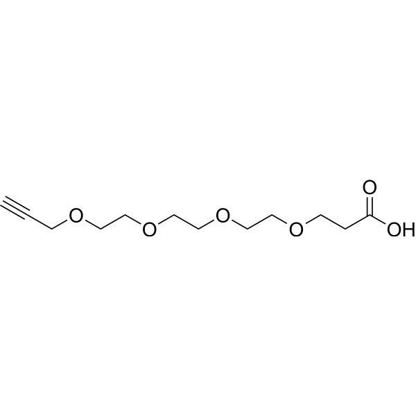
HY-130481
Propargyl-PEG4-acidPROTAC Linker -
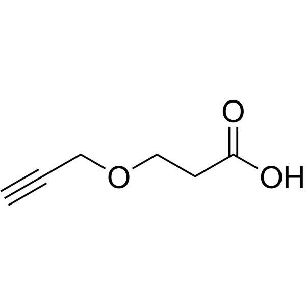
HY-130504
Propargyl-PEG1-acidPROTAC Linkers -
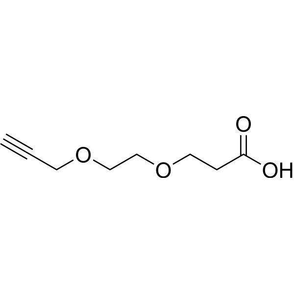
HY-118764
Propargyl-PEG2-acidPROTAC/ADC Linker -
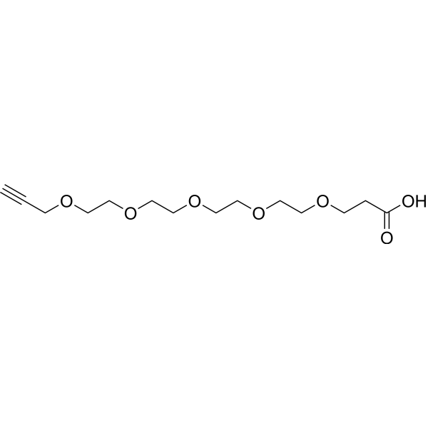
HY-101157
Propargyl-PEG5-acidADC Linker -
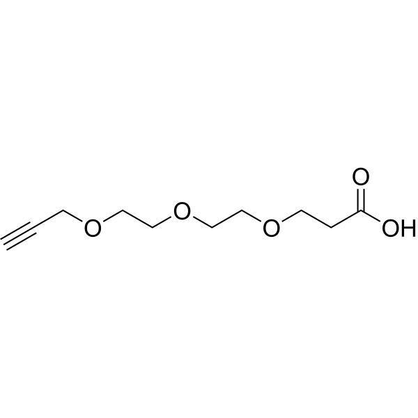
HY-126975
Propargyl-PEG3-acidADC/PROTAC Linker -
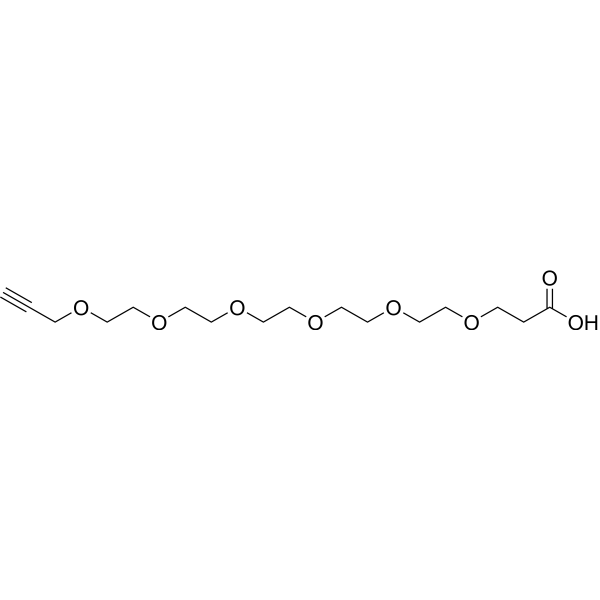
HY-130386
Propargyl-PEG6-acidADC/PROTAC Linker -
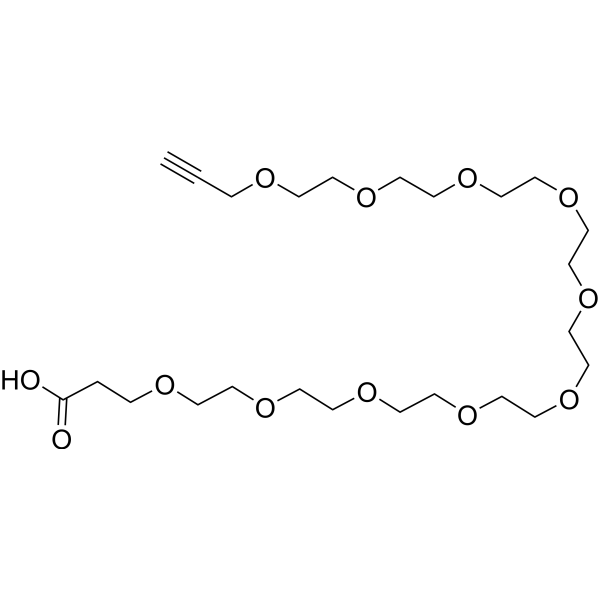
HY-140020
Propargyl-PEG10-acidPEG Derivative -
HY-130383
Propargyl-PEG7-acidADC/PROTAC Linker -

HY-101157R
Propargyl-PEG5-acid (Standard)ADC Linker
All PROTAC Linkers Isoforms
More

Biological Activity
Description
IC50 & Target
[1]|
Cleavable Linker |
PEGs |
In Vitro
PROTACs contain two different ligands connected by a linker; one is a ligand for an E3 ubiquitin ligase and the other is for the target protein. PROTACs exploit the intracellular ubiquitin-proteasome system to selectively degrade target proteins.
ADCs are comprised of an antibody to which is attached an ADC cytotoxin through an ADC linker.
MedChemExpress (MCE) has not independently confirmed the accuracy of these methods. They are for reference only. Further protocols information, click here.


Chemical Information
-
CAS No. 2055014-94-1
-
Appearance Liquid
-
Molecular Weight 436.49
-
Formula C20H36O10
-
Color Colorless to light yellow
-
SMILES
C#CCOCCOCCOCCOCCOCCOCCOCCOCCC(O)=O
-
Shipping
Room temperature in continental US; may vary elsewhere.
-
Storage
-20°C, stored under nitrogen
* In solvent : -80°C, 6 months; -20°C, 1 month (stored under nitrogen)

Protocols
-
EdU Incorporation Assay (Click Chemistry-Based DNA Synthesis Measurement)
The EdU incorporation assay measures DNA synthesis by adding the thymidine analog 5-ethynyl-2′-deoxyuridine to cells or tissues, where it is incorporated into newly synthesized DNA during S phase. Incorporated EdU is detected by copper-catalyzed azide-alkyne cycloaddition, in which a fluorescent azide covalently reacts with the ethynyl group on EdU, allowing S-phase cells to be detected by fluorescence microscopy, flow cytometry, or high-content imaging. EdU detection does not require DNA denaturation or anti-BrdU antibody access, which preserves sample structure and improves compatibility with immunostaining and multiparameter cytometry compared with BrdU-based detection. EdU can be cytotoxic in a cell-type- and exposure-dependent manner, so pulse duration, concentration, and continuous-labeling designs should be validated for each cell type.
-
Gram Staining of Tissue Sections
Gram staining of tissue sections is a histochemical technique used to differentiate Gram-positive and Gram-negative bacteria within histological specimens based on differences in bacterial cell wall structure and dye retention, adapted from classical bacteriological Gram staining into tissue-compatible “histological Gram stain” variants. In tissue applications, modifications of the Brown-Hopps and Brown-Brenn methods are commonly used to improve differentiation of microorganisms embedded within host connective tissue and to reduce overstaining or loss of Gram-negative signal, which are known limitations of earlier approaches. The principle relies on crystal violet-iodine complex retention in Gram-positive organisms and subsequent decolorization and counterstaining steps that allow contrast visualization of Gram-negative organisms against tissue background.


Purity & Documentation
-
Data Sheet (262 KB)
-
SDS (394 KB)
- English - EN (394 KB)
- Français - FR (394 KB)
- Deutsch - DE (394 KB)
- Norwegian - NO (394 KB)
- Español - ES (394 KB)
- Swedish - SV (394 KB)
- Italian - IT (394 KB)
- Korean - KR (394 KB)
- Portuguese - PT (394 KB)
-
Handling Instructions (2659 KB)
References

Calculators
Concentration (start) × Volume (start) = Concentration (final) × Volume (final)


Propargyl-PEG8-acid Related Classifications
HY-130379 Related Classifications
 Powered by Bioz