



SCR-1481B1

Based on 1 Customer Validation
SCR-1481B1 (Metatinib anhydrous; c-Met inhibitor 2) is an inhibitor of the N-terminal fragment of Gasdermin D (GSDMD), with an IC50 of 1.04 μM and a Ka of 0.42 μM in mice. SCR-1481B1 effectively blocks GSDMD-NT oligomerization and pore formation, inhibits GSDMD-mediated pyroptosis (Pyroptosis), preserves mitochondrial integrity, and reduces proinflammatory cytokine secretion. SCR-1481B1 also inhibits angiogenesis, exerts GSDMD-independent antitumor effects, and enhances the infiltration of antitumor immune cells. SCR-1481B1 is also an inhibitor targeting to c-MET and VEGFR2, can be used in studies related to melanoma and sepsis.
For research use only. We do not sell to patients.
- Purity : 99.99%
- CAS No.: 1174161-69-3
- Formula: C24H18ClF2N4O7P
- Molecular Weight:578.85
-
Storage:
4°C, sealed storage, away from moisture and light
* In solvent : -80°C, 6 months; -20°C, 1 month (sealed storage, away from moisture and light)


To place orders, for customer services and technical support, please contact: MedChemExpress USA
Tel: 609-228-6898 E-mail: [email protected] [email protected]


-
 Biological Activity
-
Chemical Information
-
Solvent & Solubility
- Protocols
- Purity & Documentation
- References
-
 Help & FAQs


-
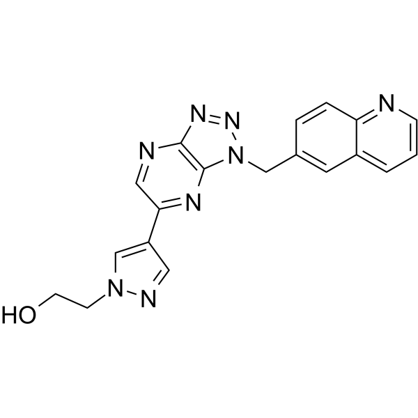
HY-12017
PF-04217903c-Met Kinase Inhibitor -
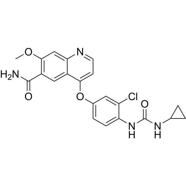
HY-10981
LenvatinibVEGFR/FGFR Inhibitor -
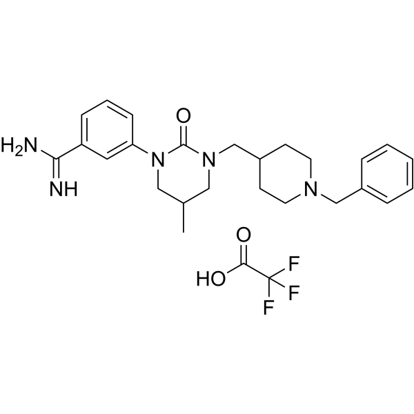
HY-114363A
SRI 31215 TFAMatriptase/Hepsin/HGFA Inhibitor -
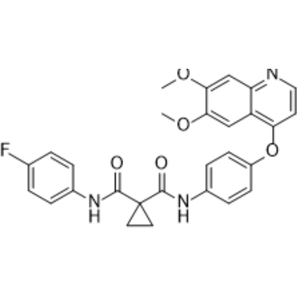
HY-13016
CabozantinibVEGFR2/MET Inhibitor -
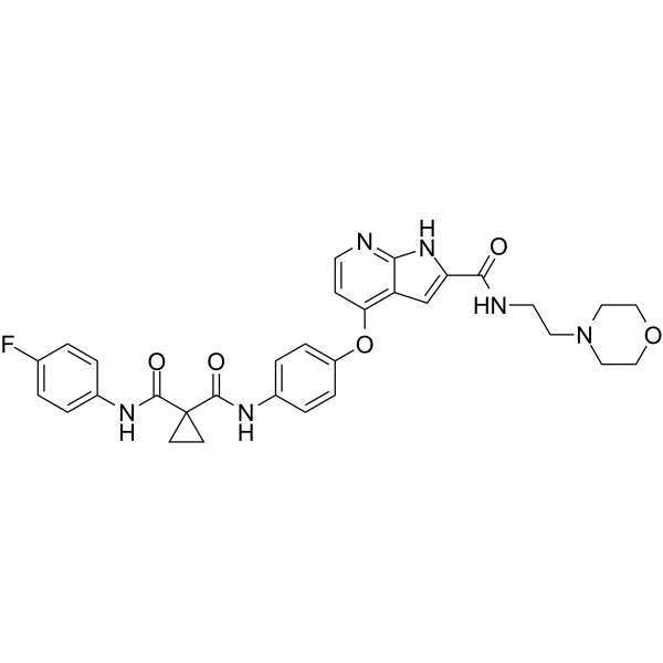
HY-10421
Tyrosine kinase inhibitorTyrosine kinase inhibitor -
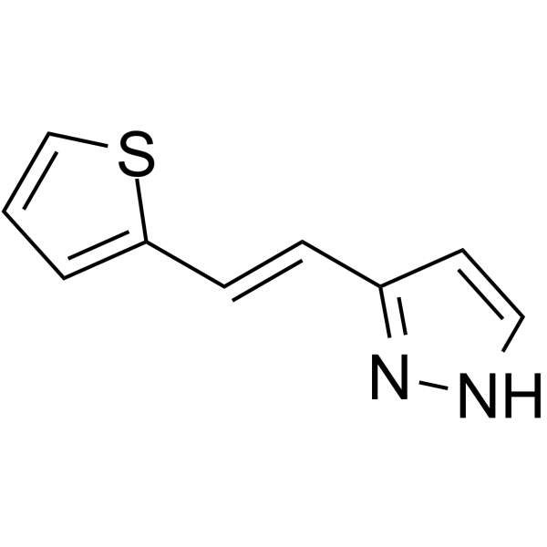
HY-137455
Terevalefimc-Met Receptor Activator -
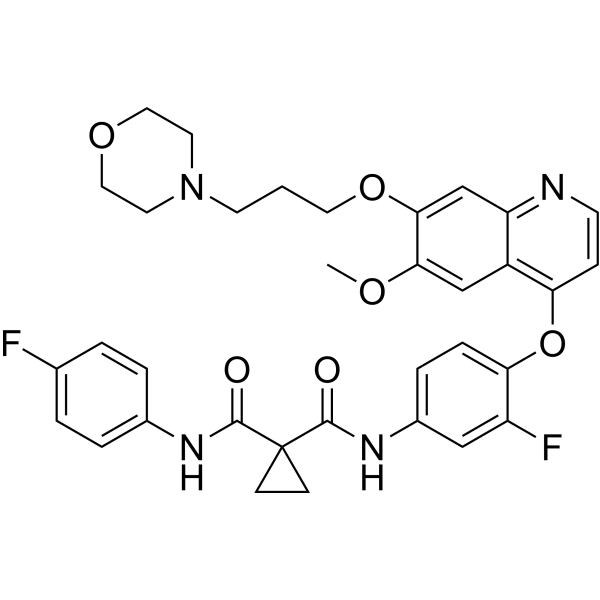
HY-10338
ForetinibMET/VEGFR2/KDR Inhibitor -
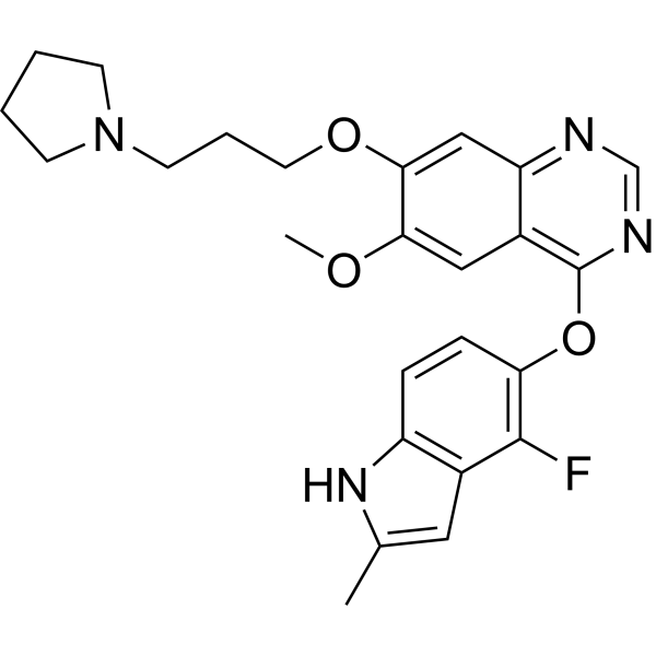
HY-10205
CediranibVEGFR2 Inhibitor -
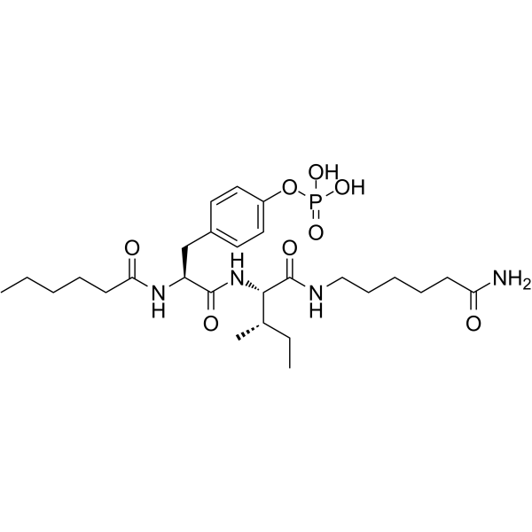
HY-132814
Fosgonimetonc-Met Agonist -
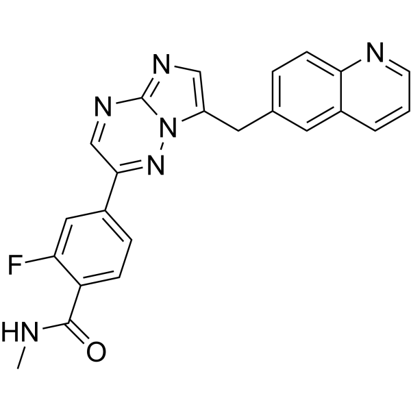
HY-13404
Capmatinibc-MET Kinase Inhibitor -
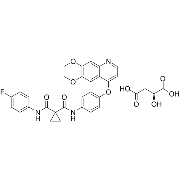
HY-12044
Cabozantinib S-malate
All VEGFR Isoforms
More

Biological Activity
Description
IC50 & Target
|
IL-1β |
VEGFR2 |
In Vitro
SCR-1481B1 (1-40 μM; 1 h pre-incubation) dose-dependently inhibits LPS plus nigericin-induced GSDMD-mediated pyroptosis in iBMDMs, reducing both LDH release and IL-1β secretion[1].
SCR-1481B1 (20 μM) inhibits LPS plus nigericin-induced GSDMD-mediated pyroptosis in human THP-1 monocytes, reducing both LDH release and IL-1β secretion[1].
SCR-1481B1 (20 μM; 4 h) inhibits mouse and human GSDMD-N oligomerization and subsequent pyroptotic cell death in HEK293T cells[1].
SCR-1481B1 (20 μM; 4 h) inhibits GSDMD oligomerization and pyroptotic cell death downstream of caspase-1/11-mediated GSDMD cleavage in HEK293T cells[1].
SCR-1481B1 (20 μM) inhibits human and mouse GSDMD-N oligomerization in the cytosol of HEK293T cells, as measured by split-luciferase reconstitution[1].
SCR-1481B1 (20 μM; 4 h pre-incubation) specifically binds to core sites at the oligomerization interface I of mouse GSDMD-N, enhancing its stability against protease degradation[1].
SCR-1481B1 (20 μM) directly binds to core sites at the oligomerization interface I of purified mouse GSDMD-N, increasing its thermal stability[1].
SCR-1481B1 (0.15-20 μM; 60 min) inhibits GSDMD-N-mediated liposome pore formation with an IC50 of 1.04 μM[1].
SCR-1481B1 (Metatinib) binds to the GSDMD dimer with a docking score of -7.28 kcal/mol[3].
MedChemExpress (MCE) has not independently confirmed the accuracy of these methods. They are for reference only. Further protocols information, click here.
In Vivo
SCR-1481B1 (10 mg/kg; i.p.; single dose) improves survival and reduces serum inflammatory cytokine levels in E. coli-induced septic mice, though it does not significantly reduce organ bacterial burden[1].
SCR-1481B1 (10 mg/kg; i.p.; every other day; starting after tumor implantation) enhances the antitumor efficacy of anti-PD-L1 antibody in a B16F10 melanoma model by increasing tumor-infiltrating immune cells and reducing T cell exhaustion, with activity independent of GSDMD[1].
MedChemExpress (MCE) has not independently confirmed the accuracy of these methods. They are for reference only.


Chemical Information
-
CAS No. 1174161-69-3
-
Appearance Solid
-
Molecular Weight 578.85
-
Formula C24H18ClF2N4O7P
-
Color White to off-white
-
SMILES
O=C(C1=CN(COP(O)(O)=O)C=C(C2=CC=C(F)C=C2)C1=O)NC3=CC=C(OC4=C(Cl)C(N)=NC=C4)C(F)=C3
-
Synonyms
Metatinib free base; c-Met inhibitor 2 free base
-
Shipping
Room temperature in continental US; may vary elsewhere.
-
Storage
4°C, sealed storage, away from moisture and light
* In solvent : -80°C, 6 months; -20°C, 1 month (sealed storage, away from moisture and light)


Solvent & Solubility
In Vitro:
DMSO : 4.55 mg/mL (7.86 mM; ultrasonic and warming and heat to 60°C; Hygroscopic DMSO has a significant impact on the solubility of product, please use newly opened DMSO)
Please refer to the solubility information to select the appropriate solvent. Once prepared, please aliquot and store the solution to prevent product inactivation from repeated freeze-thaw cycles.
Storage method and period of stock solution: -80°C, 6 months; -20°C, 1 month (sealed storage, away from moisture and light). When stored at -80°C, please use it within 6 months. When stored at -20°C, please use it within 1 month.
Please refer to the solubility information to select the appropriate solvent. Once prepared, please aliquot and store the solution to prevent product inactivation from repeated freeze-thaw cycles.
Storage method and period of stock solution: -80°C, 6 months; -20°C, 1 month (sealed storage, away from moisture and light). When stored at -80°C, please use it within 6 months. When stored at -20°C, please use it within 1 month.
Concentration (start) × Volume (start) = Concentration (final) × Volume (final)

Protocols
-
Human pluripotent stem cell endothelial-cell differentiation
Human pluripotent stem cell endothelial differentiation is based on stepwise developmental patterning: early activation of WNT/GSK3β inhibition promotes mesodermal or vascular progenitor entry, followed by endothelial specification using VEGF-related signaling, BMP4, FGF2, Notch modulation, or cAMP depending on the published protocol. Endothelial differentiation is read out by acquisition of CD31, CD34, VE-cadherin/CD144, KDR/VEGFR2, vWF, Tie2, NOS3, acetylated LDL uptake, tube/network formation, barrier function, and in vivo vessel-forming capacity where tested.
-
Cell Cytotoxicity Assay
Cytotoxicity assays are usually based on the assessment of cell membrane damage, which can also be indirectly detected by measuring cell viability. Detection methods include MTT assay, CKK-8 assay, LDH assay and ATP assay, etc.
-
Vascular/Branching Fractal Analysis
Vascular/branching fractal analysis quantifies the geometric complexity of vessel trees or vascular networks from segmented 2D images, commonly by converting vessels into binary and/or skeletonized maps and estimating fractal dimension using box-counting or related approaches. Fractal dimension is interpreted as an image-derived readout of vascular branching complexity, space filling, or density, and has been applied to retinal photographs, fluorescein angiography, OCT angiography, capillary perfusion maps, and in vitro Matrigel angiogenesis networks. The assay readout is generated from vessel-positive pixels after image preprocessing, vessel segmentation, binarization, and optional skeletonization; reported outputs include fractal dimension, vessel density, branchpoint density, endpoint density, vessel length density, tortuosity, and generation-based branching metrics when VESGEN-style analysis is used. The biological interpretation is limited to quantitative vascular patterning and s
-
LPS-Induced Endotoxemia/Systemic Inflammation
Lipopolysaccharide (LPS)-induced endotoxemia is a widely used in vivo model of acute systemic inflammation in which LPS, a Gram-negative bacterial endotoxin, activates innate immune signaling primarily through TLR4, leading to rapid and transient induction of pro-inflammatory cytokines such as TNF-α, IL-6, and IL-1β in circulation and tissues. This cytokine surge is commonly used as a measurable readout of systemic inflammatory activation and immune dysregulation, and is typically assessed within hours after intraperitoneal LPS administration in mouse models of endotoxemia. The model captures key features of systemic inflammatory response syndrome, including cytokine release, immune cell activation, and downstream tissue responses, and has been used to evaluate anti-inflammatory interventions such as cytokine modulation, lipid mediators, and immune cell-targeting therapies.
-
Endothelial Tube Formation Assay
Endothelial tube formation assay evaluates the ability of endothelial cells to attach, migrate, align, and organize into capillary-like networks when cultured on gelled basement membrane extract or Matrigel; the readout is the morphology and quantity of tube-like networks, which reflects an in vitro endothelial morphogenesis step related to angiogenesis. Basement membrane extract/Matrigel provides laminin-rich extracellular matrix cues that support endothelial differentiation into capillary-like structures, but it can contain biologically active growth factors, so growth-factor-reduced matrix is preferred when testing defined angiogenic stimulators or inhibitors.
-
Research Protocol for Inflammation-related Diseases
The NLRP3 inflammasome is a cytosolic innate immune signaling platform that integrates priming signals and danger-signal activation to promote caspase-1 activation, maturation of IL-1β and IL-18, and gasdermin D-mediated pyroptotic cell death. The core experimental logic is to determine whether inflammatory disease phenotypes are driven by increased NLRP3 expression, ASC-containing inflammasome assembly, caspase-1 cleavage, GSDMD cleavage, and extracellular release of IL-1β/IL-18 rather than by nonspecific cell injury alone. The pathway is strongly linked to inflammation-related disease phenotypes because monosodium urate crystals activate NALP3/NLRP3 inflammasome signaling in gout-like crystal inflammation, cholesterol crystals activate NLRP3 inflammasomes in atherogenesis models, and DSS-induced intestinal inflammation has been reported to involve NLRP3 inflammasome activity. However, experimental colitis studies also show context-dependent protective effects of NLRP3 inflammasome co
-
Endothelial Cell Migration/Angiogenic Sprouting Assay
Endothelial cell migration and angiogenic sprouting assays are in vitro (and partially ex vivo-adapted) functional models that quantify the ability of endothelial cells to undergo coordinated migration, extracellular matrix invasion, and multicellular organization into capillary-like sprouts in response to pro-angiogenic stimuli such as VEGF, bFGF, or conditioned microenvironments. These assays are used to model early angiogenic events including tip-cell formation, directional migration, and lumen-like sprout extension, which collectively reflect angiogenic activation and vascular morphogenesis processes observed in vivo.
-
Pyroptosis Solutions
Pyroptosis is a lytic inflammatory cell-death pathway executed by gasdermin pores, most classically through inflammasome-mediated activation of caspase-1, cleavage of gasdermin D, membrane pore formation, LDH release, and secretion of IL-1β and IL-18. The canonical pathway is commonly modeled by priming cells with an inflammatory signal such as LPS to induce pro-IL-1β and inflammasome components, followed by an activation signal such as ATP or nigericin to activate NLRP3, ASC speck formation, caspase-1 cleavage, GSDMD cleavage, cytokine release, and pyroptotic membrane rupture. The non-canonical pathway is triggered when cytosolic LPS activates mouse caspase-11 or human caspase-4/5, leading to GSDMD cleavage and pyroptosis, and this can secondarily activate NLRP3-dependent IL-1β release. Pyroptosis is linked to inflammatory injury, infection, cancer, liver disease, ocular disease, placental inflammation, and other disease phenotypes, but unresolved questions include which gasdermin fam


Purity & Documentation
-
Data Sheet (294 KB)
-
SDS (396 KB)
- English - EN (396 KB)
- Français - FR (396 KB)
- Deutsch - DE (396 KB)
- Norwegian - NO (396 KB)
- Español - ES (396 KB)
- Swedish - SV (396 KB)
- Italian - IT (396 KB)
- Korean - KR (396 KB)
- Portuguese - PT (396 KB)
-
Handling Instructions (2659 KB)
References
[1]. Hu Y, et al. Identification of two repurposed drugs targeting GSDMD oligomerization interface I to block pyroptosis. Cell Chem Biol. 2024;31(12):2024-2038.e7. [Content Brief]
[3]. Lestari B, et al. Discovery of pyroptosis-inducing natural products in neuroblastomas: computational studies with experimental validation. BMC Complement Med Ther. 2025;25(1):279. Published 2025 Jul 19. [Content Brief]
[4]. Cheng K, et al. Safety, Efficacy, and Pharmacokinetics of Metatinib Tromethamine Tablet in Patients with Advanced Refractory Solid Tumors: A Phase I Clinical Trial. Oncologist. 2021 Aug;26(8):649-e1313. [Content Brief]


Complete Stock Solution Preparation Table
Please refer to the solubility information to select the appropriate solvent. Once prepared, please aliquot and store the solution to prevent product inactivation from repeated freeze-thaw cycles.
Storage method and period of stock solution: -80°C, 6 months; -20°C, 1 month (sealed storage, away from moisture and light). When stored at -80°C, please use it within 6 months. When stored at -20°C, please use it within 1 month.
| Optional Solvent | Concentration Solvent Mass | 1 mg | 5 mg | 10 mg | 25 mg |
|---|---|---|---|---|---|
| DMSO | 1 mM | 1.7276 mL | 8.6378 mL | 17.2756 mL | 43.1891 mL |
| 5 mM | 0.3455 mL | 1.7276 mL | 3.4551 mL | 8.6378 mL |


SCR-1481B1 Related Classifications
HY-18711 Related Classifications
 Powered by Bioz