2660 Results for "

Response

" in MedChemExpress (MCE) Product Catalog:
Products (2660)

2660 Results for "Response" in MCE Product Catalog:

Cat. No.: HY-118594R
CAS No.: 632-93-9
Research Areas:  

Metabolic Disease

Diethyl 2,4,6-trimethyl-1,4-dihydropyridine-3,5-dicarboxylate (Standard) is the analytical standard of Diethyl 2,4,6-trimethyl-1,4-dihydropyridine-3,5-dicarboxylate (HY-118594). This product is intended for research and analytical applications. Diethyl 2,4,6-trimethyl-1,4-dihydropyridine-3,5-dicarboxylate is an orally active porphyrin inducer and ferrochelatase inhibitor. Diethyl 2,4,6-trimethyl-1,4-dihydropyridine-3,5-dicarboxylate can induce small bile duct obstruction in mice, resulting in blocked bile excretion and causing cholestasis. Long-term use of Diethyl 2,4,6-trimethyl-1,4-dihydropyridine-3,5-dicarboxylate can cause damage to bile duct epithelial cells, inflammatory responses, and liver fibrosis. Diethyl 2,4,6-trimethyl-1,4-dihydropyridine-3,5-dicarboxylate can be used to simulate the pathological features of cholestatic liver diseases such as sclerosing cholangitis (PSC).
loading...
    loading...
Cat. No.: HY-L250
61 compounds

In the progression of various diseases, metabolic reprogramming has emerged as a key hallmark. Lactate, as an important metabolic signaling molecule, is widely involved in tumorigenesis, immune regulation, and inflammatory responses. Particularly within the tumor microenvironment, the abnormal accumulation of lactate not only affects cellular energy metabolism but also promotes disease progression by modulating immune cell functions and mediating protein lactylation, thereby participating in epigenetic regulation and signaling networks. Therefore, systematic investigation of lactate metabolic pathways and their associated metabolites is of great significance for understanding disease mechanisms and developing novel therapeutic strategies.

The MCE lactic acid metabolite compound library contains 61 compounds and is constructed around key metabolic pathways involving lactate production, transport, and utilization. This library systematically includes core intermediates from glycolysis, the tricarboxylic acid (TCA) cycle, and the lactate cycle. Focusing on disease-associated metabolic reprogramming, it is suitable for research in oncology, inflammation, and metabolic disorders. The library can be used to elucidate the roles of lactate in tumor microenvironment regulation, immune evasion, and epigenetic modifications (such as protein lactylation). In addition, it provides high-quality small-molecule resources for drug screening, facilitating the discovery of potential modulators targeting key enzymes (such as LDH) or transporters (such as MCTs) involved in lactate metabolism.

Cat. No.: HY-L076
641 compounds

Drug-induced liver injury (DILI; also known as drug-induced hepatotoxicity) is caused by medications (prescription or OTC), herbal and dietary supplements (HDS), or other xenobiotics that result in abnormalities in liver tests or in hepatic dysfunction that cannot be explained by other causes. Drugs are an important cause of liver injury. Drug-induced hepatic injury is the most common reason cited for withdrawal of an approved drug.

DILI is thought to occur via several different mechanisms. Among these are direct impairment of the structural (e.g., mitochondrial dysfunction) and functional integrity of the liver; production of a metabolite that alters hepatocellular structure and function; production of a reactive drug metabolite that binds to hepatic proteins to produce new antigenic drug-protein adducts, which are targeted by hosts’ defenses (the hapten hypothesis); and initiation of a systemic hypersensitivity response (i.e., drug allergy) that damages the liver.

MCE Drug-induced Liver Injury (DILI) Compound Library contains a unique collection of 641 hepatotoxicity causing compounds and is a powerful tool to research DILI and other drug toxicities. This library can be used to understand the mechanisms of DILI, identify biomarkers for early DILI prediction, and allow timely recognition during drug development, thus finally achieving successful DILI prevention and assessment in the pre-marketing phase.

Cat. No.: HY-170524
CAS No.: 3052313-73-9
Research Areas:  

Infection

TDI-015051 is a highly selective, orally active antiviral agent that targets the coronavirus NSP14 guanine-N7 methyltransferase. TDI-015051 binds to substrates in a non-competitive manner and forms a stable ternary complex, precisely blocking the capping and methylation processes of viral mRNA. TDI-015051 potently inhibits a variety of coronaviruses (including SARS-CoV-2 and MERS). By impairing viral replication and translation and inducing a moderate type I interferon-mediated immune response, it significantly reduces pulmonary viral load and exhibits a synergistic effect with Nirmatrelvir (HY-138687). In addition, TDI-015051 does not inhibit non-coronavirus methyltransferases, and the drug-resistant mutations it induces impair viral fitness, demonstrating excellent antiviral properties and safety. TDI-015051 can be used for research on COVID-19 and the replication mechanism of coronaviruses .The IC50 values of TDI-015051 against SARS-CoV-2, α-hCoV-NL63, α-hCoV-229E, β-hCoV-MERS are 0.15 nM, 1.7 nM, 2.6 nM and 3.6 nM, respectively, and the Ka value against SARS-CoV-2 is 0.061 nM .
loading...
    loading...
Cat. No.: HY-176421
CAS No.: 3070438-85-3
PROTAC PI3K/110β degrader-1 is a VHL-recruiting PROTAC degrader targeting PI3K/110β, with DC50 values of 0.416 μM (MCF-7) and 19.66 μM (A549), respectively. PROTAC PI3K/110β degrader-1 recruits VHL to induce proteasomal degradation of PI3K/110β. It activates endoplasmic reticulum stress (ERS)-mediated mitochondrial apoptosis via the PERK/ATF4/CHOP unfolded protein response (UPR) pathway, downregulates p-AKT and Bcl-2, upregulates Bax, cleaved-caspase-9 and cleaved-caspase-3, inhibits the expression and activity of P-gp, and exerts synergistic anti-tumor effects with Doxorubicin (Adriamycin, ADM) (HY-15142A) and Cisplatin (DDP) (HY-17394). PROTAC PI3K/110β degrader-1 can be used in research related to multidrug-resistant cancers .
loading...
    loading...
Cat. No.: HY-182360
Cytisine-Platinum(IV) Prodrug-1 is a Pt(IV) prodrug incorporating the natural compound Cytisine (HY-N0175) with antiproliferative activity against tumor cells. Cytisine-Platinum(IV) Prodrug-1 promotes calcium transfer across the IP3R1-GRP75-VDAC1 axis to drive mitochondrial calcium overload. Cytisine-Platinum(IV) Prodrug-1 initiates unfolded protein response via PERK, eIF2α, ATF4, and CHOP to modulate Bcl-2 and Bax, triggering apoptosis. Cytisine-Platinum(IV) Prodrug-1 induces mitochondrial dysfunction, ROS production, reduced ATP synthesis, DNA damage, and S-phase cell cycle arrest. Cytisine-Platinum(IV) Prodrug-1 activates the cGAS-STING pathway, reduces PD-L1 expression, drives immunogenic cell death. Cytisine-Platinum(IV) Prodrug-1 exhibits high physiological stability, efficient cellular accumulation, and enhanced platinum-DNA binding, and inhibits tumor growth in mouse models with reduced systemic toxicity. Cytisine-Platinum(IV) Prodrug-1 can be used for the research of lung cancer .
loading...
    loading...
Cat. No.: HY-P990186

Target:  

MHC

Research Areas:  

Others

Anti-Mouse MHC Class II (I-Ak, I-Ar, I-Af, I-As,I-Ag7) Antibody (10-3.6.2) is a mouse-derived IgG2c κ type antibody inhibitor, targeting to mouse MHC Class II. Anti-Mouse MHC Class II (I-Ak, I-Ar, I-Af, I-As,I-Ag7) Antibody (10-3.6.2) reacts with mouse MHC Class II haplotypes I-Ak, I-Ar, I-Af, I-As, and I-Ag7. Anti-Mouse MHC Class II (I-Ak, I-Ar, I-Af, I-As,I-Ag7) Antibody (10-3.6.2) does not react with I-Ab, I-Ad, I-Ap, or I-Aq haplotypes. Anti-Mouse MHC Class II (I-Ak, I-Ar, I-Af, I-As,I-Ag7) Antibody (10-3.6.2) blocks MHC Class II and inhibits antigen proliferation responses. Anti-Mouse MHC Class II (I-Ak, I-Ar, I-Af, I-As,I-Ag7) Antibody (10-3.6.2) can be used for the research of immunology .
loading...
    loading...
Cat. No.: HY-W020780
CAS No.: 724722-89-8
Synonyms: mPEG5000-Maleimide
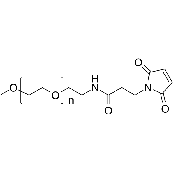
mPEG5000-Mal (mPEG5000-Maleimide) is a PEG-derived selective covalent binding agent for sulfhydryl groups (RSGs), which can form irreversible thioether bonds with sulfhydryl groups under near-neutral conditions via the maleimide group. The mechanism of action of mPEG5000-Mal can be divided into two categories: firstly, as an enzyme modifier, it binds to target proteins through hydrophobic interactions, hydrogen bonds, and van der Waals forces, altering the protein's secondary structure; secondly, as a nanoparticle surface modifier, it covalently binds to sulfhydryl groups on the surface of red blood cells, changing the surface properties and morphology of the red blood cells, leading to their phagocytosis by macrophages of the reticuloendothelial system. mPEG5000-Mal can react with free cysteine in proteins, increasing the apparent molecular weight of the modified protein by 10-15 kDa for detection purposes. mPEG5000-Mal can enhance the thermal stability and catalytic activity of enzymes, and improve the macrophage targeting of nanoparticles, enabling targeted drug delivery. mPEG5000-Mal can be applied in enzyme engineering research in the food industry and in oncology, assisting radiotherapy by inhibiting tumor-associated macrophage infiltration and enhancing anti-tumor immune responses .
loading...
    loading...
Cat. No.: HY-L249
6,182 compounds

Protein lactylation, an emerging post-translational modification identified in recent years, plays a critical role in linking cellular metabolic reprogramming, epigenetic regulation, and signaling networks. Based on a systematic framework encompassing lactate metabolism, lactylation, and downstream signaling pathways, this compound library comprehensively targets multiple regulatory layers, including histone modification enzymes (such as p300 and HDACs), key glycolytic enzymes (such as PKM2, LDHA, and GAPDH), transcriptional regulators (such as STAT3, HMGB1, and p53), as well as central signaling pathway nodes including HIF-1α, NF-κB, and PI3K-AKT-mTOR. This integrated design enables a comprehensive representation of the regulatory roles of lactylation across the “metabolism–epigenetics–signaling” axis.

MCE has assembled a collection of 6,182 known bioactive compounds and potential functional molecules, making this library suitable for a wide range of applications, including high-throughput drug screening, inhibitor identification, and mechanistic studies. It can be used to systematically evaluate the functional roles of lactylation in biological processes such as tumor metabolism, immune regulation, and inflammatory responses, and to efficiently identify small-molecule candidates with regulatory potential, thereby facilitating the development of innovative therapeutics targeting the interplay between metabolism and epigenetic regulation.

Cat. No.: HY-153552
CAS No.: 2758337-19-6
Target:  

FAP

Research Areas:  

Cancer

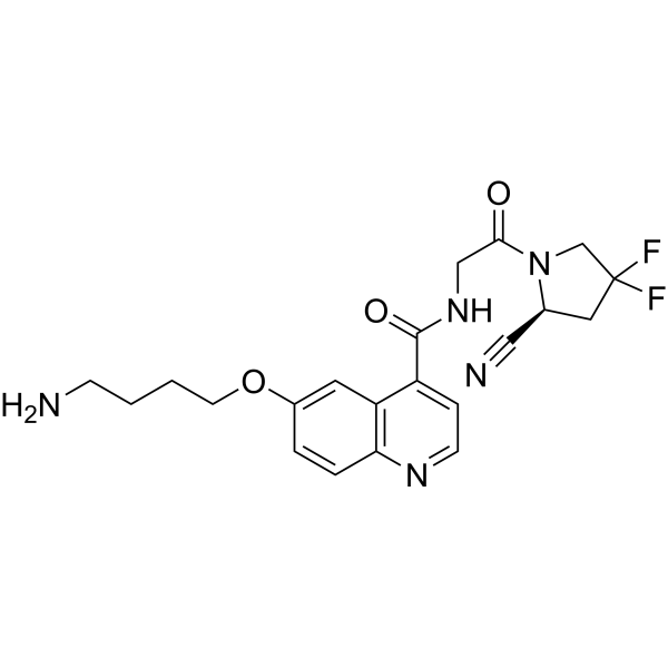
NH2-UAMC1110 is an aminobutoxy derivative of the fibroblast activation protein (FAP) inhibitor UAMC1110 (HY-100684), and is a precursor compound for the synthesis of FAP inhibitor probes, not directly used in bioactivity experiments. For example, NH2-UAMC1110 is involved in the synthesis of the radiotracer FAPI-QS, which exhibits high tumor selectivity and high dose-response, and has been used for tumor diagnosis. NH2-UAMC1110 introduces an active amino group into its structure, enabling it to form covalent bonds with various molecules (such as DOTA, DATA5m, radionuclide chelators, etc.), thereby synthesizing molecular imaging probes or targeted compounds with the ability to target FAP. NH2-UAMC1110 specifically binds to the FAP active site, inhibiting its proline-selective serine protease activity (including dipeptidyl peptidase and endopeptidase activity), blocking FAP-mediated tissue remodeling processes. Its key activity is high targeting and high affinity, and its core function is to be coupled with bifunctional chelators (such as DOTA, DATA5m) as a targeting module. NH2-UAMC1110 can be applied to diagnostic imaging studies of tumors expressing FAP (such as colorectal cancer, pancreatic cancer, etc.), and also provides molecular tools for targeted research of FAP-related diseases with high FAP expression, such as fibrosis and arthritis .
loading...
    loading...
Cat. No.: HY-P990252
Anti-Mouse Delta-like protein 4/DLL4 Antibody (HMD4-2) is an anti-mouse Delta-like protein 4/DLL4 IgG monoclonal antibody. Anti-Mouse Delta-like protein 4/DLL4 Antibody (HMD4-2) can reduce angiogenesis and density by blocking the DLL4-Notch signaling pathway. Anti-Mouse Delta-like protein 4/DLL4 Antibody (HMD4-2) reduces inflammatory response by decreasing NF-κB activity and pro-inflammatory factors (IL-1β, iNOS, IL-6) levels. Anti-Mouse Delta-like protein 4/DLL4 Antibody (HMD4-2) can inhibit Th17 cell differentiation and IL-17A production. Anti-Mouse Delta-like protein 4/DLL4 Antibody (HMD4-2) can reduce macrophage infiltration and alleviate insulin resistance. Anti-Mouse Delta-like protein 4/DLL4 Antibody (HMD4-2) can be used for researches on inflammation, metabolic conditions and cancer such as atherosclerosis, pancreatic cancer and asthma .
loading...
    loading...
Cat. No.: HY-P702970
Purity:  ≥ 90%, as determined by reducing SDS-PAGE.
Synonyms: MEF2A; Myocyte Enhancer Factor 2A Isoform 5; Myocyte Enhancer Factor 2A; Myocyte Enhancer Factor 2A Isoform 3; RSRFC4; Myocyte Enhancer Factor 2A Isoform 4; RSRFC9; Myocyte Enhancer Factor 2A Isoform 1; Serum Response Factor-Like Protein 1; Myocyte Enhancer Factor 2A Isoform 2; Myocyte-Specific Enhancer Factor 2A; Alternative Protein MEF2A; MADS Box Transcription Enhancer Factor 2, Polypeptide A (Myocyte Enhancer Factor 2A), Isoform CRA_d; ADCAD1; MADS Box Transcription Enhancer Factor 2, Polypeptide A (Myocyte Enhancer Factor 2A), Isoform CRA_b; Mef2; MADS Box Transcription Enhancer Factor 2, Polypeptide A (Myocyte Enhancer Factor 2A), Isoform CRA_a; MEF2; MADS Box Transcription Enhancer Factor 2, Polypeptide A (Myocyte Enhancer Factor 2A)
Species:  
Human
Source:  
Sf9 insect cells
loading...
    loading...
Cat. No.: HY-123996
CAS No.: 20041-64-9
3-Ethoxy-5,6-dibromosalicylaldehyde is an IRE1/ERN1 inhibitor, with an IC50 of 0.12 μM, a Ki of 71-88 nM, and a Kd of 100 nM against the ribonuclease activity of hIRE1α, as well as an IC50 of 4.8 μM against yeast Ire1. It shows selectivity toward IRE1 ribonuclease. 3-Ethoxy-5,6-dibromosalicylaldehyde blocks the IRE1/ERN1-mediated unfolded protein response (UPR) signaling pathway, including XBP-1 mRNA splicing, induction of XBP1 target genes, and activation of MAPK8/9/10, but does not alter the phosphorylation level of IRE1α or the PERK/ATF6 pathway. 3-Ethoxy-5,6-dibromosalicylaldehyde inhibits chikungunya virus replication, induces growth arrest, apoptosis and clonogenic inhibition in pancreatic cancer cells, reduces FB1 (Fumonisin B1) (HY-N6719)-induced autophagy and cell death, regulates PGG-induced senescence and apoptosis, and alleviates Sorafenib (HY-10201)-induced vacuolization and damage in hepatic stellate cells. 3-Ethoxy-5,6-dibromosalicylaldehyde can be used in research related to chikungunya virus infection, pancreatic cancer, FB1-induced nephrotoxicity, liver cancer, breast cancer, lung cancer and liver fibrosis .
loading...
    loading...
Cat. No.: HY-D3103
CAS No.: 1027058-07-6
Target:  

Fluorescent Dye

Research Areas:  

Others

CMTDP is a Fluorescent probe for detecting local polarity changes around the Cys34 domain of bovine serum albumin during pH-induced N→B conformational transition. It is a thiol-specific and polarity-sensitive probe; it covalently labels the free cysteine residue at position 34 (Cys34) of bovine serum albumin, and its fluorescence maximum emission wavelength shifts in response to solvent polarity, but not to pH and temperature. When labeled to BSA, as the local polarity around the Cys34 domain increases with rising pH, the probe shows a bathchromic shift in fluorescence emission wavelength and a decrease in fluorescence intensity, which allows quantification of the local polarity change via the relationship between emission wavelength shift and dielectric constant. CMTDP itself has absorption peaks at ~392 and 508 nm, and CMTDP-labeled BSA has excitation maxima at 376 and 470 nm at pH 6.0, with the shorter-wavelength excitation peak red-shifting as pH increases while the longer one remains stable; when excited at 470 nm, the fluorescence emission maximum shifts from 570 nm at pH 6.0 to 577 nm at pH 9.1, while free CMTDP at pH 7.4 has an emission maximum at 621 nm. The excitation/emission wavelengths for CMTDP-labeled BSA are Ex/Em = 376/570 nm at pH 6.0, Ex/Em = ~376+/573 nm at pH 7.4, Ex/Em = ~376+/575 nm at pH 8.0, Ex/Em = ~376+/577 nm at pH 9.1, and for free CMTDP at pH 7.4, Ex/Em (when excited at 470 nm) = 470/621 nm[1].
loading...
    loading...
Cat. No.: HY-W127487
CAS No.: 479050-96-9
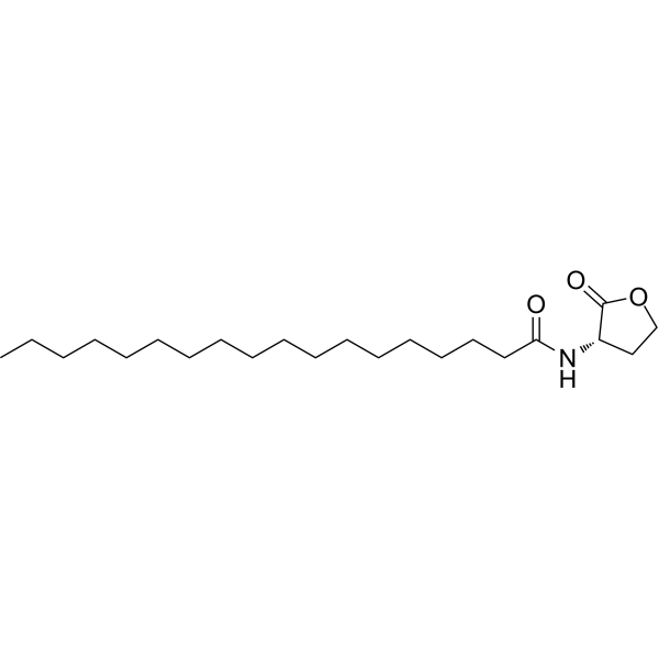
Quorum sensing is a regulatory system used by bacteria to control gene expression in response to increased cell density. This regulatory process manifests itself in a variety of phenotypes, including biofilm formation and virulence factor production. Coordinated gene expression is achieved through the production, release and detection of small diffusible signaling molecules called autoinducers. N-acylated homoserine lactones (AHLs) comprise a class of such autoinducers, each of which generally consists of a fatty acid coupled to a homoserine lactone (HSL). Modulation of bacterial quorum-sensing signaling systems to suppress pathogenesis represents a new approach to antimicrobial research for infectious diseases. AHLs differ in acyl length (C4-C18), C3 substitution (hydrogen, hydroxyl, or oxo group), and the presence or absence of one or more carbon-carbon double bonds in the fatty acid chain. These differences confer signaling specificity through the affinity of the LuxR family of transcriptional regulators. C18-HSL, one of four lipophilic long acyl side chain AHLs produced by the LuxI AHL synthase homolog SinI, is involved in quorum-sensing signaling in strains of Rhizobium meliloti (a nitrogen-fixing bacterial symbiont of the legume M. sativa) . C18-HSL and other hydrophobic AHLs tend to localize in the relatively lipophilic environment of bacterial cells and cannot diffuse freely across the cell membrane. Long-chain N-acyl homoserine lactones can be exported from cells by efflux pumps, or can be transported between communicating cells by extracellular outer membrane vesicles.
loading...
    loading...
Cat. No.: HY-D3133
CAS No.: 2410296-16-9
Target:  

Fluorescent Dye

Research Areas:  

Others

HS-CyBz is a Fluorescent probe for H₂S detection, enabling ratiometric optical/photoacoustic dual-modality in/ex vivo imaging. Its detection mechanism relies on nucleophilic substitution of its benzoic ester group by HS⁻, which releases an enolic meso-hydroxyltricarboheptamethine cyanine that then undergoes keto-enol tautomerization to form Cy-ketone; this tautomerization causes distinct shifts in absorption and emission spectra, producing a ratiometric response that reduces interferences from tissue scattering, autofluorescence, and probe concentration. In its initial state, HS-CyBz has an excitation wavelength of 595 nm, with emission bands centered at 805 nm (main) and 630 nm (minor); upon reaction with H₂S, the 805 nm emission band decreases while the 630 nm band drastically increases, and its absorption spectrum shows a sharp band at 775 nm and a shoulder band at 708 nm, which decrease upon H₂S reaction with a minor increase at 850 nm and an isosbestic point at 825 nm. For in vivo optical imaging, excitation at 560 nm is used with emission channels at 620 nm and 790 nm, while in vivo photoacoustic imaging uses excitation at 775 nm and 825 nm. The detection limit of HS-CyBz for H₂S is 0.5 μM, and it shows high selectivity, with only H₂S inducing a distinct enhancement of the emission ratio F₆₃₀/F₈₀₅ and PA ratio PA₈₂₅/PA₇₇₅, while other biochemical species including cations, anions, reactive oxygen species, biothiols, and carboxylesterase trigger only minor changes and do not interfere with H₂S sensing. Tail intravenous injection of HS-CyBz leads to accumulation in the liver of mice, and it can be used to verify endogenous H₂S upregulation triggered by S-adenosyl-L-methionine via ratiometric optical/photoacoustic imaging .
loading...
    loading...
Cat. No.: HY-N0660
CAS No.: 55466-05-2
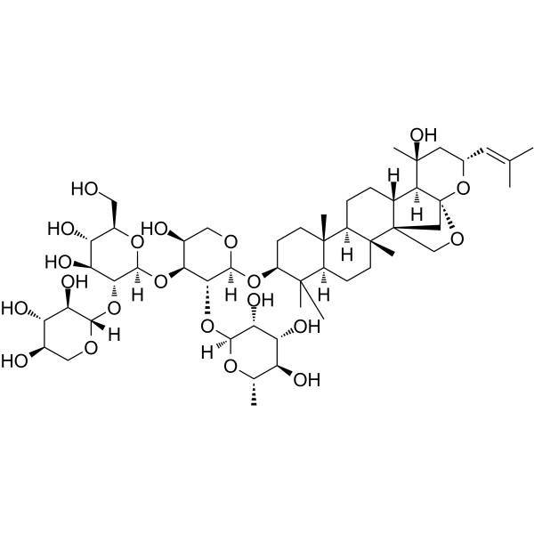
Jujuboside B is a bioactive saponin component isolated from Ziziphi Spinosae Semen (sour jujube seed), with oral efficacy and blood-brain barrier permeability. Jujuboside B induces acute leukemia cell death and drives necroptosis apoptosis by activating the RIPK1/RIPK3/MLKL pathway. Jujuboside B upregulates the expression of NOXA, PARP and caspase-3, activates AMPK, inhibits the proliferation of breast cancer cells, and induces cell apoptosis and autophagy. Jujuboside B inhibits angiogenesis and tumor growth by blocking the VEGFR-2 signaling pathway. Jujuboside B alleviates liver injury in mice by regulating the Nrf2-STING signaling pathway . Jujuboside B alleviates liver injury by regulating anti-inflammatory responses and downregulating the expression of 11β-HSD2. Jujuboside B induces ferroptosis and overcomes radioresistance in non-small cell lung cancer via the PPARγ-ATF3-Gpx4 signaling pathway. Jujuboside B exerts inhibitory effects on platelet aggregation. Jujuboside B inhibits febrile seizures by suppressing the activity of AMPA receptors. Jujuboside B reverses chronic unpredictable mild stress-promoted tumor progression by blocking the PI3K/Akt and MAPK/ERK pathways and dephosphorylating CREB signaling. Jujuboside B is applicable to related studies on acute leukemia, breast cancer, PM2.5-induced lung injury, hepatotoxicity, liver injury, colorectal cancer, non-small cell lung cancer, thromboembolic diseases, cardiovascular diseases associated with high platelet aggregation, febrile seizures, and depressive-like phenotypes .
loading...
    loading...
Cat. No.: HY-N0660R
CAS No.: 55466-05-2
Jujuboside B (Standard) is the analytical standard of Jujuboside B. This product is intended for research and analytical applications. Jujuboside B is a bioactive saponin component isolated from Ziziphi Spinosae Semen (sour jujube seed), with oral efficacy and blood-brain barrier permeability. Jujuboside B induces acute leukemia cell death and drives necroptosis apoptosis by activating the RIPK1/RIPK3/MLKL pathway. Jujuboside B upregulates the expression of NOXA, PARP and caspase-3, activates AMPK, inhibits the proliferation of breast cancer cells, and induces cell apoptosis and autophagy. Jujuboside B inhibits angiogenesis and tumor growth by blocking the VEGFR-2 signaling pathway. Jujuboside B alleviates liver injury in mice by regulating the Nrf2-STING signaling pathway . Jujuboside B alleviates liver injury by regulating anti-inflammatory responses and downregulating the expression of 11β-HSD2. Jujuboside B induces ferroptosis and overcomes radioresistance in non-small cell lung cancer via the PPARγ-ATF3-Gpx4 signaling pathway. Jujuboside B exerts inhibitory effects on platelet aggregation. Jujuboside B inhibits febrile seizures by suppressing the activity of AMPA receptors. Jujuboside B reverses chronic unpredictable mild stress-promoted tumor progression by blocking the PI3K/Akt and MAPK/ERK pathways and dephosphorylating CREB signaling. Jujuboside B is applicable to related studies on acute leukemia, breast cancer, PM2.5-induced lung injury, hepatotoxicity, liver injury, colorectal cancer, non-small cell lung cancer, thromboembolic diseases, cardiovascular diseases associated with high platelet aggregation, febrile seizures, and depressive-like phenotypes.
loading...
    loading...
Cat. No.: HY-125209A
CAS No.: 2253744-57-7
Research Areas:  

Cancer

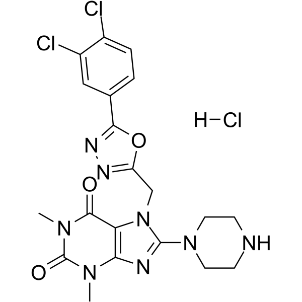
TH5427 hydrochloride is a NUDT5 inhibitor with a human target IC50 of 29 nM, ~690-fold selectivity over MTH1 in vitro, and selective functional inhibition over other NUDIX hydrolases including NUDT9 .TH5427 hydrochloride binds to the active site of NUDT5, blocking enzymatic activity related to ADP-ribose metabolism and PAR-derived ATP synthesis .TH5427 hydrochloride blocks progestin-dependent nuclear ATP synthesis, impairs progestin-induced chromatin remodeling, inhibits histone H1 displacement, disrupts progestin-dependent gene regulation, and abrogates progestin-dependent proliferation in breast cancer cells .TH5427 hydrochloride functions as a versatile probe to study nuclear ATP dynamics and ADP-ribose-related metabolism in cells .TH5427 hydrochloride engages NUDT5 at physiological temperatures, as demonstrated by Drug Affinity Responsive Target Stability (DARTS) assay .TH5427 hydrochloride stabilizes NUDT5 against thermal denaturation in cell lysates and intact cells, as shown by cellular thermal shift assay (CETSA) .TH5427 hydrochloride functionally inhibits NUDT5 activity, leading to downstream effects on oxidative DNA damage and DNA replication in triple-negative breast cancer (TNBC) cells .TH5427 hydrochloride suppresses proliferation of TNBC cells without inducing cell death or apoptosis, slows DNA replication in TNBC cells, promotes accumulation of oxidative DNA lesions, and triggers DNA damage response in TNBC cells .TH5427 hydrochloride suppresses growth of TNBC cells in vitro, inhibits growth of TNBC xenograft tumors in nude mice in vivo, and shows greater potency against TNBC cell lines compared to ER-positive and normal-like breast cell lines .TH5427 hydrochloride can be used for the research of breast cancer and triple-negative breast cancer .
loading...
    loading...
Cat. No.: HY-P86921
Synonyms: AIF 1 antibody; AIF-1 antibody; Aif1 antibody; AIF1 protein antibody; AIF1_HUMAN antibody; Allograft inflammatory factor 1 antibody; Allograft inflammatory factor 1 splice variant G antibody; allograft inflammatory factor-1 splice variant Hara-1 antibody; balloon angioplasty responsive transcription antibody; BART 1 antibody; AIF 1 antibody; AIF-1 antibody; Aif1 antibody; AIF1 protein antibody; AIF1_HUMAN antibody; Allograft inflammatory factor 1 antibody; Allograft inflammatory factor 1 splice variant G antibody; allograft inflammatory factor-1 splice variant Hara-1 antibody; balloon angioplasty responsive transcription antibody; BART 1 antibody; G1 antibody; G1 putative splice variant of allograft inflamatory factor 1 antibody; IBA 1 antibody; IBA1 antibody; interferon gamma responsive transcript antibody; Interferon responsive transcript 1 antibody; interferon responsive transcript factor 1 antibody; Ionized calcium binding adapter molecule 1 antibody; Ionized calcium-binding adapter molecule 1 antibody; ionized calcium-binding adapter molecule antibody; IRT 1 antibody; IRT1 antibody; Microglia Response factor antibody; MRF1 antibody; Protein g1 antibody;

Host:  

Rabbit

Application:  

mIHC

Reactivity:  

Mouse

loading...
    loading...

