9 Results for "

free cysteine residue

" in MedChemExpress (MCE) Product Catalog:
Products (9)

9 Results for "free cysteine residue" in MCE Product Catalog:

46
46 Publications Verification
Cat. No.: HY-D0843
CAS No.: 128-53-0
Synonyms: NEM
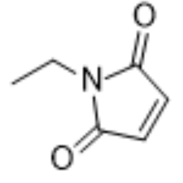
N-Ethylmaleimide (NEM) derives from maleic acid, it can alkylates free sulfhydryl. N-Ethylmaleimide is an irreversible cysteine protease inhibitor. N-ethylmaleimide specific inhibits phosphate transport in mitochondria. N-Ethylmaleimide inhibits prolyl endopeptidase with an IC50 value of 6.3 μM. N-Ethylmaleimide can be used to modify cysteine residues in proteins and peptides .
loading...
    loading...
28
28 Cited Publications
Cat. No.: HY-135056
CAS No.: 201860-17-5
Purity:  95.36%
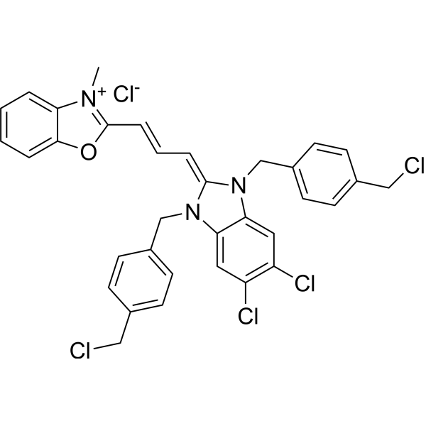
Mito-Tracker Green is a green fluorescent dye that selectively accumulates in the mitochondrial matrix. MitoTracker Green FM covalently binds mitochondrial proteins by reacting with free mercaptan of cysteine residues, allowing staining of mitochondrial membrane potential independent of membrane potential. Excitation/emission wavelength 490/523 nm.
loading...
    loading...
19
19 Cited Publications
Cat. No.: HY-D1783
CAS No.: 873315-86-7
Synonyms: MTDR FM
Target:  

Fluorescent Dye

Research Areas:  

Cancer

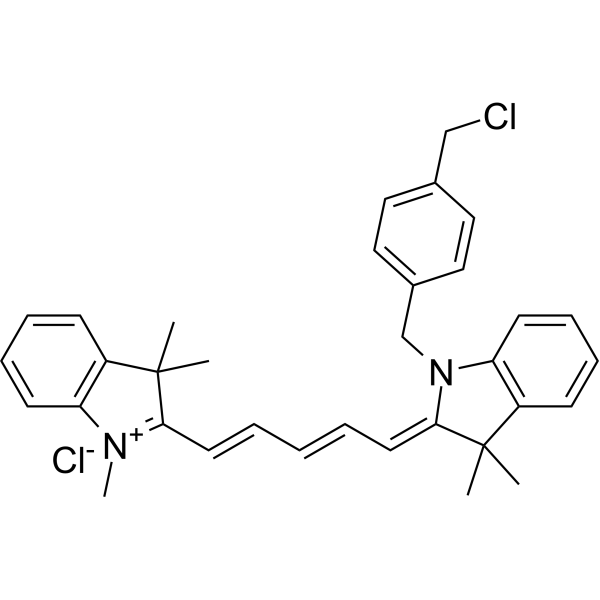
MitoTracker Deep Red (MTDR) FM fluorescent dye that selectively accumulates in the mitochondrial matrix. MitoTracker Deep Red FM covalently binds mitochondrial proteins by reacting with free mercaptan of cysteine residues, allowing staining of mitochondrial membrane potential independent of membrane potential. Excitation/emission wavelength 644/665 nm . MitoTracker Deep Red dyes have an excitation/emission wavelength of 633/650-750 nm .
The Ex/Em of MitoTracker Deep Red (MTDR) FM is 644/665 nm.
loading...
    loading...
Cat. No.: HY-DY1032
CAS No.: 201860-17-5
Mito-Tracker Green (solution) is a green fluorescent dye that selectively accumulates in the mitochondrial matrix. MitoTracker Green FM covalently binds mitochondrial proteins by reacting with free mercaptan of cysteine residues, allowing staining of mitochondrial membrane potential independent of membrane potential. Excitation/emission wavelength 490/523 nm.
Solvent and concentration: DMSO: 1 mM
loading...
    loading...
Cat. No.: HY-120128
CAS No.: 16599-33-0
Purity:  97.90%
Research Areas:  

Neurological Disease

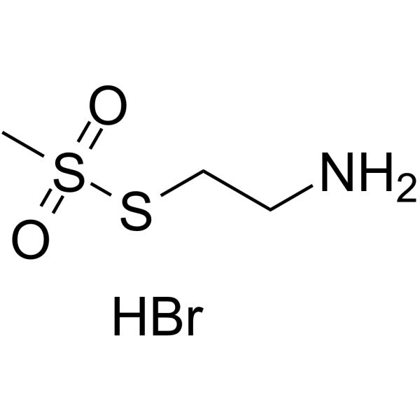
MTSEA hydrobromide is a sulfhydryl-reactive compound that modifies free cysteine residues to produce a positively charged side chain approximately the size of lysine .
loading...
    loading...
Cat. No.: HY-152919
CAS No.: 2387738-43-2
Target:  

ADC Linkers

Research Areas:  

Cancer

Mal-amide-PEG8-Val-Cit-PAB-OH is a cleavable ADC linker featuring a maleimide, a hydrophilic PEG spacer, a Val-Cit dipeptide, and a PAB functional group. Maleimide is used to covalently bind free thiols on the cysteine residues of proteins. The Val-Cit dipeptide is cleaved by cellular proteases within the cell to allow for efficient payload delivery with the help of the PAB structure.
loading...
    loading...
Cat. No.: HY-DY1105
CAS No.: 873315-86-7
Synonyms: MTDR FM (solution)
Target:  

Fluorescent Dye

Research Areas:  

Others

MitoTracker Deep Red FM (solution) (MTDR FM (solution)) selectively accumulates in the mitochondrial matrix. MitoTracker Deep Red FM covalently binds mitochondrial proteins by reacting with free mercaptan of cysteine residues, allowing staining of mitochondrial membrane potential independent of membrane potential. The Ex/Em of MitoTracker Deep Red (MTDR) FM is 644/665 nm.
Solvent and concentration: DMSO: 1 mM
loading...
    loading...
Cat. No.: HY-D3436
Target:  

Fluorescent Dye

Research Areas:  

Cancer

Pt-Luc is a bioluminescent probe used for real-time, non-invasive monitoring of Pt (IV) prodrug reduction in living cells and animal models. The detection mechanism of Pt-Luc relies on the intracellular reduction mediated by active cysteine residues in proteins. Pt-Luc can penetrate the membrane of living cells; after entering cells, active cysteine thiolates in high-molecular-weight protein components reduce the Pt (IV) complex, thereby cleaving the caged aminoluciferin substrate. The released free aminoluciferin is then oxidized by luciferase in the presence of ATP to generate a bioluminescent signal, whose intensity is directly correlated with the reduction degree of Pt (IV) .
loading...
    loading...
Cat. No.: HY-D3103
CAS No.: 1027058-07-6
Target:  

Fluorescent Dye

Research Areas:  

Others

CMTDP is a Fluorescent probe for detecting local polarity changes around the Cys34 domain of bovine serum albumin during pH-induced N→B conformational transition. It is a thiol-specific and polarity-sensitive probe; it covalently labels the free cysteine residue at position 34 (Cys34) of bovine serum albumin, and its fluorescence maximum emission wavelength shifts in response to solvent polarity, but not to pH and temperature. When labeled to BSA, as the local polarity around the Cys34 domain increases with rising pH, the probe shows a bathchromic shift in fluorescence emission wavelength and a decrease in fluorescence intensity, which allows quantification of the local polarity change via the relationship between emission wavelength shift and dielectric constant. CMTDP itself has absorption peaks at ~392 and 508 nm, and CMTDP-labeled BSA has excitation maxima at 376 and 470 nm at pH 6.0, with the shorter-wavelength excitation peak red-shifting as pH increases while the longer one remains stable; when excited at 470 nm, the fluorescence emission maximum shifts from 570 nm at pH 6.0 to 577 nm at pH 9.1, while free CMTDP at pH 7.4 has an emission maximum at 621 nm. The excitation/emission wavelengths for CMTDP-labeled BSA are Ex/Em = 376/570 nm at pH 6.0, Ex/Em = ~376+/573 nm at pH 7.4, Ex/Em = ~376+/575 nm at pH 8.0, Ex/Em = ~376+/577 nm at pH 9.1, and for free CMTDP at pH 7.4, Ex/Em (when excited at 470 nm) = 470/621 nm[1].
loading...
    loading...

