



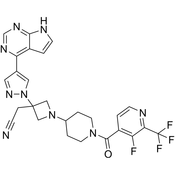
Itacitinib
Based on 23 publication(s) in Google Scholar
Itacitinib (INCB039110) is an orally active selective inhibitor of JAK1 with an IC50 value of 2 nM for human JAK1. Itacitinib inhibits IFN-γ-mediated phosphorylation of STAT1 and downstream pro-inflammatory signaling pathways. Itacitinib reduces the frequency and number of splenic neutrophils in mouse models, downregulates the levels of pro-inflammatory cytokines and chemokines, and inhibits pro-inflammatory gene expression pathways. Itacitinib improves survival rate and clinical scores in mouse models of hemophagocytic lymphohistiocytosis (HLH), and also suppresses metastasis in NSCLC models with high Rab1A expression. Itacitinib can be used for research on hemophagocytic lymphohistiocytosis and non-small cell lung cancer.
For research use only. We do not sell to patients.
- Purity : 99.97%
- CAS No.: 1334298-90-6
- Formula: C26H23F4N9O
- Molecular Weight:553.51
-
Storage:Powder -20°C, 3 years , 4°C, 2 years ; In solvent -80°C, 1 year , -20°C, 6 months

To place orders, for customer services and technical support, please contact: MedChemExpress USA
Tel: 609-228-6898 E-mail: [email protected] [email protected]


-
 Biological Activity
-
Chemical Information
-
Solvent & Solubility
- Protocols
- Purity & Documentation
- References
-
 Help & FAQs


-
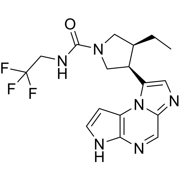
HY-19569
UpadacitinibJAK1 Inhibitor -
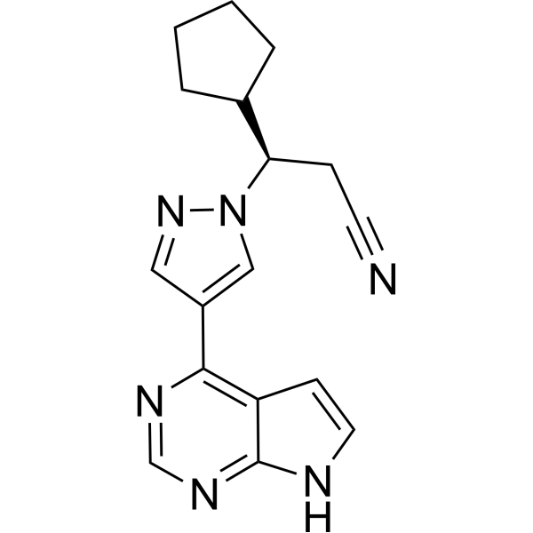
HY-50856
RuxolitinibJAK1/2 Inhibitor -
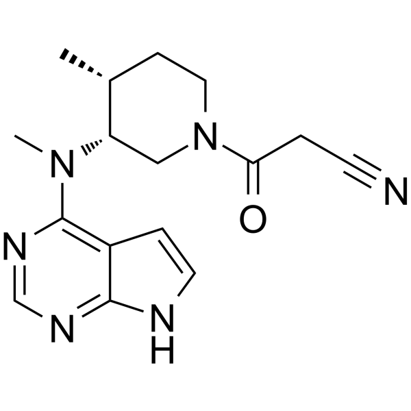
HY-40354
TofacitinibJAK1/JAK3 Inhibitor -
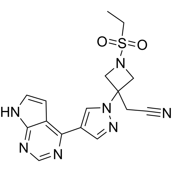
HY-15315
BaricitinibJAK1/2 Inhibitor -
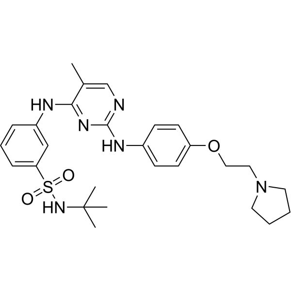
HY-10409
FedratinibJAK2 Inhibitor -
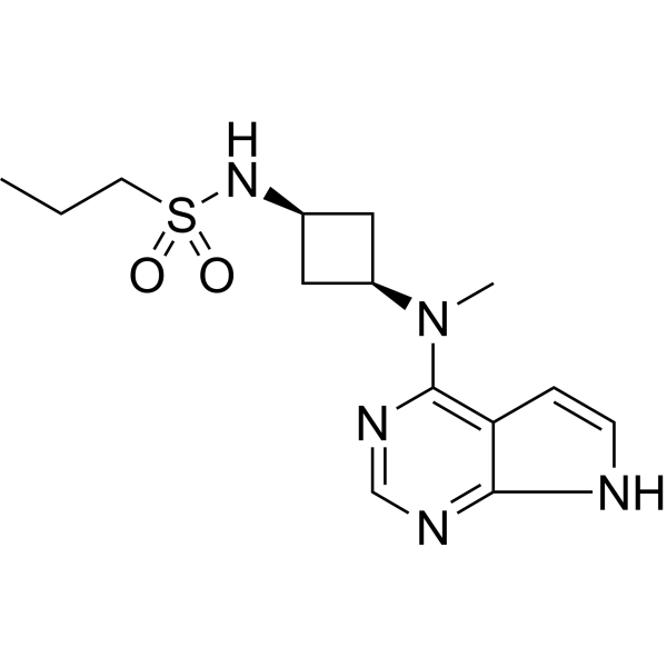
HY-107429
AbrocitinibJAK1 Inhibitor -
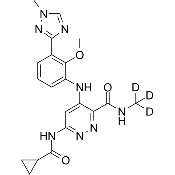
HY-117287
DeucravacitinibTYK2 Inhibitor -
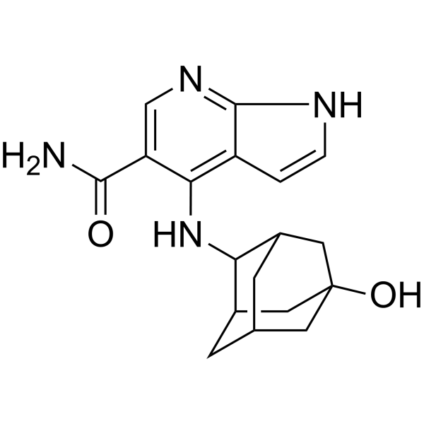
HY-19568
PeficitinibJAK Inhibitor -
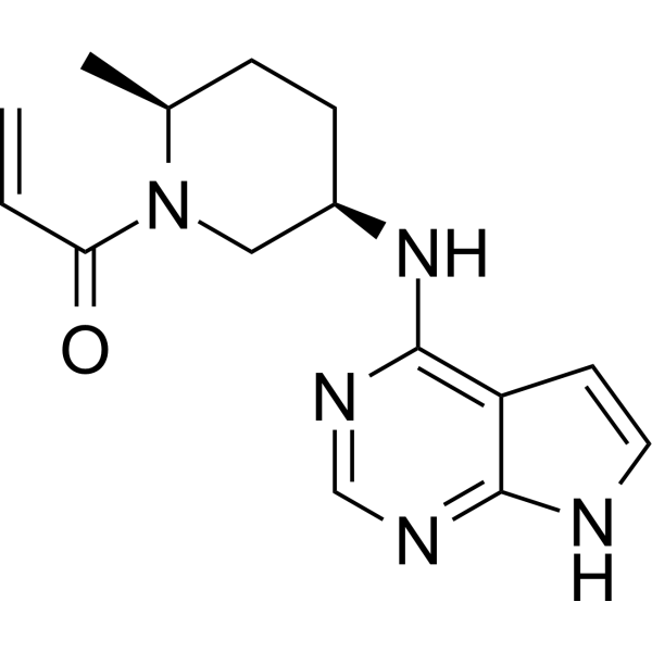
HY-100754
RitlecitinibJAK3 Inhibitor -
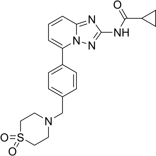
HY-18300
FilgotinibJAK1 Inhibitor -
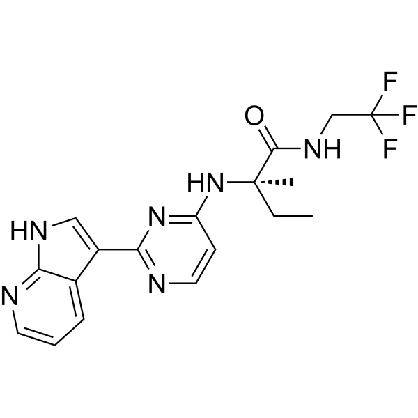
HY-12469
DecernotinibJAK Inhibitor -
HY-15270
BMS-911543JAK2 Inhibitor
Publications Citing Use of MedChemExpress (MCE) Itacitinib
More- Nature. 2025 Jun;642(8066):201-211. [Abstract]
- Circulation. 2024 Oct 15;150(16):1302-1305. [Abstract]
- Nat Commun. 2025 Sep 29;16(1):8560. [Abstract]
- Oncogene. 2024 Oct;43(41):3062-3077. [Abstract]
- Leukemia. 2019 Aug;33(8):1964-1977. [Abstract]
- J Med Chem. 2024 Jun 27;67(12):10012-10024. [Abstract]
- JCI Insight. 2021 Apr 8;6(7):e142205. [Abstract]
- J Autoimmun. 2019 May:99:39-47. [Abstract]
- Mol Syst Biol. 2024 Jan;20(1):28-55. [Abstract]
- CNS Neurosci Ther. 2025 Sep;31(9):e70609. [Abstract]
- Biochem Pharmacol. 2025 Oct:240:117120. [Abstract]
- Biochem Pharmacol. 2020 Aug:178:114103. [Abstract]
- EMBO Rep. 2019 Jun;20(6):e47202. [Abstract]
- Cell Rep Methods. 2023 Oct 23;3(10):100599. [Abstract]
- Int Immunopharmacol. 2024 Aug 20:137:112523. [Abstract]
- J Immunol. 2023 Feb 1;210(3):229-244. [Abstract]
- J Leukoc Biol. 2022 Nov;112(5):1343-1356. [Abstract]
- Technol Cancer Res Treat. 2025 Jan-Dec:24:15330338251406931. [Abstract]
- Fundam Clin Pharmacol. 2021 Oct;35(5):919-929. [Abstract]
- Eur J Drug Metab Pharmacokinet. 2021 Sep;46(5):625-635. [Abstract]
- SSRN. 2026 May 21.
- bioRxiv. 2026 Mar 7.
- bioRxiv. 2025 February 21.


Biological Activity
Description
|
JAK1 2 nM (IC50) |
STAT1 |
In Vitro
Itacitinib adipate (50-2500 nM; 1 h pretreatment) dose-dependently inhibits IFN-γ-induced STAT1 phosphorylation in bone marrow-derived macrophages, with significant suppression observed at 2500, 500, and 50 nM concentrations[1].
Itacitinib (INCB039110) is a potent and selective JAK1 inhibitor with an IC50 value of 2 nM for human JAK1. Its selectivity for JAK1 is more than 20 times that for JAK2, and its selectivity for JAK3 and TYK2 is more than 100 times[4].
MedChemExpress (MCE) has not independently confirmed the accuracy of these methods. They are for reference only. Further protocols information, click here.
In Vivo
Itacitinib adipate (120 mg/kg; p.o.; twice daily; for 30 consecutive days) is well tolerated in untreated wild-type mice and effectively inhibits IFN-γ-induced STAT1 phosphorylation in vivo[1].
Itacitinib adipate (120 mg/kg; p.o.; twice daily; for 5 or 26 consecutive days) partially improves survival rate and clinical scores in a primary HLH mouse model, and exerts only mild effects on disease-related immune parameters and transcriptional profiles[1].
Itacitinib adipate (25 mg/kg; intraperitoneal injection; administration schedule consists of 9 cycles, with 5 consecutive days of dosing followed by 2 days of withdrawal per cycle, lasting for 2 months) significantly inhibits the metastasis of non-small cell lung cancer with high Rab1A expression in the xenograft model derived from the A549-Rab1A-OE cell line, reduces bioluminescence flux and the number of metastatic organs; it also significantly reduces the number of metastatic organs in the non-small cell lung cancer xenograft model derived from the H358 cell line[2].
MedChemExpress (MCE) has not independently confirmed the accuracy of these methods. They are for reference only.
-
Animal Model:C57BL/6J wildtype (sex- and age-matched, 8 to 12 weeks old; secondary HLH induced by intraperitoneal injection of CpG 1826 (HY-146245) and anti-IL-10 receptor antibody)[1]
-
Dosage:120 mg/kg
-
Administration:p.o.; twice daily; 5 or 6 days
-
Result:Markedly extended survival versus vehicle.
Lowered clinical scores with equivalent efficacy to ruxolitinib.
Exerted no beneficial effects on splenomegaly or thrombocytopenia, and aggravated anemia relative to vehicle.
Sharply lowered the proportion and count of splenic neutrophils (CD80+ neutrophils included).
Dropped serum CXCL10, IL-12(p70), IL-6, GM-CSF, MCP-1 and MIP1a concentrations significantly.
-
Animal Model:C57BL/6J Prf1tm1Sdz/J (perforin-deficient, sex- and age-matched, 8 to 12 weeks old; primary HLH induced by intraperitoneal infection with LCMV)[1]
-
Dosage:120 mg/kg
-
Administration:p.o.; twice daily; 5 or 26 days
-
Result:Partially elevated survival relative to vehicle.
Reduced clinical disease scores relative to vehicle.
Alleviated splenomegaly at day 9 post-infection, with no impact on anemia or thrombocytopenia.
Failed to lower serum IFN-γ, TNF and sCD25 concentrations.
Did not alter total, CD44+ effector or gp33-specific splenic CD8 T cell populations, nor suppress IFN-γ secretion from these cells.
Repressed proinflammatory (IL-2 STAT5, IFN-α, IFN-γ response) and heme metabolism pathways in splenic CD8 T cells.
Suppressed proinflammatory IL-6 JAK STAT3, IFN-α and IFN-γ response pathways while boosting Myc-driven proliferation and oxidative phosphorylation metabolism in splenic monocytes; each cell subset only presented 3 distinct differentially expressed genes versus vehicle.
-
Animal Model:C57BL/6J wildtype (sex- and age-matched, 8 to 12 weeks old)[1]
-
Dosage:120 mg/kg
-
Administration:p.o.; twice daily; 30 days
-
Result:Showed no significant untoward effects on clinical score, body weight, bone marrow cellularity, or peripheral blood cell counts compared to vehicle-treated controls.
Significantly decreased the percentage of pSTAT1+ F4/80+ peritoneal macrophages and the fold-change mean fluorescence intensity of pSTAT1 compared to vehicle-treated mice after IFN-γ stimulation.
-
Animal Model:BALB/c nude (male, 6-8 weeks old, inoculated via tail vein with luciferase-expressing A549-Rab1A-OE cells)
[2] -
Dosage:25 mg/kg
-
Administration:i.p.; 9 cycles of 5 consecutive days on medication followed by 2 consecutive days off; over 2 months
-
Result:Reduced total bioluminescence flux from a median of ~7.0 lg[p/s] in controls to ~6.0 lg[p/s].
Decreased the number of metastatic organs from a median of ~6 organs in controls to ~2 organs.
-
Animal Model:BALB/c nude (male, 6-8 weeks old, inoculated via tail vein with H358 cells)[2]
-
Dosage:25 mg/kg
-
Administration:i.p.; 9 cycles of 5 consecutive days on medication followed by 2 consecutive days off; over 2 months
-
Result:Decreased the number of metastatic organs from a median of ~6 organs in controls to ~2 organs.
Clinical Trial
| NCT Number | Sponsor | Condition | Start Date |
Phase

|
|---|---|---|---|---|
| NCT01329991 | Plexxikon| | 2011-05 | PHASE1 |


Chemical Information
-
CAS No. 1334298-90-6
-
Appearance Solid
-
Molecular Weight 553.51
-
Formula C26H23F4N9O
-
Color White to light yellow
-
SMILES
N#CCC1(N2N=CC(C3=C4C(NC=C4)=NC=N3)=C2)CN(C5CCN(C(C6=C(F)C(C(F)(F)F)=NC=C6)=O)CC5)C1
-
Synonyms
INCB039110
-
Shipping
Room temperature in continental US; may vary elsewhere.
-
Storage
Powder -20°C 3 years 4°C 2 years In solvent -80°C 1 year -20°C 6 months


Publications (23)
-
Journal Impact Factor
-
Most Recent
-
Nature
2025 Jun;642(8066):201-211. PMID: 40269158 -
Circulation
Sustained but Decoyed Activation of the JAK1-STAT Pathway by Aberrant Protein Aggregation Exacerbates Proteotoxicity. [Abstract]2024 Oct 15;150(16):1302-1305. PMID: 39401278 -
Nat Commun
A loss-of-function human ADAR variant activates innate immune response and promotes bowel inflammation. [Abstract]2025 Sep 29;16(1):8560. PMID: 41022715 -
Oncogene
RBM12 drives PD-L1-mediated immune evasion in hepatocellular carcinoma by increasing JAK1 mRNA translation. [Abstract]2024 Oct;43(41):3062-3077. PMID: 39187545 -
Leukemia
Stem cell persistence in CML is mediated by extrinsically activated JAK1-STAT3 signaling. [Abstract]2019 Aug;33(8):1964-1977. PMID: 30842608 -
J Med Chem
Functional and Structural Characterization of Clinical-Stage Janus Kinase 2 Inhibitors Identifies Determinants for Drug Selectivity. [Abstract]2024 Jun 27;67(12):10012-10024. PMID: 38843875 -
JCI Insight
2021 Apr 8;6(7):e142205. PMID: 33830087 -
J Autoimmun
Autophagy promotes aortic adventitial fibrosis via the IL-6/Jak1 signaling pathway in Takayasu's arteritis. [Abstract]2019 May:99:39-47. PMID: 30765261 -
Mol Syst Biol
Illuminating phenotypic drug responses of sarcoma cells to kinase inhibitors by phosphoproteomics. [Abstract]2024 Jan;20(1):28-55. PMID: 38177929 -
CNS Neurosci Ther
Network Pharmacology-Based and Experimental Validation Elucidate the Target Mechanism of Vinorine in Ameliorating Secondary Brain Injury After Intracerebral Hemorrhage. [Abstract]2025 Sep;31(9):e70609. PMID: 40994248 -
Biochem Pharmacol
Dual inhibition of EGR1/STAT3 transcriptional hubs suppresses macrophage-driven liver fibrosis: A multi-omics-guided drug repurposing strategy. [Abstract]2025 Oct:240:117120. PMID: 40623460 -
Biochem Pharmacol
Combined anti-fibrotic and anti-inflammatory properties of JAK-inhibitors on macrophages in vitro and in vivo: Perspectives for scleroderma-associated interstitial lung disease. [Abstract]2020 Aug:178:114103. PMID: 32562787 -
EMBO Rep
Tau accumulation triggers STAT1-dependent memory deficits by suppressing NMDA receptor expression. [Abstract]2019 Jun;20(6):e47202. PMID: 31085626 -
Cell Rep Methods
RECOVER identifies synergistic drug combinations in vitro through sequential model optimization. [Abstract]2023 Oct 23;3(10):100599. PMID: 37797618 -
Int Immunopharmacol
APLNR inhibited nasopharyngeal carcinoma growth and immune escape by downregulating PD-L1. [Abstract]2024 Aug 20:137:112523. PMID: 38909500 -
J Immunol
IL-10 Negatively Controls the Primary T Cell Response of Tilapia by Triggering the JAK1/STAT3/SOCS3 Axis That Suppresses NF-κB and MAPK/ERK Signaling. [Abstract]2023 Feb 1;210(3):229-244. PMID: 36548476 -
J Leukoc Biol
2022 Nov;112(5):1343-1356. PMID: 35588262 -
Technol Cancer Res Treat
Combining Radiation and anti-PD-L1 Enhances the Antitumor Activity in Colorectal Cancer via IFN-γ-Dependent Activation of STAT1. [Abstract]2025 Jan-Dec:24:15330338251406931. PMID: 41406067 -
Fundam Clin Pharmacol
2021 Oct;35(5):919-929. PMID: 33523504 -
Eur J Drug Metab Pharmacokinet
Differential Inhibition of Equilibrative Nucleoside Transporter 1 (ENT1) Activity by Tyrosine Kinase Inhibitors. [Abstract]2021 Sep;46(5):625-635. PMID: 34275128 -
-
-


Solvent & Solubility
In Vitro:
DMSO : ≥ 30 mg/mL (54.20 mM; Hygroscopic DMSO has a significant impact on the solubility of product, please use newly opened DMSO)
* "≥" means soluble, but saturation unknown.
Please refer to the solubility information to select the appropriate solvent. Once prepared, please aliquot and store the solution to prevent product inactivation from repeated freeze-thaw cycles.
Storage method and period of stock solution: -80°C, 1 year; -20°C, 6 months. When stored at -80°C, please use it within 1 year. When stored at -20°C, please use it within 6 months.
Please refer to the solubility information to select the appropriate solvent. Once prepared, please aliquot and store the solution to prevent product inactivation from repeated freeze-thaw cycles.
Storage method and period of stock solution: -80°C, 1 year; -20°C, 6 months. When stored at -80°C, please use it within 1 year. When stored at -20°C, please use it within 6 months.
Concentration (start) × Volume (start) = Concentration (final) × Volume (final)
In Vivo:
Select the appropriate dissolution method based on your experimental animal and administration route.
- For the following dissolution methods, please ensure to first prepare a clear stock solution using an In Vitro approach and then sequentially add co-solvents:
- To ensure reliable experimental results, the clarified stock solution can be appropriately stored based on storage conditions. As for the working solution for In Vivo experiments, it is recommended to prepare freshly and use it on the same day.
- The percentages shown for the solvents indicate their volumetric ratio in the final prepared solution. If precipitation or phase separation occurs during preparation, heat and/or sonication can be used to aid dissolution.
Add each solvent one by one: 10% DMSO 40% PEG300 5% Tween-80 45% Saline
Solubility: ≥ 2.5 mg/mL (4.52 mM); Clear solution
This protocol yields a clear solution of ≥ 2.5 mg/mL (saturation unknown).
Taking 1 mL working solution as an example, add 100 μL DMSO stock solution (25.0 mg/mL) to 400 μL PEG300, and mix evenly; then add 50 μL Tween-80 and mix evenly; then add 450 μL Saline to adjust the volume to 1 mL.
Preparation of Saline: Dissolve 0.9 g sodium chloride in ddH₂O and dilute to 100 mL to obtain a clear Saline solution.
Add each solvent one by one: 10% DMSO 90% (20% SBE-β-CD in Saline)
Solubility: ≥ 2.5 mg/mL (4.52 mM); Clear solution
This protocol yields a clear solution of ≥ 2.5 mg/mL (saturation unknown).
Taking 1 mL working solution as an example, add 100 μL DMSO stock solution (25.0 mg/mL) to 900 μL 20% SBE-β-CD in Saline, and mix evenly.
Preparation of 20% SBE-β-CD in Saline (4°C, storage for one week): 2 g SBE-β-CD powder is dissolved in 10 mL Saline, completely dissolve until clear.
In Vivo Dissolution Calculator
Please enter the basic information of animal experiments:
-
-
-
-
Recommended: Prepare an additional quantity of animals to account for potential losses during experiments.
Please enter your animal formula composition:
-
%DMSO +
Recommended: Keep the proportion of DMSO in working solution below 2% if your animal is weak.
-
%+
-
+%Tween-80 + +
-
%Saline +
The co-solvents required include: DMSO, . All of co-solvents are available by MedChemExpress (MCE). , Tween 80. All of co-solvents are available by MedChemExpress (MCE).
Working solution concentration: 0.22 mg/mL
Method for preparing stock solution: mg drug dissolved in μL DMSO. Stock solution concentration: mg/mL.
1. Take μL DMSO stock solution;
2. Add μL .
μL , mix evenly;
3. Then add μL Tween 80, mix evenly;
4. Then add μL
Please ensure that the stock solution in the first step is dissolved to a clear state, and add co-solvents in sequence. You can use ultrasonic heating (ultrasonic cleaner, recommended frequency 20-40 kHz), vortexing, etc. to assist dissolution.

Protocols
-
Kinase activity and phosphorylation assays
Kinase activity assays measure the ability of kinases to transfer phosphate groups from ATP to specific substrates, while phosphorylation assays detect the presence and levels of phosphorylated proteins. Common methods include radiolabeled ATP incorporation (e. g. ,), ADP release detection via bioluminescence (e. g. ,[3]), enzyme-linked immunosorbent assays (ELISA) for phospho-specific epitopes (e. g. ,[6]), and microtiter-based formats for high-throughput screening (e. g. ,[8]). The ADP-Glo assay quantifies kinase activity by measuring ADP produced during phosphorylation using a luciferase-based system. Radiometric assays involve autoradiography or scintillation counting after incorporation of 32P-labeled ATP into substrate proteins. ELISA-based approaches rely on phospho-specific antibodies to detect activated kinases in cell lysates or purified samples.
-
RT-PCR
Reverse transcription technology uses RNA as a template to synthesize DNA. RT-PCR is simple, specific and sensitive, and can be used to detect gene expression levels and expression differences in cells; detect RNA virus content; clone cDNA sequences of specific genes.
-
Western Blot
Western blotting (WB) is a commonly used experimental method in molecular biology, biochemistry, and immunogenetics for identifying and quantifying target proteins. It combines gel electrophoresis with immunoassay, enabling researchers to analyze protein expression, post-translational modifications, and molecular weight.
-
RNA extraction experimental
By lysing cells, releasing RNA, and removing impurities such as proteins and DNA, high-purity RNA products are finally obtained. The commonly used traditional method is the guanidine isothiocyanate/phenol/chloroform method (Trizol), which is suitable for a variety of animal materials including animal tissues, microorganisms, cultured cells, etc., and most plant materials.
-
Cell Cytotoxicity Assay
Cytotoxicity assays are usually based on the assessment of cell membrane damage, which can also be indirectly detected by measuring cell viability. Detection methods include MTT assay, CKK-8 assay, LDH assay and ATP assay, etc.
-
LPS-Induced Endotoxemia/Systemic Inflammation
Lipopolysaccharide (LPS)-induced endotoxemia is a widely used in vivo model of acute systemic inflammation in which LPS, a Gram-negative bacterial endotoxin, activates innate immune signaling primarily through TLR4, leading to rapid and transient induction of pro-inflammatory cytokines such as TNF-α, IL-6, and IL-1β in circulation and tissues. This cytokine surge is commonly used as a measurable readout of systemic inflammatory activation and immune dysregulation, and is typically assessed within hours after intraperitoneal LPS administration in mouse models of endotoxemia. The model captures key features of systemic inflammatory response syndrome, including cytokine release, immune cell activation, and downstream tissue responses, and has been used to evaluate anti-inflammatory interventions such as cytokine modulation, lipid mediators, and immune cell-targeting therapies.
-
Protocol for Kinase activity and phosphorylation assays
Kinase activity assays measure transfer of phosphate from ATP to a protein or peptide substrate, generating phosphorylated substrate, ADP, or incorporated radiolabeled phosphate as the readout; phosphorylation assays measure site-specific phosphorylation in cells or tissues as a proxy for kinase-pathway activation, inhibition, or substrate regulation. Phosphorylation can be detected by phospho-specific Western blot, immunoprecipitation kinase assay, phospho-immunofluorescence, phospho-flow cytometry, luminescent ADP detection, radiolabeled ATP incorporation, or reporter-based pathway assays, and these readouts can be applied to cancer cells, primary neurons, mouse tumors, organoids, inflammatory macrophages, ferroptosis studies, and mitophagy studies when the kinase target is biologically relevant.
-
Research Protocol for Inflammation-related Diseases
The NLRP3 inflammasome is a cytosolic innate immune signaling platform that integrates priming signals and danger-signal activation to promote caspase-1 activation, maturation of IL-1β and IL-18, and gasdermin D-mediated pyroptotic cell death. The core experimental logic is to determine whether inflammatory disease phenotypes are driven by increased NLRP3 expression, ASC-containing inflammasome assembly, caspase-1 cleavage, GSDMD cleavage, and extracellular release of IL-1β/IL-18 rather than by nonspecific cell injury alone. The pathway is strongly linked to inflammation-related disease phenotypes because monosodium urate crystals activate NALP3/NLRP3 inflammasome signaling in gout-like crystal inflammation, cholesterol crystals activate NLRP3 inflammasomes in atherogenesis models, and DSS-induced intestinal inflammation has been reported to involve NLRP3 inflammasome activity. However, experimental colitis studies also show context-dependent protective effects of NLRP3 inflammasome co
-
Real Time qPCR (Q-PCR)
Real-time quantitative PCR (qPCR) quantifies an amplifiable nucleic-acid target by monitoring fluorescence during PCR cycling rather than measuring product only after amplification. The increase in fluorescence tracks accumulation of PCR product, and the quantification cycle (Cq; historically also Ct/CP) is related to the initial amount of target: samples containing more starting target generally reach the defined fluorescence threshold in fewer cycles.
-
Orthotopic Cell-Line Xenograft
Orthotopic cell-line xenograft models involve implantation of human cancer cell lines into the anatomically corresponding organ of immunodeficient mice to reproduce tumor growth within a native microenvironment, enabling more clinically relevant tumor behavior compared with subcutaneous models. These models are widely used because orthotopic placement better recapitulates tumor progression, including invasion and metastatic spread, which are often underrepresented in heterotopic implantation systems. Compared with conventional xenografts, orthotopic implantation is described as more technically complex but provides improved simulation of tumor-microenvironment interactions and metastatic behavior, making it particularly valuable for translational oncology research. Surgical orthotopic implantation approaches have been emphasized as enabling faithful reproduction of clinical cancer features, including metastasis and disease progression patterns that align with the tumor’s organ of origi


Purity & Documentation
-
Data Sheet (289 KB)
-
SDS (481 KB)
- English - EN (481 KB)
- Français - FR (481 KB)
- Deutsch - DE (481 KB)
- Norwegian - NO (481 KB)
- Español - ES (481 KB)
- Swedish - SV (481 KB)
- Italian - IT (481 KB)
- Korean - KR (481 KB)
- Portuguese - PT (481 KB)
-
Handling Instructions (2659 KB)
References
[1]. Keenan C, et al. Differential effects of itacitinib, fedratinib, and ruxolitinib in mouse models of hemophagocytic lymphohistiocytosis. Blood. 2024 Jun 06;143(23):2386-2400. [Content Brief]
[3]. Carmona-Rocha E, et al. New and Emerging Oral/Topical Small-Molecule Treatments for Psoriasis. Pharmaceutics. 2024 Feb 06;16(2):239. [Content Brief]
[4]. Lescoat A, et al. Combined anti-fibrotic and anti-inflammatory properties of JAK-inhibitors on macrophages in vitro and in vivo: Perspectives for scleroderma-associated interstitial lung disease. Biochem Pharmacol. 2020 Aug;178:114103. [Content Brief]


Complete Stock Solution Preparation Table
Please refer to the solubility information to select the appropriate solvent. Once prepared, please aliquot and store the solution to prevent product inactivation from repeated freeze-thaw cycles.
Storage method and period of stock solution: -80°C, 1 year; -20°C, 6 months. When stored at -80°C, please use it within 1 year. When stored at -20°C, please use it within 6 months.
| Optional Solvent | Concentration Solvent Mass | 1 mg | 5 mg | 10 mg | 25 mg |
|---|---|---|---|---|---|
| DMSO | 1 mM | 1.8067 mL | 9.0333 mL | 18.0665 mL | 45.1663 mL |
| 5 mM | 0.3613 mL | 1.8067 mL | 3.6133 mL | 9.0333 mL | |
| 10 mM | 0.1807 mL | 0.9033 mL | 1.8067 mL | 4.5166 mL | |
| 15 mM | 0.1204 mL | 0.6022 mL | 1.2044 mL | 3.0111 mL | |
| 20 mM | 0.0903 mL | 0.4517 mL | 0.9033 mL | 2.2583 mL | |
| 25 mM | 0.0723 mL | 0.3613 mL | 0.7227 mL | 1.8067 mL | |
| 30 mM | 0.0602 mL | 0.3011 mL | 0.6022 mL | 1.5055 mL | |
| 40 mM | 0.0452 mL | 0.2258 mL | 0.4517 mL | 1.1292 mL | |
| 50 mM | 0.0361 mL | 0.1807 mL | 0.3613 mL | 0.9033 mL |


Itacitinib Related Classifications
HY-16997 Related Classifications
 Powered by Bioz